

 PDF
PDF ePub
ePub Citation
 Print
Print


Abbreviations
IL-6
interleukin 6
TGF-β
transforming growth factor beta
TNF-α
tumor necrosis factor alpha
ELISA
enzyme-linked immunosorbent assay
Th17
T-helper 17
CTL
cytotoxic T lymphocyte
VEGF
vascular endothelial growth factor
MHC
major histocompatibility complex
HRP
streptavidin-horseradish peroxidase
TNM
tumor-node-metastasis
TILs
tumor-infiltrating lymphocytes
INTRODUCTION
CD4+ T cells have an important role in organizing the host immune responses through their ability in coordinating the functions of the immune system. Approximately two decades ago, Robert Coffman and Tim Mosmann showed that CD4+ T cells differentiate into T-helper (Th) 1 or Th2 subsets with reciprocal functions and patterns of cytokine secretion (1). The classification noted was recently modified by addition of Th17 as a third subset of CD4+ T cells (2,3). Recent studies have described role and function of Th17 cells in the infiltration and recruitment of inflammatory cells against intercellular parasites and fungi (4). Recently, a role for this subset was reported in many inflammation, autoimmune diseases and cancers (5,6).
Th17 cells are identified by producing of interleukin 17 (IL-17), IL-21, IL22, IL-26 (7). Recent studies demonstrated that Th17 cell differentiation is induced by several factors, such as IL-21, IL-23, IL-1β, transforming growth factor beta (TGF-β), IL-6, and tumor necrosis factor alpha (TNF-α) (8,9). Together, TGF-β and IL-6 stimulate differentiation of Th17 cells from CD4+ T cells (10). In contrast, TGF-β in combination retinoic acid inhibits Th17 cell differentiation and promotes T regulatory cells (11). IL-6 induces the production of IL-21, which subsequently favors Th17 differentiation in an autocrine manner (12).
Although Th17 cells play a critical role in the host defense by IL-17 production, there are considerable attentions in recent year primarily in autoimmunity and more importantly in cancer. Recent studies demonstrated that IL-17 acts as an interface between the inflammatory response and cell mediated immunity in cancer and infectious diseases. IL-17 by enhancing of IL-6, IL-1β and TNF-α production can promote the inflammatory reactions (13). Therefore, the role of IL-17 in recruiting inflammatory cells and potentiating inflammation can be pivotal.
Although there are multiple studies in IL-17 in enhancing inflammatory reactions, reports on the role of this cytokine in tumor development are contradictory (14). IL-17-secreting Th17 promotes inflammation and thus may promote both tumor growth and tumor regression. IL-17 appears to have protumor role in inflammation-associated cancer that relies on its proangiogenic property of surrounding endothelial cells and fibroblasts (15). It seems vascular endothelial growth factor (VEGF) and TGF-β contribute to tumor progression by IL-17 (16,17). Wang et al. also revealed that IL-17 can increase growth and proliferation of cervical cancer cells via IL-6 (18) or has a potential to act as a prognostic biomarker for the progression of colorectal cancer (19). In contrast, antitumor functions of IL-17 have also been noted. For instance, IL-17 has been shown to induce IL-6 and IL-12 to stimulate tumor-specific cytotoxic lymphocyte development (20). Furthermore, Muranski et al. showed that the release of IL-17 by Th17-polarized cells can eradicate established tumors more effective than by Th1 cells (21). Other study has revealed that IL-17 can stimulate CD8+ cytotoxic T lymphocyte (CTL) responses via IL-2 and major histocompatibility complex (MHC) I against melanoma (22).
While bladder cancer is reported to be more prevalent among Iranian seniors with ages of 60 to 70 years, officials report the high incidence of the condition in younger ages (23). The roles of IL-17, IL-6 and TGF-β in bladder cancer pathogenesis are not fully understood. Therefore, in the present study, we have examined the serum concentration of IL-17 in patients with bladder cancer and compared it with the healthy control subjects. In addition, we examined the relationship between cytokine serum levels and the patients' clinical or pathological status.
MATERIALS AND METHODS
Subjects
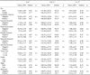
The participants in this study were 40 patients all with transitional cell carcinoma of bladder with mean age, 63±11 years, confirmed by histological studies by urologists and pathologists. The patients were referred to our laboratory from the hospitals of Shiraz University of Medical Sciences, Shiraz Iran. All patients provided their informed consent to take part in this study. Peripheral venous blood samples (2 ml) were collected by venipuncture before any clinical intervention. Data on age, tumor histology, tumor size, tumor invasion, clinical stage, histological grade, and presence of other organ metastases were obtained from the hospital records of the patients. Clinical stage was determined with the tumor-nodemetastasis classification: Primary tumor (T1=tumor invades subepithelial connective tissue, T2=tumor invades muscle, T3=tumor invades perivesical tissue and T4=tumor invades prostate, uterus or vagina and pelvic wall or abdominal wall). Lymph nodes (N1=involvement in a single lymph node 2 cm or less in greatest dimension, N2=involvement in a single lymph node 2~5 cm and N3=involvement in a lymph node more than 5 cm. Metastasis (M0=no metastasis and M1=metastasis). TNM classification and tumor diameter were obtained from pathology reports by two pathologists. In addition, tumors were graded according to the world health organization (WHO) classification criteria as well, moderately or poorly differentiated. Table I demonstrates the distribution of patients regarding different clinical criteria. The high-grade and metastatic bladder cancer patients were a small group of patients and statistically they were not able to be compared with low-grade and non-metastatic patients. Blood samples from 38 healthy individuals with mean age, 61±3 years without history of malignancies or autoimmune disorders were also obtained as control group. During sample collection, it was ensured that subjects had neither infection nor any acute or chronic disease. The mean age of the patients and healthy control were 63 and 61 years respectively. The protocol for the present study was approved by the Ethics Committee of the Shiraz University of Medical Sciences (Shiraz, Iran).
Enzyme-linked immunosorbent assay (ELISA)
The amounts of IL-6, TGF-β and IL-17 in the patients and controls sera were measured at the same time by the same technician, using ELISA-kits (eBiosciences, San Diego, CA, USA). Briefly, premixed standards were reconstituted in peripheral blood smear (PBS) (pH:7.2), generating a stock concentration of 200, 500 and 1,000 pg/ml for IL-6, IL-17 and TGF-β, respectively. Sensitivity for IL-17 was 4 pg/ml and minimal crossreactivity IL-17 to the recombinant human IL-17AF heterodimer is observed at 0.4%. Sensitivity for IL-6 and TGF-β were 2 and 8 pg/ml, respectively. The standard stocks were serially diluted in Reagent Diluent to generate 7 points for the standard curves. Diluted Capture Antibody was added to a 96-well, flat-bottomed, polystyrene microtiter plate, at final volume of 100µl. Plates were sealed and incubated overnight at room temperature, then washed with Wash Buffer. Premixed standards or samples (100µl) were added to each well containing washed beads, covered with an adhesive strip and incubated for overnight at -4 C. After incubation and washing, 100µl of the premixed Detection Antibody was added to each well and the plate was covered with a new adhesive strip and incubated for 2 hours at room temperature. After incubation and washing, Streptavidin- horseradish peroxidase (HRP) was added to each well (100µl). The incubation was terminated after 20 min at room temperature and the plates were kept away from direct light. After washing, the beads were re-suspended in 100µl of Substrate Solution. Then, 50µl of Stop Solution was added to each well, and the optical density of each well was immediately determined using a microplate reader set to 450 nm. The results were expressed in pg/ml.
Statistical analysis
The serum levels of IL-6, TGF-β and IL-17 in the peripheral blood was evaluated to the corresponding values from control samples using nonparametric kruskal-wallis and Mann-Whitney tests by SPSS software v. 11.5 (SPSS, Chicago, IL,173 USA). Finally, correlations between different cell populations were evaluated using Spearman's rank correlation coefficient. Relative levels was plotted and evaluated by means of Prism 4 software (Inc; San Diego CA, USA, 2003). p<0.05 was regarded as significant in all statistical analysis.
RESULTS
IL-6 serum level
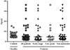
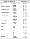
Serum level of IL-6 in patients with bladder cancer did not differ compared to the control group (10.24±2.31 vs. 6.51±0.45 pg/ml; p=0.16). These results are summarized in Table II. Although IL-6 mean in patients was higher than controls, its median was lower than healthy individuals but that was not significantly different. Also, among the patients in the early disease stages (stages I and II), low grade and non-metastasis status, the serum level of IL-6 was not different compare to controls (11.66±3.00, 5.75±1.49 and 14.82±4.26 vs. 6.51±0.45 pg/ml; p=0.33, p=0.28 and p=0.5, respectively) (Fig. 1). In addition no correlation was found between IL-6 serum level and clinicopathogical data of the cases (Table III). IL-6 was not significantly related to depth of tumor; and it is not seen significantly difference between negative and positive invasion status (p>0.05) (Table III). It seems IL-6 is not dependent to gender because there was any significant difference in IL-6 serum level between male and female patients.
TGF-β serum level
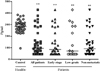
Comparison of the findings shows significantly less expression of TGF-β in all patients, early disease stages (stages I and II), low grade and non-metastatic than in healthy volunteers (Fig. 2) (153.59±24.11, 160.14±29.90, 153.24±33.77 and 150.91±25.25 vs. 283.96±8.60 pg/ml; p<0.0001, p<0.0001, p=0.002 and p<0.0001, respectively). These results are summarized in Table II. Serum level between male and female patients was similar. Like IL-6, there was no correlation between TGF-β serum level with different clinicopathological parameters (Table III). TGF-β differences between the positive and negative invasions are listed in Table III. TGF-β in patients with positive invasion to adjacent environments, was not significantly difference with other group with negative invasion (p>0.05) (Table III).
IL-17 serum level
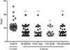
It was shown that serum level of IL-17 among patients is significantly bellow compared to control group (7.32±0.55 vs. 13.30±1.45 pg/ml; p<0.0001) (Fig. 3). We noted significant difference in the early disease stages (stages I and II), low grade and non-metastatic status compared to control volunteers (7.32±0.65, 7.07±0.73 and 7.14±0.56 vs. 13.30±1.45 pg/ml; p<0.0001, p<0.0001, and p<0.0001, respectively). These results are summarized in Table II. Nevertheless, no correlation was found between IL-17 serum level with non of clinicopathological parameters in patient group (Table III). In addition, patients in none of general characteristics showed significant difference in IL-17 serum level. In addition, IL-17 between male and female patients was similar (Table III).
DISCUSSION
In this study, serum levels of IL-17, IL-6 and TGF-β in peripheral blood of patients with bladder cancer were analyzed. The data indicated lower levels of IL-17 and TGF-β, but not IL-6, in patients compared to controls. In addition, the status of immunity was evaluated. The effects of different clinical and pathologic status on the immune response of patients were also investigated.
There are several studies in IL-17 expression in cancers. For example, Zhang et al. reported IL-17 and IL-23 expression increase in gastric tumor tissues that it may be due to Th17 cell differentiation in gastric cancer (24). According to other study, IL-17 that released by Th17, may increases inflammation and promote tumor growth because of proangiogenic factors (25). Zhu et al. announced IL-17, which is increased in breast cancer tumor, is mostly restricted to macrophages that results tumor progression and invasion (26). The expression of IL-17 mRNA was evaluated by Wang et al., who detected increased expression of IL-17 and IL-6 in tumor cells. These authors suggested that IL-17 might promote tumor cell growth through IL-17-stimulated IL-6, both in tumor cells and nonmalignant stromal cells (18). It also seems VEGF and TGF-β contribute to tumor progression by IL-17 (16). Study on ovarian cancer showed high expression of IL-17 with a significant role in tumor growth via angiogenesis (27). Although Radosavljevic et al. detected no significant IL-17 serum protein in colorectal cancer patients, IL-17 protein expression in tumor tissue was heterogeneous and it seems IL-17 be a valuable serum tumor marker in patients with colorectal carcinoma (19). There is another study that showed a prominence of Treg and Th17 cell populations in the Tumor-infiltrating lymphocytes (TILs) associated bladder cancer. It indicated that tumor-infiltrating Th17 cells expressed high levels of homing molecules CCR4 and CCR6, which might be associated with Th17 cell migration and retention within the tumor. Also, it suggested that Th17 cells, together with Treg cells, might contribute to the immunopathogenesis of bladder cancer (28). According to other study, IL-17 and IL-21 were shown to be elevated in oral and gastric squamous cell carcinomas and they can be used as a prognostic marker (29). Also Kwon et al. demonstrated no significant difference between IL-6 in control and colorectal cancer patients while Ravishankaran et al. proved against in breast cancer (30,31). Chen et al. revealed that IL-6 was over-expressed in the bladder cancer specimens compared with non-malignant tissues at both mRNA and protein levels. Their findings showed that IL-6 could be a significant predictor for clinical stage and prognosis of bladder cancer (32).
The above studies imply that IL-17 is a multifunctional cytokine which it can augment tumor growth and progression, and detection of IL-17 at the protein or transcript level is base of this concept. An important aspect, in relation to the functional activity of Th17 and emergence IL-17, is the reciprocal effect of T regulatory cells with Th17 in modification and regulation of the immune response (33,34). Thus, effect of Th17 should be considered in relation with Treg function in growth or regression of the tumors. According to this concept, finding of present study showed a reduced serum level of IL-17 and TGF-β in peripheral blood of patients that are mostly in the early stages of bladder carcinoma, which it can be interpreted as a reflection of reduced Th17 responses. Therefore, systemic recruitment of the host's immune cells to the site of a malignant transformation in bladder tissue is influenced by reduced IL-17-procucing Th17 cells. On the other hand, reduced IL-17 and TGF-β may be because of probable chemotherapy and radiotherapy.
Although there are several reports on role of inflammation and initiation of cancer, most evidences strongly support the importance of inflammation in tumorigenesis (35,36). Therefore, lower inflammation-induced IL-6 and IL-17 can be associated with lack of angiogenesis. As explained, while cancer progresses particularly in late stage phases, with increasing of Th17 development as a result of increased IL-6 and TGF-β, cancer may deteriorate.
TGF-β is known to be released both by tumor and T regulatory cells during the late stages of most solid cancers (37). Reduced TGF-β serum level in our study shows probable chemotherapy drugs suppress tumor and Treg cells.
In conclusion, tumor can effect on immune system such as cytokine profile according to current study indicates that the most patients are in low grade and early stage of bladder cancer and produce significantly low levels of IL-17 and TGF-β but not IL-6. These findings perhaps indicate probable chemotherapy and radiotherapy effects on suppress IL-17-procucing Th17 cells and TGF-β-producing tumor cells in early stage but with tumor progress, increased IL-17-producing Th17 is expected. Therefore, along with increased TGF-β in late stage of cancer, angiogenic factors cause cancer deterioration. So, current cytokine profile clinically can be used as an indicator in following of malignancies progress and immune response to cancer. Also, It is probably IL-17 and TGF-β can be used as imminent targets by monoclonal antibodies for bladder cancer immunotherapy in late stage (III, IV) that immune system is not competent versus cancer. Finally, a better understanding of the underlying mechanism regulating the Th17 Treg balance may lead to better therapeutic strategies for cancers, especially bladder cancer.

 XML Download