Journal List > Immune Netw > v.14(1) > 1033421

Park, Cho, and Park: NF-κB Activation in T Helper 17 Cell Differentiation

Abstract

CD28/T cell receptor ligation activates the NF-κB signaling cascade during CD4 T cell activation. NF-κB activation is required for cytokine gene expression and activated T cell survival and proliferation. Recently, many reports showed that NF-κB activation is also involved in T helper (Th) cell differentiation including Th17 cell differentiation. In this review, we discuss the current literature on NF-κB activation pathway and its effect on Th17 cell differentiation.

Abbreviations

RHD

rel homology domain

TAD

transactivation domain

TCR

T cell receptor

PI3K

phosphoinositide 3-kiase

PDK1

phosphoinositide-dependent kinase 1

CBM

Carma1-Bcl10-Malt1

GLK

GCK-like kinase

Th

T helper

RA

rheumatoid arthritis

IBD

inflammatory bowel disease

SLE

systemic lupus erythematous

PGE2

prostaglandin E2

INTRODUCTION

NF-κB is activated during immune responses and is important for the expression of immune response related genes including cytokine, chemokine, and adhesion molecule genes (1-3). The NF-κB family is composed of RelA, RelB, c-Rel, p50 (NF-κB1), and p52 (NF-κB2) subunits. The NF-κB transcription factor binds to κB sites as dimers, either homodimers or heterodimers. The NF-κB protein contains N-terminal Rel homology domain (RHD), which makes contact with DNA and supports subunit dimerization. Of the NF-κB subunits, only RelA, RelB, and c-Rel have transactivation domain (TAD) at C-terminus and this TAD domain is important for initiation of target gene transcription (1-3). However, p50 and p52 lack TAD domain. Thus, p50 and p52 can positively regulate gene expression through heterodimerization with TAD containing NF-κB subunits or other regulators (Fig. 1).
NF-κB complexes are inactive in most cells, and these complexes are located in the cytoplasm in a complex with inhibitory IκB proteins (IκBα, IκBβ, IκBε, IκBζ p100, p105, Bcl3, and IκBns). NF-κB pathway activating signals including cytokine receptor signals and antigen receptor signals activate the IκB kinase (IKK) complex, which phosphorylates IκB. This phosphorylation induces IκB degradation, which leads to NF-κB complex translocation to the nucleus. Once in the nucleus, the NF-κB complex activates target gene transcription (1-3).

NF-κB PATHWAY IN T CELL ACTIVATION

Antigen recognition by T cell receptor (TCR) induces activation of many transcription factors including NF-κB, NF-AT, and AP-1 (2,4), which are important for the induction of proliferation of activated T cells and their differentiation into Th1, Th2, Th17 and other Th cells (2,5). Ligation of CD28 co-receptor, along with the TCR, is essential for full activation of T cells (6,7). Especially, CD28 co-receptor ligation is required for efficient activation of NF-κB. It is well known that CD28 greatly enhances phosphoinositide 3-kinase (PI3K) activity and this activity is required for many cellular responses including cell survival, and cell proliferation (8). The CD28 cytoplasmic tail contains a PI3K binding motif such as YMNM motif (9). PI3K has been reported to bind to the YMNM phosphotyrosine, which leads to PI3K activation. CD28-mediated PI3K activation is involved in phosphoinositide-dependent kinase 1 (PDK1) and AKT recruitment into the immunological synapse (10-12), which activates PDK1 and AKT (13). These processes are important for PKCθ and Carma1-Bcl10-Malt1 (CBM) complex-mediated NF-κB activation during T cell activation (13-15). However, the concept of NF-κB activation by YNMN-mediated PI3K activation has been challenged because YNMN motif mutation had no significant effect on T cell proliferation and IL-2 production (16,17). The report suggested that another region of the CD28 cytoplasmic tail is responsible for PDK1 activation and subsequently this induces PKCθ-mediated NF-κB activation (17). A recent in vivo infection experiment suggested that another CD28 cytoplasmic tail region is responsible for NF-κB activation (10). Even though the exact CD28 cytoplasmic tail region for NF-κB activation is controversial, CD28-mediated PDK1 activation and subsequent PKCθ-mediated NF-κB activation pathway is well supported by previous studies (13,17,18).
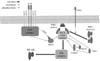
During T cell activation, CD28 recruits PDK1 into the immunological synapse where the recruited PDK1 is converted into competent states for binding to down-stream molecules such as PKCθ and Carma1 (13,19). During this process, phosphorylation of threonine 513th on PDK1 is important for the conversion of PDK1 heterotypic dimer to homotypic dimer, which enables the formation of the PDK1-PKCθ-CBM complex. In addition, recently, GCK-like kinase (GLK) was suggested as the kinase for PKCθ phosphorylation at threonine 538th (20). Thus, it is possible that PDK1 works as a scaffold for the complex formation. Subsequently, IκB kinase (IKK) is activated by the signaling complex (2), and the IKK complex activates NF-κB during T cell activation (Fig. 2).

Th17 CELLS

CD4 T cells play an essential role in the adaptive immune response. During the adaptive immune response, activated CD4 T cells differentiate into T helper (Th) 1, Th2 or Th17 effector cells. Th1 cells are important in host defense against intracellular pathogens and Th2 cells are involved in allergic immune responses and defense against parasite infections. Th17 cells play an important role in host defense against extracellular pathogens and fungal infections (21,22). Furthermore, Th1 and Th17 cells are important in intestinal immune responses. During CD4 T cell differentiation, IL-6 and TGF-β are important for Th17 cell differentiation. IL-23 is also involved in Th17 cell differentiation (23). In addition to the direct effect of IL-23 on Th17 cell differentiation, IL-23 stimulates intestinal TCRγδ T cells, invariant natural killer T cells (iNKT), and intestinal innate-like T cells to secrete cytokines related to Th17 differentiation (24). In addition to TGF-β and IL-23, IL-6 is also important for Th17 differentiation. However, while TGF-β negatively regulates human Th17 cell differentiation, this cytokine is important for Th17 cell differentiation in murine Th17 differentiation (25-27). During Th17 cell differentiation, the transcription factors RORγt, RORγ, RORα, IRF4, and STAT3 are important for effector T cell differentiation. However, IFN-γ, IL-2, and IL-4 negatively regulate Th17 cell differentiation (28,29).

Th17 CELLS IN AUTOIMMUNE DISEASE

Differentiated Th17 cells produce proinflammatory cytokines such as IL-17A, IL-17F, IL-21, TNF, and GM-CSF (5,30,31). These cytokines are important in host defense against extracellular bacteria through acute immune responses (32). In addition, Th17 cells are involved in the development of autoimmune diseases including rheumatoid arthritis (RA), inflammatory bowel disease (IBD), and multiple sclerosis (26,33).
It has been suggested that unbalanced immune responses can induce inflammatory diseases such as IBD. The detailed mechanism of IBD induction, including Crohn's disease and ulcerative colitis, has not been clarified (34-36); however, uncontrolled T cell activation and biased effector T cell (Th1, Th2, and Th17 cells) differentiation have been suggested as causative factors. Unbalanced production of Th17-related cytokines is also involved in the induction of IBD and other autoimmune disease. Th17 cells produce cytokines including IL-17A, IL-17F, IL-21, and IL-22 (37,38). IL-17 is the representative cytokine produced by Th17 cells and is involved in RA, asthma, and systemic lupus erythematous (SLE) development. A number of studies have investigated the role of IL-17A in intestinal inflammation, and showed that IL-17A is overproduced in patients with Crohn's disease and ulcerative colitis (39-42). In addition, IL-17 family cytokines are also increased in patients with autoinflammatory diseases including RA, asthma, and SLE (43-45). IL-17 and IL-23R genomic DNA sequence analysis found polymorphic regions related to IBD induction (46,47). In addition, IL-21 produced by Th17 cells was found to be involved in exacerbation of IBD (48,49). Furthermore, IL-21 gene deleted mice are resistant to Th1/Th17 cell-mediated colitis induction (50).

EXTRINSIC EFFECT OF NF-κB ACTIVATION ON Th17 CELL DIFFERENTIATION

The NF-κB pathway regulates antigen presenting cell functions and affects CD4 T cell differentiation into Th effector cells (51,52). Dendritic cells are the most important antigen presenting cells for Th cell differentiation. RelA deficiency reduced IL-1α, IL-1β, and IL-6 production from dendritic cells in response to LPS stimulation (53). In fact, these cytokines are involved in Th17 cell differentiation (25). In inflammatory responses, prostaglandin E2 (PGE2), an endogenous lipid mediator, enhances the production of IL-1β and TNF-α from bone marrow derived dendritic cells. In addition, PGE2 reduces the level of IL-12, but increases the level of IL-23 production. In addition to changes in cytokine production, PGE2 affects the expression of TLR-4, 2, and 9, IL-1R-associated kinase 1 (IRAK1), IKK, and p38 activator (MKK3), which are important components of the NF-κB activation pathway in dendritic cells. In addition, PGE2 reduces the level of IL-12, but increases the level of IL-23 production. In addition to changes in cytokine production, PGE2 affects the expression of TLR-4, 2, and 9, IL-1R-associated kinase 1 (IRAK1), IKK, and p38 activator (MKK3), which are important components of the NF-κB activation pathway in dendritic cells. Moreover, PGE2 treatment of bone marrow derived dendritic cells increases Th17 cell differentiation in vitro (54). Thus, it is suggested that NF-κB modulation in antigen presenting cells can also affect Th17 cell differentiation.

INTRINSIC EFFECT OF NF-κB ACTIVATION ON Th17 CELL DIFFERENTIATION

Binding of RORγt and RORγ to IL-17A and IL-17F promoters regulates their expression, which is important for Th17 differentiation. It has been shown that NF-κB subunits such as p65 and c-Rel are localized in the RORγt and RORγ promoter regions and affect RORγt and RORγ gene expression. Thus, it has been suggested that NF-κB activation is important for Th17 differentiation (55). Also, IKKβ can stimulate PKCθ-mediated STAT3 promoter activation. This promoter activation is essential for Th17 cells differentiation (56). During T cell activation, Malt1 is an important component of the NF-κB activation pathway through regulation of IKK activation. In in vitro conditions, naïve T cell differentiation into Th17 cells is decreased by Malt1 deficiency (57). In addition, Carma1, which is adaptor protein for TCR-mediated NF-κB activation, is needed for expressions of IL-17A, IL-17F, IL-21, IL-22, IL-23R, and CCR6. Carma1 deficiency also blocks Th17 cell development because chromatin loci of Th17 effector molecules cannot form open conformation, but transcription factors, which are needed for Th17 cells development, were normally expressed (58). In addition, overexpression of calpastatin minimal domain, which is a natural inhibitor of calpain, also decreased Th17 cell differentiation through stabilization of IκBα and subsequent inhibition of NF-κB activation (59). In addition, one recent report described an important role for IκBζ in Th17 differentiation, and showed that IκBζ acts with the nuclear orphan receptors RORγ and RORα to promote IL-17A gene expression (60). Thus, many data support the importance of NF-κB activation in Th17 cell differentiation.
However, recently, negative results were also reported. IL-2 is secreted from activated T cells, and acts as a negative regulator of Th17 cell differentiation (61). In the above report, c-Rel was suggested as a positive regulator of Th17 cell differentiation. c-Rel deficiency in T cells decreases IL-2 production from activated T cells. However, this condition did not affect Th17 cell differentiation. In the report, even though IL-2 was added in Th17 cell differentiation conditions, it did not affect Th17 cell differentiation. IRF-4 is a positive regulator of Th17 cell differentiation; however, it was not affected by c-Rel deficiency. Thus, the report argued that c-Rel is not involved in Th17 cell differentiation (62). It has been reported that USP18 regulates the TAK1-TAB1 complex, which is known as NF-κB pathway. USP18 deficient T cells showed NF-κB hyperactivation, and subsequently increased the level of IL-2 secretion. This dysregulation of NF-κB reduced Th17 cell differentiation (63).

CONCLUSION

NF-κB activation is important during T cell activation and for cytokine gene expression in antigen presenting cells. NF-κB activation-mediated Th17 cell cytokine gene expression is important for Th17 cell differentiation; however, different experimental systems showed different roles of antigen receptor-mediated NF-κB activation in Th17 cell differentiation (Fig. 3). Thus, deciphering the role of NF-κB in each of the Th17 cell differentiation conditions, such as different disease states, is an area of great interest.

Figures and Tables

Figure 1
NF-κB family. The mammalian NF-κB protein family consists of five members: p65 (RelA), RelB, c-Rel, NF-κB2 (precursor, p100; mature form, p52), and NF-κB1 (precursor, p105; mature form, p50).
in-14-14-g001
Figure 2
T cell receptor-mediated NF-κB activation. T cell receptor complex and CD28-mediated PDK1 activation are important for signaling complex formation composed of PKCθ, Carma1, Bcl10, and Malt1. This signaling complex leads to IKK complex activation and subsequently activates NF-κB during T cell activation by antigen.
in-14-14-g002
Figure 3
Effect of NF-κB activation on Th17 cell differentiation. NF-κB activation in antigen presenting cells is important for production of Th17 cell differentiation cytokines. Some research groups report that NF-κB activation in CD4 T cells positively regulates Th17 cell differentiation while others showed that NF-κB activation does not affect or negatively regulates Th17 cell differentiation.
in-14-14-g003

Abbreviations

RHD

rel homology domain

TAD

transactivation domain

TCR

T cell receptor

PI3K

phosphoinositide 3-kiase

PDK1

phosphoinositide-dependent kinase 1

CBM

Carma1-Bcl10-Malt1

GLK

GCK-like kinase

Th

T helper

RA

rheumatoid arthritis

IBD

inflammatory bowel disease

SLE

systemic lupus erythematous

PGE2

prostaglandin E2

ACKNOWLEDGEMENTS

This work was supported by a grant of the Korean Health Technology R&D Project, Ministry of Health & Welfare, Republic of Korea (A111838) and by the National Research Foundation of Korea funded by the Ministry of Education, Science and Technology (NRF-2013R1A1A2010995).

Notes

The authors have no financial conflict of interest.

References

1. Hayden MS, Ghosh S. Signaling to NF-kappaB. Genes Dev. 2004; 18:2195–2224.
2. Hayden MS, Ghosh S. NF-kappaB in immunobiology. Cell Res. 2011; 21:223–244.
3. Hayden MS, West AP, Ghosh S. NF-kappaB and the immune response. Oncogene. 2006; 25:6758–6780.
4. Schulze-Luehrmann J, Ghosh S. Antigen-receptor signaling to nuclear factor kappa B. Immunity. 2006; 25:701–715.
5. Oh H, Ghosh S. NF-kappaB: roles and regulation in different CD4(+) T-cell subsets. Immunol Rev. 2013; 252:41–51.
crossref
6. Alegre ML, Frauwirth KA, Thompson CB. T-cell regulation by CD28 and CTLA-4. Nat Rev Immunol. 2001; 1:220–228.
crossref
7. Kane LP, Lin J, Weiss A. It's all Rel-ative: NF-kappaB and CD28 costimulation of T-cell activation. Trends Immunol. 2002; 23:413–420.
8. Frauwirth KA, Riley JL, Harris MH, Parry RV, Rathmell JC, Plas DR, Elstrom RL, June CH, Thompson CB. The CD28 signaling pathway regulates glucose metabolism. Immunity. 2002; 16:769–777.
crossref
9. Pages F, Ragueneau M, Rottapel R, Truneh A, Nunes J, Imbert J, Olive D. Binding of phosphatidylinositol-3-OH kinase to CD28 is required for T-cell signalling. Nature. 1994; 369:327–329.
crossref
10. Pagan A, Pepper JM, Chu HH, Green JM, Jenkins MK. CD28 promotes CD4+ T cell clonal expansion during infection independently of its YMNM and PYAP motifs. J Immunol. 2012; 189:2909–2917.
11. Sanchez-Lockhart M, Marin E, Graf B, Abe R, Harada Y, Sedwick CE, Miller J. Cutting edge: CD28-mediated transcriptional and posttranscriptional regulation of IL-2 expression are controlled through different signaling pathways. J Immunol. 2004; 173:7120–7124.
crossref
12. Yokosuka T, Kobayashi W, Sakata-Sogawa K, Takamatsu M, Hashimoto-Tane A, Dustin ML, Tokunaga M, Saito T. Spatiotemporal regulation of T cell costimulation by TCR-CD28 microclusters and protein kinase C theta translocation. Immunity. 2008; 29:589–601.
crossref
13. Park SG, Schulze-Luehrman J, Hayden MS, Hashimoto N, Ogawa W, Kasuga M, Ghosh S. The kinase PDK1 integrates T cell antigen receptor and CD28 coreceptor signaling to induce NF-kappaB and activate T cells. Nat Immunol. 2009; 10:158–166.
crossref
14. Narayan P, Holt B, Tosti R, Kane LP. CARMA1 is required for Akt-mediated NF-kappaB activation in T cells. Mol Cell Biol. 2006; 26:2327–2336.
crossref
15. Matsumoto R, Wang D, Blonska M, Li H, Kobayashi M, Pappu B, Chen Y, Wang D, Lin X. Phosphorylation of CARMA1 plays a critical role in T Cell receptor-mediated NF-kappaB activation. Immunity. 2005; 23:575–585.
crossref
16. Garcon F, Patton DT, Emery JL, Hirsch E, Rottapel R, Sasaki T, Okkenhaug K. CD28 provides T-cell costimulation and enhances PI3K activity at the immune synapse independently of its capacity to interact with the p85/p110 heterodimer. Blood. 2008; 111:1464–1471.
crossref
17. Dodson LF, Boomer JS, Deppong CM, Shah DD, Sim J, Bricker TL, Russell JH, Green JM. Targeted knock-in mice expressing mutations of CD28 reveal an essential pathway for costimulation. Mol Cell Biol. 2009; 29:3710–3721.
crossref
18. Villalba M, Bi K, Hu J, Altman Y, Bushway P, Reits E, Neefjes J, Baier G, Abraham RT, Altman A. Translocation of PKC[theta] in T cells is mediated by a nonconventional, PI3-K- and Vav-dependent pathway, but does not absolutely require phospholipase C. J Cell Biol. 2002; 157:253–263.
crossref
19. Kang JA, Jeong SP, Park D, Hayden MS, Ghosh S, Park SG. Transition from heterotypic to homotypic PDK1 homodimerization is essential for TCR-mediated NF-kappaB activation. J Immunol. 2013; 190:4508–4515.
crossref
20. Chuang HC, Lan JL, Chen DY, Yang CY, Chen YM, Li JP, Huang CY, Liu PE, Wang X, Tan TH. The kinase GLK controls autoimmunity and NF-kappaB signaling by activating the kinase PKC-theta in T cells. Nat Immunol. 2011; 12:1113–1118.
crossref
21. Romagnani S. Lymphokine production by human T cells in disease states. Annu Rev Immunol. 1994; 12:227–257.
crossref
22. Korn T, Bettelli E, Oukka M, Kuchroo VK. IL-17 and Th17 Cells. Annu Rev Immunol. 2009; 27:485–517.
crossref
23. Ahern PP, Izcue A, Maloy KJ, Powrie F. The interleukin-23 axis in intestinal inflammation. Immunol Rev. 2008; 226:147–159.
crossref
24. Cua DJ, Tato CM. Innate IL-17-producing cells: the sentinels of the immune system. Nat Rev Immunol. 2010; 10:479–489.
crossref
25. Laurence A, O'Shea JJ. T(H)-17 differentiation: of mice and men. Nat Immunol. 2007; 8:903–905.
crossref
26. Acosta-Rodriguez EV, Napolitani G, Lanzavecchia A, Sallusto F. Interleukins 1beta and 6 but not transforming growth factor-beta are essential for the differentiation of interleukin 17-producing human T helper cells. Nat Immunol. 2007; 8:942–949.
crossref
27. Chen Z, Tato CM, Muul L, Laurence A, O'Shea JJ. Distinct regulation of interleukin-17 in human T helper lymphocytes. Arthritis Rheum. 2007; 56:2936–2946.
crossref
28. Harrington LE, Hatton RD, Mangan PR, Turner H, Murphy TL, Murphy KM, Weaver CT. Interleukin 17-producing CD4+ effector T cells develop via a lineage distinct from the T helper type 1 and 2 lineages. Nat Immunol. 2005; 6:1123–1132.
crossref
29. Park H, Li Z, Yang XO, Chang SH, Nurieva R, Wang YH, Wang Y, Hood L, Zhu Z, Tian Q, Dong C. A distinct lineage of CD4 T cells regulates tissue inflammation by producing interleukin 17. Nat Immunol. 2005; 6:1133–1141.
crossref
30. Littman DR, Rudensky AY. Th17 and regulatory T cells in mediating and restraining inflammation. Cell. 2010; 140:845–858.
crossref
31. El-Behi M, Ciric B, Dai H, Yan Y, Cullimore M, Safavi F, Zhang GX, Dittel BN, Rostami A. The encephalitogenicity of T(H)17 cells is dependent on IL-1- and IL-23-induced production of the cytokine GM-CSF. Nat Immunol. 2011; 12:568–575.
crossref
32. Cooke A. Th17 cells in inflammatory conditions. Rev Diabet Stud. 2006; 3:72–75.
crossref
33. Kramer JM, Gaffen SL. Interleukin-17: a new paradigm in inflammation, autoimmunity, and therapy. J Periodontol. 2007; 78:1083–1093.
crossref
34. Kaser A, Zeissig S, Blumberg RS. Inflammatory bowel disease. Annu Rev Immunol. 2010; 28:573–621.
crossref
35. Chebotar IV, Zaslavskaia MI, Konyshkina TM, Maianskii AN. IgG- and C3-dependent adhesion of neutrophils in systems with allogeneic and xenogeneic ligands. Biull Eksp Biol Med. 1991; 112:403–404.
36. Xavier RJ, Podolsky DK. Unravelling the pathogenesis of inflammatory bowel disease. Nature. 2007; 448:427–434.
crossref
37. Zhou L, Ivanov II, Spolski R, Min R, Shenderov K, Egawa T, Levy DE, Leonard WJ, Littman DR. IL-6 programs T(H)-17 cell differentiation by promoting sequential engagement of the IL-21 and IL-23 pathways. Nat Immunol. 2007; 8:967–974.
crossref
38. Dong C. TH17 cells in development: an updated view of their molecular identity and genetic programming. Nat Rev Immunol. 2008; 8:337–348.
crossref
39. Kobayashi T, Okamoto S, Hisamatsu T, Kamada N, Chinen H, Saito R, Kitazume MT, Nakazawa A, Sugita A, Koganei K, Isobe K, Hibi T. IL23 differentially regulates the Th1/Th17 balance in ulcerative colitis and Crohn's disease. Gut. 2008; 57:1682–1689.
crossref
40. Fujino S, Andoh A, Bamba S, Ogawa A, Hata K, Araki Y, Bamba T, Fujiyama Y. Increased expression of interleukin 17 in inflammatory bowel disease. Gut. 2003; 52:65–70.
crossref
41. Zhang Z, Zheng M, Bindas J, Schwarzenberger P, Kolls JK. Critical role of IL-17 receptor signaling in acute TNBS-induced colitis. Inflamm Bowel Dis. 2006; 12:382–388.
crossref
42. Park SG, Mathur R, Long M, Hosh N, Hao L, Hayden MS, Ghosh S. T regulatory cells maintain intestinal homeostasis by suppressing gammadelta T cells. Immunity. 2010; 33:791–803.
crossref
43. Kotake S, Udagawa N, Takahashi N, Matsuzaki K, Itoh K, Ishiyama S, Saito S, Inoue K, Kamatani N, Gillespie MT, Martin TJ, Suda T. IL-17 in synovial fluids from patients with rheumatoid arthritis is a potent stimulator of osteoclastogenesis. J Clin Invest. 1999; 103:1345–1352.
crossref
44. Wong CK, Ho CY, Ko FW, Chan CH, Ho AS, Hui DS, Lam CW. Proinflammatory cytokines (IL-17, IL-6, IL-18 and IL-12) and Th cytokines (IFN-gamma, IL-4, IL-10 and IL-13) in patients with allergic asthma. Clin Exp Immunol. 2001; 125:177–183.
crossref
45. Wong CK, Lit LC, Tam LS, Li EK, Wong PT, Lam CW. Hyperproduction of IL-23 and IL-17 in patients with systemic lupus erythematosus: implications for Th17-mediated inflammation in auto-immunity. Clin Immunol. 2008; 127:385–393.
crossref
46. Kim SW, Kim ES, Moon CM, Park JJ, Kim TI, Kim WH, Cheon JH. Genetic polymorphisms of IL-23R and IL-17A and novel insights into their associations with inflammatory bowel disease. Gut. 2011; 60:1527–1536.
crossref
47. Glas J, Stallhofer J, Ripke S, Wetzke M, Pfennig S, Klein W, Epplen JT, Griga T, Schiemann U, Lacher M, Koletzko S, Folwaczny M, Lohse P, Goke B, Ochsenkuhn T, Muller-Myhsok B, Brand S. Novel genetic risk markers for ulcerative colitis in the IL2/IL21 region are in epistasis with IL23R and suggest a common genetic background for ulcerative colitis and celiac disease. Am J Gastroenterol. 2009; 104:1737–1744.
crossref
48. Monteleone G, Monteleone I, Fina D, Vavassori P, Del Vecchio Blanco G, Caruso R, Tersigni R, Alessandroni L, Biancone L, Naccari GC, MacDonald TT, Pallone F. Interleukin-21 enhances T-helper cell type I signaling and interferon-gamma production in Crohn's disease. Gastroenterology. 2005; 128:687–694.
crossref
49. Sarra M, Monteleone I, Stolfi C, Fantini MC, Sileri P, Sica G, Tersigni R, Macdonald TT, Pallone F, Monteleone G. Interferon-gamma-expressing cells are a major source of interleukin-21 in inflammatory bowel diseases. Inflamm Bowel Dis. 2010; 16:1332–1339.
crossref
50. Stolfi C, Rizzo A, Franze E, Rotondi A, Fantini MC, Sarra M, Caruso R, Monteleone I, Sileri P, Franceschilli L, Caprioli F, Ferrero S, MacDonald TT, Pallone F, Monteleone G. Involvement of interleukin-21 in the regulation of colitis-associated colon cancer. J Exp Med. 2011; 208:2279–2290.
crossref
51. Ouaaz F, Arron J, Zheng Y, Choi Y, Beg AA. Dendritic cell development and survival require distinct NF-kappaB subunits. Immunity. 2002; 16:257–270.
crossref
52. O'Keeffe M, Grumont RJ, Hochrein H, Fuchsberger M, Gugasyan R, Vremec D, Shortman K, Gerondakis S. Distinct roles for the NF-kappaB1 and c-Rel transcription factors in the differentiation and survival of plasmacytoid and conventional dendritic cells activated by TLR-9 signals. Blood. 2005; 106:3457–3464.
53. Gerondakis S, Siebenlist U. Roles of the NF-kappaB pathway in lymphocyte development and function. Cold Spring Harb Perspect Biol. 2010; 2:a000182.
54. Khayrullina T, Yen JH, Jing H, Ganea D. In vitro differentiation of dendritic cells in the presence of prostaglandin E2 alters the IL-12/IL-23 balance and promotes differentiation of Th17 cells. J Immunol. 2008; 181:721–735.
crossref
55. Ruan Q, Kameswaran V, Zhang Y, Zheng S, Sun J, Wang J, DeVirgiliis J, Liou HC, Beg AA, Chen YH. The Th17 immune response is controlled by the Rel-RORgamma-RORgamma T transcriptional axis. J Exp Med. 2011; 208:2321–2333.
crossref
56. Kwon MJ, Ma J, Ding Y, Wang R, Sun Z. Protein kinase C-theta promotes Th17 differentiation via upregulation of Stat3. J Immunol. 2012; 188:5887–5897.
crossref
57. Brustle A, Brenner D, Knobbe CB, Lang PA, Virtanen C, Hershenfield BM, Reardon C, Lacher SM, Ruland J, Ohashi PS, Mak TW. The NF-kappaB regulator MALT1 determines the encephalitogenic potential of Th17 cells. J Clin Invest. 2012; 122:4698–4709.
58. Molinero LL, Cubre A, Mora-Solano C, Wang Y, Alegre ML. T cell receptor/CARMA1/NF-kappaB signaling controls T-helper (Th) 17 differentiation. Proc Natl Acad Sci USA. 2012; 109:18529–18534.
crossref
59. Iguchi-Hashimoto M, Usui T, Yoshifuji H, Shimizu M, Kobayashi S, Ito Y, Murakami K, Shiomi A, Yukawa N, Kawabata D, Nojima T, Ohmura K, Fujii T, Mimori T. Overexpression of a minimal domain of calpastatin suppresses IL-6 production and Th17 development via reduced NF-kappaB and increased STAT5 signals. PloS one. 2011; 6:e27020.
60. Okamoto K, Iwai Y, Oh-Hora M, Yamamoto M, Morio T, Aoki K, Ohya K, Jetten AM, Akira S, Muta T, Takayanagi H. IkappaBzeta regulates T(H)17 development by cooperating with ROR nuclear receptors. Nature. 2010; 464:1381–1385.
crossref
61. Stockinger B. Good for Goose, but not for Gander: IL-2 interferes with Th17 differentiation. Immunity. 2007; 26:278–279.
crossref
62. Visekruna A, Huber M, Hellhund A, Bothur E, Reinhard K, Bollig N, Schmidt N, Joeris T, Lohoff M, Steinhoff U. c-Rel is crucial for the induction of Foxp3(+) regulatory CD4(+) T cells but not T(H)17 cells. Eur J Immunol. 2010; 40:671–676.
crossref
63. Liu X, Li H, Zhong B, Blonska M, Gorjestani S, Yan M, Tian Q, Zhang DE, Lin X, Dong C. USP18 inhibits NF-kappaB and NFAT activation during Th17 differentiation by deubiquitinating the TAK1-TAB1 complex. J Exp Med. 2013; 210:1575–1590.
crossref
TOOLS
Similar articles