

 PDF
PDF ePub
ePub Citation
 Print
Print


INTRODUCTION
Follicular dendritic cells (FDCs) are stromal cells found in the primary and secondary follicles of the peripheral lymphoid organs (1). They are observed ectopically in the chronic inflammatory sites such as synovial tissues of rheumatoid arthritis (2). In addition to the well-known function of presenting native antigens to B cells in the germinal centers (GC) of the secondary lymphoid tissues, they are required for the survival, proliferation, and differentiation of B cells in the GC (3). Although less is known, the cellular interactions between FDC and T cells are also recognized (4). However, the cellular interactions between FDC and lymphocytes are poorly understood at the molecular level partly due to the paucity of experimental models. We have established an experimental system of GC reactions by employing FDC-like cells, HK cells (5). Using this model, we revealed that human FDCs produce prostaglandins (PGs) to regulate the cellular responses of B and T cells (4,6,7).
PG is a lipid mediator produced by the enzymatic reactions of cyclooxygenases (COXs). The immunoregulatory roles for PG are emerging (8). We have recently demonstrated that production of prostaglandin E2 and I2 is coupled with COX-2 in HK cells (9). Since we reported the inhibitory activity of IL-4 in PG production by HK cells (4), our laboratory has focused to elucidate the molecular mechanism of inhibitory IL-4 activity. IL-4 is produced by GC T cells (10).
Histone deacetylase (HDAC) is an enzyme responsible for removal of acetyl groups from histone proteins to regulate chromatin structure and gene expression. HDAC has been demonstrated to act as a negative regulator of proinflammatory gene expression in human cells (11). Thus, HDAC inhibitors are considered to stimulate proinflammatory gene expression. Regarding COX-2 expression in human cells, trichostatin A (TSA) treatment of a gastric tubular adenocarcinoma cells resulted in increased COX-2 mRNA expression (12). The presence of TSA in a bronchial epithelial cell line increased COX-2 gene expression (11), suggesting that downregulation of HDAC activity leads to the transcriptional activation of COX-2. In contrast, TSA inhibited LPS-induced COX-2 expression in human umbilical vein endothelial cells (13). Therefore, the roles of HDAC inhibitors should be investigated extensively in various experimental systems to clarify their physiological significance.
In this study, we examined the effect of HDAC inhibitors on the protein expression of COX-1 and COX-2 in HK cells. HDAC inhibitors dose-dependently attenuated COX-2 expression while they exhibited opposing effects on COX-2 expression in peripheral blood monocytes. Since IL-4 displayed a broad inhibition of COX-2 expression in HK cells, our results suggest a potential involvement of HDACs in IL-4-regulated PG production in FDC. Furthermore, our findings provide insight into the biological consequences of cellular interactions between T cells and FDC during GC reactions.
MATERIALS AND METHODS
Culture of HK cells and monocytes
HK cells and peripheral blood monocytes were prepared as described previously (14). Cells were maintained in RPMI-1640 (Irvine Scientific, Santa Ana, CA) containing 10% fetal calf serum (Hyclone, Logan, UT), 2 mM L-glutamine (Invitrogen, Carlsbad, CA), 100 U/ml penicillin G (Sigma-Aldrich, St. Louis, MO), and 100 µg/ml streptomycin (Invitrogen). LPS, trichostatin A (TSA), and sodium butyrate (NaB) were purchased from Sigma-Aldrich. Recombinant IL-4 was prepared in our laboratory (15). TNF-α and TGF-β were purchased from R&D Systems (Minneapolis, MN). The viability of HK cells was determined colorimetrically using Cell Counting Kit-8 (CCK-8) reagents (Dojindo Molecular Technologies, Inc., Santa Clara, CA) according to the manufacturer's instructions.
Immunoblotting
The whole cell lysates of HK cells or monocytes were subject to immunoblotting as previously described (14). The protein concentrations of the each fraction were assayed with a bicinchoninic acid (BCA) assay. Used antibodies were anti-COX-1, anti-COX-2 (Cayman Chemical, Ann Arbor, MI), anti-β-actin (Sigma-Aldrich), and horseradish peroxidase (HRP)-conjugated anti-mouse IgG (Jackson Immunoresearch, West Grove, PA). The membranes were incubated with SuperSignal West Pico Chemiluminescent Substrate (Pierce, Rockford, IL) and exposed to X-ray films.
RESULTS AND DISCUSSION
IL-4 inhibits COX-2 expression in HK cells stimulated with various stimuli
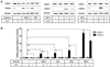
Since our first demonstration of the inhibitory activity of IL-4 in PG production by HK cells stimulated with TGF-β, LPS, or TNF-α (4), we have explored the underlying molecular mechanisms. Based on the critical role for COX-2 in PG production, we examined whether COX-2 expression would be inhibited by IL-4. Indeed, IL-4 repressed mRNA and protein levels of COX-2 that were stimulated by TGF-β (16) and LPS (14). In this study, we further examined whether TNF-α-induced COX-2 expression would undergo the similar inhibitory mechanism by IL-4. The optimal concentrations of cytokines and LPS were determined by examining their effect on COX-2 expression. Consistent with previous results, TGF-β or LPS increased COX-2 expression levels by approximately 6-fold, which was significantly suppressed by IL-4 almost to basal levels (Fig. 1). Enhancing effect of TNF-α on COX-2 was dramatic because approximately 30-fold increase of COX-2 levels was obtained by TNF-α treatment. IL-4 also inhibited TNF-α-induced COX-2 induction, which was reduced by 30%. Similar results were observed with another cytokine IFN-γ. However, IL-4 alone did not increase COX-2 expression levels. The augmentation by TGF-β, LPS, TNF-α, and IFN-γ was specific to COX-2 because COX-1 levels were not significantly modulated by these stimuli. Considering that IL-4 is the typical Th2 or Tfh cytokine expressed by GC T cells (17), the common inhibitory activity of IL-4 in COX-2 expression that are stimulated by different stimuli, i.e., LPS, TGF-β, TNF-α, and IFN-γ, implies a dominant regulatory role of Th2 or Tfh cells in PG production over Th1 (TNF-α and IFN-γ), Treg (TGF-β), or bacterial infections (LPS). The importance of Treg in GC reactions has been recently suggested by different investigators (18,19). Since the prolonged production of PG may contribute to the transition of acute inflammation into chronic one, IL-4 appears to be highly important to the resolution of acute inflammation. In line with this speculation, IL-4 was shown to abrogate TNF-α-mediated PGE2 production in synovial fibroblasts (20).
HDAC inhibitors display opposing effects on HK cells and monocytes in COX-2 expression
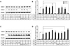
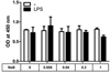
In as much as IL-4 inhibited COX-2 expression that was induced by various stimuli, we reasoned that IL-4 may regulate COX-2 expression by a global mechanism such as chromatin remodeling. Chromatin structure is modified by alterations of acetylation, methylation, or phosphorylation (11). As an initial approach to elucidate the inhibitory mechanism of IL-4 in PG production, we examined the effect of HDAC inhibitors on COX-2 expression levels. Considering their wide-spread uses as HDAC inhibitors (21), we chose TSA and NaB and analyzed their effects on the protein expression levels of COX-2 that were induced by LPS. Both TSA and NaB decreased COX-2 expression levels in dose-dependent manners (Fig. 2A and B). Low concentrations of TSA and NaB up to 10 nM and 0.1 mM, respectively, were ineffective whereas 100 nM and 1 mM of TSA and NaB, respectively, markedly suppressed COX-2 to basal levels. However, COX-1 levels were not significantly altered by these inhibitors. In order to exclude the possibility that the inhibitory effect of HADC inhibitors on COX-2 had resulted from their potential cytotoxic effect on HK cells, we examined whether NaB would affect HK cell growth. The viability of HK cells was not modulated by up to 1 mM of NaB. The growth of LPS-stimulated HK cells was also refractory to NaB treatment (Fig. 3), suggesting that the reduced COX-2 levels observed in the presence of HDAC inhibitors was not due to their cytotoxic effect.
Since FDCs are derived from mesenchymal stem cells whereas monocytes from hematopoietic stem cells (22), we examined the effect of TSA and NaB on COX-2 expression in peripheral blood monocytes. Interestingly, both TSA and NaB did not inhibit but rather augmented COX-2 expression (Fig. 2C and D). TSA and NaB slightly but significantly enhanced expression levels of COX-2 at 10 nM and 1 mM, respectively. We previously demonstrated a difference between FDCs and monocytes. COX-2 expression in monocytes is down-regulated by IL-10 but not in FDC/HK (14). This study reveals another difference between FDC and monocytes since they display opposing responses to HDAC inhibitors in COX-2 expression.
Our findings that both IL-4 and HDAC inhibitors suppress COX-2 expression imply post-translational modifications of histone proteins as an inhibitory mechanism exerted by IL-4. Whether IL-4 indeed modifies chromatin structures around the COX-2 gene or not, it should be clarified by future studies. Given the growing interests in HDAC inhibitors in cancer chemotherapy (20), their complicated activities in various cell types raise cautions on their clinical use. Those reagents may have serious effects on inflammation and innate immune responses.
Conclusion remarks
This study is an extension of our previous reports that demonstrated an inhibitory activity of IL-4 in PG production. The distinct effect of HDAC inhibitors to suppress COX-2 expression suggests that PG production by FDC may be regulated via chromatin remodeling and implies that such a mechanism may be triggered by IL-4. Therefore, our results provide insight into the role of cellular interactions between T cells and FDC during GC reactions.
 XML Download