

 PDF
PDF ePub
ePub Citation
 Print
Print


Abbreviations
APCs
antigen-presenting cells
DCs
dendritic cells
CTLs
cytotoxic T lymphocytes
PLGA
poly[lactide-co-glycolide]
FDA
food and drug administration
γ-PGA-Phe
poly(γ-glutamic acid)-graft-L-phenylalanine ethyl ester
PAMPs
pathogen associated molecular patterns
OVA
ovalbumin
IFN-γ
interferon-gamma
DOTAP
1,2-dioleoyl-3-trimethylammonium-propane
BMDCs
bone marrow DCs
ROS
reactive oxygen species
ERK
extracellular signal-regulated kinase
TLR
toll-like receptor
CLRs
c-type lectin receptors
AuNP
gold nanoparticles
INTRODUCTION
Nanoparticle based delivery platforms hold great potential for cancer immunotherapy (1-3). Over the past decade, nanosized carriers such as virus-like particles (4), liposomes (5), polymeric nanoparticles (6), and non-degradable nanospheres (7) have attracted attention as potential delivery carriers for vaccine antigens. These carriers can stabilize vaccine antigens and independently act as adjuvants. In addition, these nanoparticle systems facilitate entry into antigen-presenting cells (APCs) by different pathways, modulating immune responses to antigens (8,9). This modulation play a critical role for the induction of protective Th1-type immune responses to intracellular pathogens.
In cancer immunotherapy, nanoparticles allow vaccine delivery to immune cells (10-12). In particular, vaccination based on a dendritic cells (DCs) platform has been used for cancer immunotherapy as a promising cell based therapeutic modulator, which successfully deliver tumor specific antigens to lymphatic organs and enhances the immune response for cytotoxic T cell (13,14). However, DCs based vaccination has been limited for cancer eradication by insufficient tumor antigen uptake by DCs. Therefore, to increase antigen uptake by DCs, nanoparticle based antigen delivery systems have been explored (5,15,16).
While nanoparticle based vaccination has been efficacious for some cancers, effective vaccine delivery systems remain to be developed for many other cancers. The use of nanoparticle systems as a vaccine carrier results in increased antigen delivery efficiency to targeted immune cells, which can play a feasible role for increasing immune responses to immune cells (17).
More recently, nanoparticle based vaccine delivery has been studied as a potential carrier system to overcome limitations that include inherent instability of soluble macromolecules, low internalization of soluble materials, and insufficient cross-presentation to cytotoxic T lymphocytes (CTLs) (18). Moreover, most nanoparticles used for immunotherapy are particularly attractive for clinical and biological applications due to their low immunogenicity, low toxicity, biocompatibility, and biodegradability. The vaccine antigen can be either encapsulated within or conjugated on the surface of nanoparticles by chemical modification (19,20). By antigen encapsulation, nanoparticles provide an effective method for delivering antigens, which may otherwise degrade rapidly upon injection or reduce the immune response. Conjugation of antigens on the surface of nanoparticles can allow presentation of the immunogen to immune systems, leading that the pathogen will provoke a similar response (16). Additionally, nanoparticles made from some composites enable not only site directed delivery of antigens but also the sustained release of antigens to maximize exposure to the immune system. Here, we review the current nanoparticle platform technologies for cancer immunotherapy.
Nanotechnology and nanomedicine
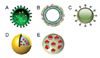
Nanotechnology encompasses the design, synthesis, characterization, and application of materials and devices less than 1 micron in diameter (21). Usually nano-sized objects are 100~10,000 times smaller than the size of mammalian cells (22). One of nanotechnologies, nanomedicine concerns the use of specifically designed materials to develop novel therapeutic and diagnostic modalities (Fig. 1) (23). In addition, nanoparticles are particularly attractive for clinical and biological applications due to their low immunogenicity, low toxicity, and biocompatibility, creating nanoparticles with high therapeutic payloads. Moreover, nanoparticle based approaches hold great potential for cancer immunotherapy as a potent vaccine carrier. The use of nanoparticles may allow the development of a broad armamentarium of targeted drugs against specific immune cells. This system will overcome delivery mediated hurdles that are difficult to address with other traditional approaches such as small molecules or monoclonal antibodies. A desirable delivery system should lead to increased concentrations of therapeutic payloads at target sites, and should ultimately raise the therapeutic index. Delivery of immunomodulatory agents across cell membranes in vivo has been achieved using specifically designed delivery systems incorporating liposomes, nanoparticles and polymeric emulsions through the chemical modification of the surface.
With the aim of achieving targeted delivery, various receptors on the surface of immune cells have been investigated as target binding sites to achieve selective delivery, which has been considered for in vivo and in vitro delivery of nanoparticles to target immune cells. The specific ligand against receptor on immune cells is overexpressed in a wide range of the immune cells, and is largely absent in normal tissues, which is a desirable feature for selective delivery.
The use of nanoparticles for delivery of immunomodulatory agents
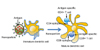
Nanoparticle based delivery system is a promising approach to enhance the efficiency of antigen delivery for cancer immunotherapy. Recent advances in nanoparticle systems for cancer immunotherapy have provided diverse groups of synthetic particles with defined cellular and biological functions (24-27). Liposomes and polymeric particles as well as virus and virus-like particles have been used to facilitate antigen delivery, with concurrent delivery of antigens and adjuvant serving to enhance immune responses to subunit vaccines. Nanoparticle based carriers have been shown sustained release of antigens at target sites, oriented antigen and/or adjuvant presentation, multivalent presentation, and specific targeting. The potential for encapsulated and sustained release of antigen within cells has been proposed to increase antigen-presentation by DCs (Fig. 2). Sustained release of antigen from particles can induce strong protection, eliminating the need for repeated doses of the vaccine (priming-boosting). Several studies have reported that particulate delivery systems could enhance the uptake of antigens and adjuvants by DCs and result in better immune responses compared to the soluble counterparts (28-30).
Shen et al. (31) assessed antigen uptake and CD8+ T cell activation in DCs treated with soluble antigen, particles with surface-modified poly[lactide-co-glycolide] (PLGA) or anitigen encapsulated PLGA nanoparticles. PLGA is a biocompatible and biodegradable material that has been approved as an in vivo substitute to polymeric matrix by the United States Food and Drug Administration (FDA) (32). Antigen encapsulation into PLGA nanoparticles resulted in increased cellular uptake of antigen and induced T cell responses. The mechanism of antigen delivery involved cross-presentation. While macropinocytosis of soluble antigen leads to poor MHC class I presentation by antigen presenting cells (APCs), phagocytosis of particle-loaded antigen enhances cross presentation, leading to potent CTL responses.
In addition, while most vaccines require addition of adjuvants to induce successful immune responses, nanoparticle based vaccines can induce immune responses without additional adjuvants. Shima et al. (33) reported that nanoparticles composed of amphiphilic poly (γ-glutamic acid)-graft-L-phenylalanine ethyl ester (γ-PGA-Phe) can be used to evaluate the effect on vaccine carriers on the antigen encapsulation behavior, cellular uptake, activation of dendritic cells, and induction of antigen-specific cellular immunity-based immune responses. These nanoparticles could efficiently encapsulate antigens and the uptake amount of the encapsulated antigen by DCs was induced. Fabrication of nanoparticles in geometries resembling pathogens and the ability to orient pathogen-relevant danger signals on the nanoparticle surface activate APCs and stimulate nanoparticle uptake. Reddy et al. (34) had developed pluronic-stabilized polypropylene sulfide nanoparticles, which activated the complement cascade, generating a danger signal in situ and potently activating DCs. Multivalent presentation of pathogen associated molecular patterns (PAMPs) by nanoparticles recreates the repetitive presentation by live pathogens, leading to an enhanced immune response through receptor cross-linking and immune-cell activation.
Physicochemical properties of nanoparticles on cellular responses
Uptake of nanoparticle loaded antigens by DCs highly depends on physicochemical properties of nanoparticles including size, shape, surface charge, hydrophobicity, and hydrophilicity. These are important parameters that influence biodistribution, cellular interactions, and cellular infiltration. Altered electrostatic or receptor-binding properties facilitate improved interaction with DCs compared to soluble antigens (35). Foged et al. (36) reported that an optimal particle size for uptake by human blood-derived DCs was under 0.5µm. Particle size also has affects the APCs. Particles traffic to the draining lymph node in a size-dependent manner. Large particles (500~2,000 nm) are taken up by peripheral APCs at the injection site, while small nanoparticles (20~200 nm) are internalized in DCs and macrophages residing in lymph nodes. Smaller nanoparticles can independently diffuse across the interstitium and penetrate the lymphatic system, while delayed transport of larger nanoparticles to lymph nodes supports a requirement for cell-based transport. Particles of 40~ 50 nm in size have been shown to elicit stronger T cell responses. Covalent conjugation of antigens including ovalbumin (OVA) and human papilloma virus peptide E7 to the nanoparticles reportedly allows for antibody and CD8+ T cell immunity and provides protective immunity upon animal challenge with OVA expressing EL4 cells. Also, the interferon-gamma (IFN-γ) secretory Th1 response is greater for OVA presented on particles 40~50 nm in size compared to other sizes. In contrast, IL-4 secretory Th2 responses are greatest when OVA is presented using particles 93~123 nm in size (37).
The stability of nanoparticle is also an important factor for the rate of drug-release. Both liposomes and polymers such as polylactides PLA and PLGA are rapidly hydrolyzed in the body. PLGA particles have slower antigen release kinetics compared to liposomes. Mice vaccinated with ex vivo stimulated splenocytes from PLGA particles displayed higher IFN-γ secretion compared to splenocytes from liposome. Therefore, kinetics of sustained release from PLGA particles compared to liposomes was thought to account for more effective in vivo CD8+ T cell responses. An anticipated advantage of sustained release is single-inoculation therapy rather than treatment involving priming and boosting injections. The importance of understanding release and degradation kinetics, as well as metabolic products is also exemplified by the potential for particle-degradation to influence the immune response.
The surface charge of nanoparticles profoundly affects the internalization capability. This is due to the negative charge of the cell membrane, which increases the affinity for positively charged molecules (38). Additionally, cationic charged nanoparticles can enhance DC uptake compared to negative charged particles through electronic binding. The inherent adjuvant of nanoparticles is exemplified by cationic liposomes composed of the cationic lipid 1,2-dioleoyl-3-trimethylammonium-propane (DOTAP), which leads to activation of mouse bone marrow DCs (BMDCs). Yan et al. (39) reported that DOTAP stimulates a concentration-dependent manner generation of reactive oxygen species (ROS) in BMDCs, which in turn lead to activation of extracellular signal-regulated kinase (ERK) and p38, cytokine/chemokine production, and expression of the B7 costimulatory molecules CD80 and CD86. However, elevated levels of DOTAP also lead to cytotoxicity and subsequent cell death. Within 2 hours, 8%, 68%, and 91% of cells treated with 50, 200, and 800µM DOTAP were double-positive for annexin V and ROS.
Particle shape is another key parameter in biodistribution, cellular uptake, and toxicity. Venkataraman et al. (40) reported a comprehensive summary of the influence of nanostructures with different shapes on important biological processes in drug delivery. Non-spherical particles possess drug loading capacities and biological behaviors that frequently deviate from their historically well-studied spherical counterparts, which have been shown to be advantageous for improving blood circulation time and organ distribution, and avoiding premature clearance by phagocytosis in various situations. Non-spherical particles are taken up by cells avidly than spherical particles with a negative correlation evident between aspect ratio and uptake rate. This was attributed to the larger average curvature radius of adsorbed non-spherical particles experienced by the cells (41).
Targeted delivery of nanoparticles
Surface modification of nanoparticles can be used to enhance the circulation time in the bloodstream, reduce nonspecific distribution, and increase target selective delivery (42). Many ligands are currently being assessed for their capacity to functionalize nanocarriers, including peptides, antibodies, proteins, polysaccharides, glycolipids, glycoproteins, and lectins. Some ligands make use of mononuclear phagocyte characteristic receptor expression and phagocytic innate processes (43). Nanoparticles can provide direct intracellular access, facilitating engagement of the intracellular Toll-like receptor (TLR)3, 7, 8, and 9 by their ligands, improving their efficacy as vaccine adjuvant (44). Targeting of specific cell populations with nanoparticles has been described as passive or active delivery. Passive targeting is influenced by intrinsic particle properties that include size, charge, and rigidity. Physiological factors influencing particle trafficking and tissue-specific accumulation include lymphatic and hemodynamic forces, diffusive mechanisms, and epithelial/endothelial permeability. On the other hand, active targeting involves the addition of ligands or surface modification of the nanoparticles exterior to direct cellular interactions by ligand-receptor binding. The presence of microbial surface antigens on particles can facilitate cellular uptake by DCs through recognition and activation of surface receptors. For example, C-type lectin receptors (CLRs) for sugar moieties like mannose and TLRs for PAMPs can lead to receptor-mediated endocytosis. Receptor binding (CLRs, TLRs, and cytokines) may also induce DC maturation, achieving both uptake and immune cell activation. Other receptor targets include integrins, CD40, and CD11c, which have been targeted using specific antibodies. The impact of ligand-receptor interactions on the cellular surface has been also considered (18). For example, targeting TLR with ligands results in DC maturation, which favors Th1 responses. Lipopolysaccharide (LPS) and pathogen-derived lipopeptides can interact with cell-surface TLRs, resulting in the production of type 1 IFN and proinflammatory cytokines, as well as enhanced surface expression of co-stimulatory molecules (45).
Multifunctional nanoparticles
The advantages of nanoparticles for cancer immunotherapy include rapid phagocytosis by immune cells and the ability to create hybrid platforms that allow diverse functions. Moreover, multifunctional nanoparticles have been explored for various in vivo applications because of their diverse and unique physicochemical and functional properties. Nanoparticles are attractive for delivering antigens into DCs because their large surface area allows the immobilization of multiple therapeutic agents. Bimodal nanostructures that contain either fluorescent chemicals or quantum dots as well as super paramagnetic iron oxide nanoparticles have been used to label DCs for both optical and magnetic resonance imaging. Cho et al. (15) reported that multifunctional super paramagnetic iron oxide nanoparticles provide exceptional contrast for lymphoid tissues and provides high-resolution in vivo images, and can target DCs to deliver tumor antigen to generate potent CTLs and CD4+ helper T cells. In addition, a nanoparticle based approach allows efficient antigen delivery to lymph nodes through lymphatic vessels in a size-dependent manner. Lee et al. (46) reported gold nanoparticles (AuNP)-based cancer vaccines to increase immune response for toll-like receptor 9 (TLR-9) activation of DCs. Antigen delivery to lymph nodes can be tracked using computed tomography (CT) imaging. Finally, proteins, peptides, and oligonucleotides can be easily attached to the surfaces of gold nanoparticles by simple chemistry.
Nanoparticles are especially advantageous since they can be used to simultaneously encapsulate various antigens. Liu et al. (32) reported a nanoparticle-based multi-adjuvant whole cell tumor vaccine for cancer immunotherapy. They demonstrated the satisfactory effects on tumor growth inhibition, metastasis inhibition, and recurrence prevention. They also applied PLGA nanoparticles as a carrier of whole cell tumor vaccine and demonstrated efficient inhibition of tumor growth. Generally, an immune response is a multi-step programed process involving, DC recruitment, antigen presentation, and T-cell activation. These nanoparticles can boost immune cells in a single step. Therefore, development of multifunctional nanoparticles has a potential for effective delivery strategies to enhance immune response for immune cells.
CONCLUSION
This review highlights the potential of nanoparticles for use as cancer vaccines to target tumor antigens and as an adjuvant to DCs for priming antigen-specific T cell responses. The use of nanoparticles to deliver immuomodulatory agents will benefits various immunological diseases. Nanoparticles can easily encapsulate target antigen, protein, peptide, or combined with chemo-drugs and provide sustained release of the therapeutic payload into immune cells after penetration, obviating the need for repeated doses of the vaccine. Surface functionalization makes it possible to orient pathogen-relevant danger signals on the particle surface and enables multivalent presentation of antigens, mimicking repetitive presentation by live pathogens and leading to enhanced antigenicity through receptor cross-linking and immune-cell activation.
Development of hybrid-nanoparticle platforms is advantageous over single-particle constructs on the basis of co-delivery of multiple therapeutic payloads to the same target cell, shielded delivery of secondary nanoparticles, and the potential for dual-site intracellular targeting and subsequent antigen processing by both the MHC class I and II pathways.
Nanoparticle platform-based immunotherapy increases the selective immune response of immunological payloads. A nanoparticle platform is a novel and highly selective delivery system for immunological payloads with the potential for broad applications in human immune disease. While nanoparticle technologies can be useful for delivery of payloads to immune cells, additional selective delivery approaches may be useful. Nevertheless, the nanoparticle-based delivery strategy has broad potential as a delivery platform in human disease and could be adapted for other presently incurable diseases.
 XML Download