

 PDF
PDF ePub
ePub Citation
 Print
Print


INTRODUCTION
Lactoferrin (LF), an 80 kDa glycoprotein and a member of a transferrin family, is secreted from many mucosal epithelial cells and neutrophils during inflammation (1). It is a key factor in the host defense system (2,3). Although the mechanisms of LF has not been fully elucidated, there are evidences that LF directly affects antigen presenting cells (APCs) in activation, migration and inflammation (4,5).
APCs such as macrophages and dendritic cells (DCs) directly enhances B cells proliferation and differentiation (6-8). BAFF, known as THANK, BLYS, TALL-1 and zTNF4, belongs to the TNF family and is derived from APCs. It plays an important role in B cell survival, maturation, and Ig class switching recombination (CSR) (9-11). Nevertheless, it is unknown if LF modulates macrophages to express BAFF, which in turn has effect on B cells.
In this study, we explored the effect of LF on BAFF expression by mouse macrophages to ask if LF indirectly regulates B cell differentiation by influencing macrophages. We found that LF can stimulate macrophages to express BAFF through the Smad3/4 pathway.
MATERIALS AND METHODS
Reagents
Bovine lactoferrin was supplied by Morinaga Milk Co., Ltd. (Zama, Japan), and anti-bovine LF antiserum was purchased from Bethyl laboratories, Inc. (Montgomery, TX, USA). Recombinant human TGF-β1, pan specific anti-TGF-β Ab, and anti-mouse BAFF Ab were purchased from R&D Systems (Minneapolis, MN, USA). ABTS was obtained from Sigma Chemical Co. (St. Louis, MO, USA).
Mice
BALB/c mice were purchased from Orient Co. Ltd (Gyeonggi-do, Korea). They were maintained on an 8:16-h light:dark cycle in an animal environmental control chamber (Daehan Biolink. Co., Incheon, Korea). Animals were fed Purina Laboratory Rodent Chow 5001 ad libitum. Eight- to twelve-week-old mice were used in this study. We followed the animal care of the institutional guidelines of Kangwon National University.
Culture of macrophages
Murine macrophage cell line RAW264.7 was purchased from ATCC (Manassas, VA, USA) and cultured in Dulbecco's modified Eagle's medium (2 mM L-glutamine; 100 U/ml penicillin; 100µg/ml streptomycin) plus 10% fetal bovine serum (HyClone Labs, Logan, UT, USA) in a humidified CO2 incubator. To prepare peritoneal macrophages, mice were injected with 3 ml of 4% thioglycolate broth in PBS intraperitoneally. After 72 h, peritoneal cells were harvested by peritoneal lavage with PBS containing 2% FBS, washed twice with warmed HBSS, resuspended in 10% FBS-DMEM, and dispensed in 100-mm culture dishes. Cells were incubated in a CO2 incubator for 2 h at 37℃, and adherent cells were used as peritoneal macrophages.
Cell viability assay
The cell viability after treatment with LF was measured by Cell Counting Kit-8 (CCK-8) reagent (Dojindo Laboratories, Tabaru, Japan). RAW264.7 cells were suspended at a final concentration of 1×10⁴ cells/well and cultured in 96-well microplate. After exposure to LF for different periods of time (0, 12, 24, 48 h), CCK-8 (10µl) was added to each well of 96-well microplate and the plate was incubated for 1 h at 37℃. Viable cells were counted by absorbance measurements at 450 nm using a VERSAmax ELISA reader (Molecular Devices, Sunnyvale, CA, USA). The viable cell number was determined using the absorbance value of a previously prepared calibration curve. All experiments were performed in triplicate on three separate occasions.
RNA preparation and RT-PCR
RNA preparation, reverse transcription, and PCR were performed as described before (12). PCR primers were synthesized by Bioneer Corp. (Seoul, Korea). The primers for mouse BAFF were forward primer 5'-GCC GCC ATT CTC AAC ATG AT-3' and reverse primer 5'-TTA GGG CAC CAA AGA AGG TG-3' (product size: 468, 409 bp). All reagents for RT-PCR were purchased from Promega (Madison, WI, USA). BAFF products were amplified 28 cycles and resolved by electrophoresis on 1.5% agarose gels. PCR was also performed with β-actin to allow for normalization of cDNA concentrations in each set of samples. Band intensities were quantified using KODAG GL 100 Image system (Biostep GmbH, Meinersdorfer, Germany).
ELISA for mouse BAFF
BAFF retained in culture supernatants was detected by ELISA. Anti-mouse BAFF Ab (2µg/ml) in bicarbonate buffer (pH 9.3) was added to 96 well microplates. After incubation overnight at 4℃, the plates were washed and blocked with 1% gelatin for 1 h. Supernatant samples (50µl) or recombinant mouse BAFF (200 ng/ml) diluted in 0.5% gelatin were added to the wells. After incubation for 1 h at 37℃, the plates were washed again, and 2µg/ml monoclonal anti-mouse BAFF Ab was added for 1 h at 37℃. Then, the plates were washed and incubated with goat anti-rat IgG-HRPO (1:500) for 1 h. After washing, 0.2 mM ABTS was added to the wells, and 10 min later, the colorimetric reaction was measured at 405 nm with a VERSAmax ELISA reader.
Plasmids and transfection
Mammalian expression vectors, Smad3 and Smad4 were generously provided by Dr. Masahiro Kawabata (13). The DN-Smad3 plasmid was provided by Dr. M. Kato (Department of Biochemistry, The Cancer Institute, Tokyo, Japan) (14).
RAW264.7 cells were transfected using FuGeneHD according to the manufacturer's protocol (Promega Corp., Madison, WI, USA).
RESULTS AND DISCUSSION
Effect of Latoferrin on BAFF expression by mouse macrophages
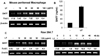
It is well known that macrophage has influence on B cell response, activation and differentiation directly (8). In particular, BAFF is believed to be the most important macrophage-derived B cell-activating factor (6). We have previously shown that TGF-β1 stimulate mouse macrophage to express BAFF (15). Though LF has immuno-modulatory effects on macrophage (5), it is not known if the action of LF on macrophages is linked to B cell activation and differentiation. To test the possibility that LF regulates BAFF expression in mouse macrophages, we examined the effect of LF on BAFF expression. First, LF increased BAFF transcription in the mouse peritoneal macrophages and sixty microgram per milliliter was optimal (Fig. 1A). We next examined the effect of LF on BAFF expression at the protein level. As shown Fig. 1B, LF increased the secretion of BAFF protein in mouse peritoneal macrophages. Consistently, LF also induced BAFF transcription in a macrophage cell line, RAW 264.7 and BAFF transcripts was detectable 12 h at the earliest after stimulation (Fig. 1C). These results clearly show that LF stimulates mouse macrophage to express BAFF.
Effect of LF on cell viability in mouse macrophage cell line
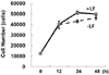
We asked whether LF-induced BAFF expression was related to cell viability. Cell numbers were virtually the same in the presence and absence of LF. LF increased viable cell number by 20% at 24 h after incubation (Fig. 2). These results indicate that the increase of BAFF by LF is not attributed to its effect on cell proliferation.
LF is not contaminated with TGF-β and acts without induction of TGF-β
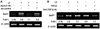
To evaluate the purity of LF used in this study, we first examined it by using anti-bovine LF Ab. As shown in Fig. 3A, goat anti-LF antiserum completely abrogated the LF-induced BAFF transcription, whereas normal goat serum had no effect. These results indicate that LF is not contaminated at all. LF used in this study was extracted from bovine milk. It is well known that TGF-β is abundant in the milk. In addition, we have previously shown that TGF-β1 stimulates mouse macrophage to express BAFF (15). Thus, it was necessary to examine if TGF-β is involved in LF-induced BAFF expression. We have treated LF with anti-TGF-β Ab before and after adding it to the cultures. We found that anti-TGFβ Ab did not inhibit the LF-induced BAFF expression (Fig. 3B). These results suggest that LF was neither contaminated with TGF-β nor acted via TGF-β.
Involvement of Smad3 and Smad4 in LF-induced BAFF expression
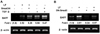
In the previous study, we have demonstrated that Smad3 and Smad4 are critical mediators of TGFβ1-induced BAFF expression in mouse macrophages (15). Since LF is also known to activate Smad pathway (16), we were interested in the involvement of Smad pathway in LF-induced BAFF expression. Overexpression of Smad3 and Smad4 significantly increased LF-induced BAFF transcription (Fig. 4A), whereas overexpression of dominant negative Smad3 (DN-Smad3) completely abolished its effect (Fig. 4B). These results are largely consistent with our early finding that TGF-β induces BAFF in such cell line (15). Therefore, these results demonstrate that LF also induces BAFF expression via Smad3 and Smad4 pathway. We are currently examining the mechnaisms by which Smads mediate LF-inducible BAFF expression in mouse macrophages.
In conclusion, our in vitro studies raise the possibility that lactoferrin cause macrophages to produce BAFF and so have an important effect on Ig isotype switching in vivo.
 XML Download