Abstract
Silica nanoparticles, which are applicable in many industrial fields, have been reported to induce cellular changes such as cytotoxicity in various cells and fibrosis in lungs. Because the immune system is the primary targeting organ reacting to internalized exogenous nanoparticles, we tried to figure out the immunostimulatory effect of silica nanoparticles in macrophages using differently sized silica nanoparticles. Using U937 cells we assessed cytotoxicity by CCK-8 assay, ROS generation by CM-H2DCFDA, intracellular Ca++ levels by staining with Fluo4-AM and IL-8 production by ELISA. At non-toxic concentration, the intracellular Ca++ level has increased immediately after exposure to 15 nm particles, not to larger particles. ROS generation was detected significantly in response to 15 nm particles. However, all three different sizes of silica nanoparticles induced IL-8 production. 15 nm silica nanoparticles are more stimulatory than larger particles in cytotoxicity, intracellular Ca++ increase and ROS generation. But IL-8 production was induced to same levels with 50 or 100 nm particles. Therefore, IL-8 production induced by silica nanoparticles may be dependent on other mechanisms rather than intracellular Ca++ increase and ROS generation.
The application of nanomaterials is becoming more popular in various fields of industry. Among nanomaterials with different chemical natures, silica nanoparticles are widely applicable nanomaterial. They can be used in chemical mechanical polishing, as additives, and recently in some medical applications (1-4). However, when applying as industrial products, the toxicity of each nanoparticle should be analyzed carefully based on their physical and chemical characteristics. Our group has studied the immunotoxicity or inflammatory effect of silver nanoparticles intensively and published papers reporting internalization mechanisms (5), inflammasome induction and further IL-1 production (6) in human blood monocytes after treatment with silver nanoparticles. Also the results of cDNA microarray analysis and protein analysis by enzyme-linked immunosorbent assay (ELISA) or western blot suggested that some stress genes, heme oxygenase-1, heat shock proteins and superoxide dismutase, have increased 2 h after exposure to silver nanoparticles in macrophages after exposure to silver nanoparticles (7). Interestingly, among inflammatory cytokines, IL-8, the neutrophil chemokine, is the earliest inducible gene (7,8). This immunostimulatory effect of silver nanoparticles was dependent on particle size and the production of reactive oxygen species (ROS). The smaller silver nanoparticles, for example, 5 nm particles showed more stimulatory effects than 20 nm or 100 nm particles did (7,8).
Silica nanoparticles have been reported to increase fibrinogen concentration and blood viscosity (9). The exposure of silica nanoparticles also resulted in DNA damage, which was size-dependent and free hydroxyl radical-mediated (10-12). In vivo study revealed that lung fibrosis was induced in rats (13). Therefore, we tried to figure out the immunostimulatory effect of silica nanoparticles and the early cellular changes when macrophages are exposed to silica nanoparticles. In detail, we assessed ROS generation, intracellular Ca++ levels and IL-8 release using differently sized silica nanoparticles and speculated their relationship with the responses of macrophage cells.
In our study 15 nm silica oxide particles used were silica oxide (99.5% purity), purchased from Sigma-Aldrich (St. Louis, MO, USA), and 50 nm or 100 nm silica oxide nanoparticles in water suspension were synthesized in the laboratory of Professor K. Lee (Department of Chemical and Biomolecular Engineering, Yonsei University). 15 nm particles were suspended in water, vortexed for 5 min, and then sonicated for 10 min in ice. Particle diameter was determined by transmission electron microscopy (TEM, model JEM-1011, JEOL, Tokyo, Japan). Agglomeration states of nanoparticles in 10% FBS RPMI 1640 medium at 1, 5, and 15 mg/ml concentrations were analyzed using DLS (Novato, CA, USA) before each experiment.
Human macrophage cells (U937) were cultured in RPMI 1640 medium containing 10% FBS and streptomycin/penicillin (each 100 IU/ml) at 37℃ in a moisturized 5% CO2 incubator. Fresh culture media was changed every two to three days to maintain cell density at approximately 2×10⁶ cells/ml. Cell viability was assessed using the colorimetric cell counting kit-8 (CCK-8) (Dojindo laboratories, Kyoto, Japan). CCK-8 is based on a colorimetric assay utilizing a highly water soluble tetrazolium salt, ST-8[2-(2-methyxy-4-nitrophenyl)-3-(4-nitrophenyl)-5-(2,4-disulfophenyl)-2H-tetrazolium, monosodium salt]. To assess viability cells they were plated in 24-well plates at a density of 1×10⁵ cells in 200 µl growth medium per well. The wells were then treated with 200 µl of silica oxide nanoparticles solutions diluted in growth medium. After 24 h, 15 µl of CCK-8 reagent was added to each well and incubated at 37℃ for 2 h. After centrifugation, 100 µl of supernatant was transferred to 96-well microtiter plates and optical densities (O.D) were measured at 450 nm. N-acetyl cysteine (NAC, Sigma-Aldrich) was pre-treated at 1 mM 30 min before nanoparticle treatment.
IL-8 levels in culture supernatant were determined by ELISA. Macrophage cells were plated in 24-well plates at 2×10⁵ cells per well in 200 µl of 10% FBS RPMI 1640 medium. Silica nanoparticles in cell culture media were added to each well, making a final volume of 400 µl per well. After 6 h, the cell culture supernatants were collected and stored at -80℃. A human cytokine IL-8 assay kit (BD Biosciences, San Jose, CA, USA) was used and the O.D. was read at 450 nm.
To assess ROS generation, macrophage cells were treated with silica nanoparticles (32.5-500 µg/ml) for 30 min. After treatment with nanoparticles, cells were stained with CM-H2DCFDA for 30 min at 37℃ in the dark. VICTOR X4 multi label plate reader (Perkin-Elmer, Norwalk, CT, USA) analysis was performed at 530 nm. Intracellular Ca++ level was estimated as following. Cells were stained with fluorescent dyes Fluo4-AM (Invitrogen, CA, USA) for 30 min and diluted in Hank's balanced salt solution at 37℃ in the dark. After treatment with fluorescent dyes, cells were treated with silica nanoparticles (32.5-250 µg/ml). After treatment with nanoparticle, cells were monitored using a Victor X4 multi label plate reader (Perkin-Elmer, Norwalk, CT, USA) set at a constant temperature of 36℃ and measured every 5 sec over a time course of 600 sec.
All data were expressed as the mean±SD. Statistical comparisons of the means were performed using two-way analysis of variance (ANOVA) with Bonferroni post-tests. Statistical analyses were performed using the software GraphPad Prism 5. The differences were considered to be significant when the p-value was less than 0.05.
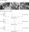
Fig. 1 shows the results of analysis by TEM and DLS. The size of silica nanoparticles are uniform (Fig. 1A) and DLS results showed that mean size is 15.2 nm in 15 nm particles, 62.9 nm in 50 nm particles and 101.4 nm in 100 nm particles (Fig. 1B). Aggregation or agglomeration was not detected (Fig. 1B). As shown in Fig. 2, cell viability was dependent on the particles size. Up to 125 µg/ml, the cells were relatively viable after treatment with 15 nm, 50 nm or 100 nm silica particles. From 250 µg/ml cell death was induced and 15 nm particles triggered cell death more significantly than 50 nm or 100 nm particles did. The LD50 of 15 nm particles was 1.29 mg/ml and that of 50 nm particles was 1.5 mg/ml. Cell death was inhibited by treatment of NAC, which suggest that ROS involves in cell death after silica nanoparticle exposure.
The production of IL-8 was induced at similar levels in response to three different sizes of silica nanoparticles (Fig. 3A and B). The IL-8 production was moderate when compared to the results of silver nanoparticles, in which the treatment of 12.5 µg/ml of 4 nm silver nanoparticles elicit 1,200 pg/ml of IL-8 production as reported in our previous paper (8). Against our expectation, the size effect could not be shown in IL-8 induction. This finding is quite different compared to the results of silver nanoparticles, in which small particles induced higher levels of IL-8 production significantly.
The levels of intracellular Ca++ showed interesting findings. In medium with Ca++ the level of intracellular Ca++ has increased immediately after exposure to only 15 nm silica nanoparticles, which was dose-dependent (Fig. 4A). 50 nm or 100 nm particles did not elicit Ca++ intracellular Ca++ increase. Intracellular Ca++ levels could be increased by influx of medium Ca++ through disrupted cell membrane or by release from intracellular endoplasmic reticulum compartments. Therefore, we assessed intracellular Ca++ levels in Ca++-free medium. The treatment with 15 nm particle did not trigger a significant intracellular Ca++ increase when cells were treated in Ca++-free medium. These results suggest that cell membrane damage rather than endoplasmic reticulum disturbance is responsible for the increase of Ca++.
Finally, ROS generation was detected after treatment with silica nanoparticles. Fig. 5 shows that 15 nm particles induced significantly more ROS than 50 nm or 100 nm particles. This ROS generation was inhibited to control levels by NAC treatment.
Our results showed that silica nanoparticles elicit intracellular Ca++ increase and ROS generation, which is size-dependent. However, the IL-8 production was not dependent on particle size. All three different sizes of silica nanoparticles induced IL-8 production although the levels of IL-8 production were not strong as silver nanoparticles. Taken together, we suggest that the immunostimulatory effect of nanoparticles may differ from nanoparticles with different chemical backgrounds although small nanoparticles are more stimulatory or toxic in general.
Figures and Tables
 | Figure 1Characterization of silica nanoparticles. (A) TEM images of silica nanoparticles show 15 nm, 50 nm or 100 nm particles are relatively uniform in sizes. (B) DLS analysis shows that the mean size is 15.2 nm for 15 nm silica nanoparticles, 62.9 nm for 50 nm particles and 101.4 nm for 100 nm particles by number. Silica nanoparticles dispersed in RPMI 1640 medium containing 10% FBS for DLS analysis. Images shown are representatives of three independent trials. |
 | Figure 2Cytotoxicity of silica nanoparticles. U937 cells were treated with different sizes of silica nanoparticles for 24 h and cytotoxicity was determined by CCK-8 assay. Student's t-test was used for statistical analysis. **p<0.01, ***p<0.001. |
 | Figure 3U937 cells were treated with silica nanoparticles and supernatant levels of IL-8 were assessed by ELISA. Data represent means±S.D. of three independent experiments. Student's t-test was used for statistical analysis. *p<0.05, ***p<0.001. |
 | Figure 4Detection of intracellular Ca++ level. U937 cells were pre-stained with Fluo4-AM for 30 min and treated with silica nanoparticles. Cells were monitored using a Victor X4 multi label plate reader and measured every 5 sec. Data shown here are representative results from two independent experiments. (A) HBSS with Ca++, (B) HBSS without Ca++. |
ACKNOWLEDGEMENTS
This research was supported by a National Research Foundation of Korea Grant funded by the Korean Government (MEST) (NRF-2009-0082417).
References
1. Feng X, Sayle DC, Wang ZL, Paras MS, Santora B, Sutorik AC, Sayle TX, Yang Y, Ding Y, Wang X, Her YS. Converting ceria polyhedral nanoparticles into single-crystal nanospheres. Science. 2006. 312:1504–1508.

2. Anas A, Jiya J, Rameez MJ, Anand PB, Anantharaman MR, Nair S. Sequential interactions of silver-silica nanocomposite (Ag-SiO(2) NC) with cell wall, metabolism and genetic stability of Pseudomonas aeruginosa, a multiple antibiotic-resistant bacterium. Lett Appl Microbiol. 2013. 56:57–62.

3. Li X, Chen Y, Wang M, Ma Y, Xia W, Gu H. A mesoporous silica nanoparticle - PEI - Fusogenic peptide system for siRNA delivery in cancer therapy. Biomaterials. 2013. 34:1391–1401.

4. Scalia S, Franceschinis E, Bertelli D, Iannuccelli V. Comparative Evaluation of the Effect of Permeation Enhancers, Lipid Nanoparticles and Colloidal Silica on in vivo Human Skin Penetration of Quercetin. Skin Pharmacol Physiol. 2012. 26:57–67.

5. Kim S, Choi IH. Phagocytosis and endocytosis of silver nanoparticles induce interleukin-8 production in human macrophages. Yonsei Med J. 2012. 53:654–657.

6. Yang EJ, Kim S, Kim JS, Choi IH. Inflammasome formation and IL-1β release by human blood monocytes in response to silver nanoparticles. Biomaterials. 2012. 33:6858–6867.

7. Lim DH, Jang J, Kim S, Kang T, Lee K, Choi IH. The effects of sub-lethal concentrations of silver nanoparticles on inflammatory and stress genes in human macrophages using cDNA microarray analysis. Biomaterials. 2012. 33:4690–4699.

8. Park J, Lim DH, Lim HJ, Kwon T, Choi JS, Jeong S, Choi IH, Cheon J. Size dependent macrophage responses and toxicological effects of Ag nanoparticles. Chem Commun (Camb). 2011. 47:4382–4384.

9. Chen Z, Meng H, Xing G, Yuan H, Zhao F, Liu R, Chang X, Gao X, Wang T, Jia G, Ye C, Chai Z, Zhao Y. Age-related differences in pulmonary and cardiovascular responses to SiO2 nanoparticle inhalation: nanotoxicity has susceptible population. Environ Sci Technol. 2008. 42:8985–8992.

10. Bhattacharya K, Naha PC, Naydenova I, Mintova S, Byrne HJ. Reactive oxygen species mediated DNA damage in human lung alveolar epithelial (A549) cells from exposure to non-cytotoxic MFI-type zeolite nanoparticles. Toxicol Lett. 2012. 215:151–160.

11. Chu Z, Huang Y, Li L, Tao Q, Li Q. Physiological pathway of human cell damage induced by genotoxic crystalline silica nanoparticles. Biomaterials. 2012. 33:7540–7546.

12. Zhang Z, Chai A. Core-shell magnetite-silica composite nanoparticles enhancing DNA damage induced by a photoactive platinum-diimine complex in red light. J Inorg Biochem. 2012. 117:71–76.
13. Borak B, Biernat P, Prescha A, Baszczuk A, Pluta J. In vivo study on the biodistribution of silica particles in the bodies of rats. Adv Clin Exp Med. 2012. 21:13–18.


 PDF
PDF ePub
ePub Citation
 Print
Print


 XML Download