

 PDF
PDF ePub
ePub Citation
 Print
Print


Abbreviations
GALT
Gut-Associated Lymphoid Tissue
GI
Gastrointestinal
MLN
Messenteric Lymph Node
AD
Atopic Dermatitis
HMP
Human Microbiome Project
IBD
Inflammatory Bowel Disease
T1D
Type 1 Diabetes
DC
Dendritic Cell
NOD
Nucleotide-binding Oligomerization Domain
TLT
Toll-Like Rceptor
Treg
Regulatory T Cells
iDO
Indoleamine 2,3-dioxygenase
SCORD
SCORing Atopic Dermatitis
INTRODUCTION
"Probiotics" are defined as live microorganisms that, when administered in adequate amounts, confer a health benefit on the host (1). Although the beneficial effect of probiotics administration was mainly known to improve the balance of intestinal microflora, recent studies showed that probiotics can also exert beneficial effects by directly modulating immune system. For example, an oral treatment of certain probiotics or a mixture of them prevented and improved symptoms of experimental inflammatory bowel disease, atopic dermatitis and asthma by down-regulating inflammatory cytokines or inducing immune regulatory mechanisms. However, these beneficial effects are strain-specific (2-4), suggesting the necessity of development of screening system to identify specific probiotics to treat specific disorders.
Atopic dermatitis (AD) is chronic, relapsing inflammatory skin disorder and a complex immune response mediated by genetic, environment factors and skin barrier dysfunction. Recently, the incidence of atopic dermatitis has remarkably increased especially in the industrial cities compared to rural areas and developing countries, which strongly support the "hygiene hypothesis" in the development of allergic immune disorders (5,6). Hygiene hypothesis has received worldwide attention from many scientists as the subject remains questioned for many years. Hygiene hypothesis states that a lack of early childhood exposure to infectious agents, symbiotic microorganisms (e.g. gut flora or probiotics), and parasites increases susceptibility to allergic diseases by suppressing natural development of the immune system (7). Even though treatment with corticosteroid is the most well used method to treat atopic dermatitis, long term usage of steroid can cause many side effects including the atrophoderma and growth retardation (8). So many studies were focused on the development of innovative prevention and treatment strategies for AD to replace the steroid treatment. Interestingly, recent studies also suggest the association of dysregulation of gut microbiota with diverse diseases including atopic dermatitis (9). Therefore manipulation of the intestinal flora with good probiotics will be complementary and alternative treatments to divert immune response from the hyper-immune states. In this review, we summarized the hygiene hypothesis and its correlation with gut microbiome, especially on the aspect of probiotics, in the development and regulation mechanisms of diverse disorders.
Microbiome and its relevance to the susceptibility of diverse diseases
Several hypotheses have been proposed as the cause of development in allergic disease, including atopic dermatitis. Among them, "hygiene hypothesis" has gained the most attentions (7). This hypothesis explains that early childhood infection caused by unhygienic contact with older siblings could prevent the development of allergic disease (10). Later, Th1/Th2 paradigm was added that a lack of early childhood infection results a decreased Th1 immune response, which disturbs the Th1/Th2 balance and leads to an abundant Th2 response, causing allergic disease (11). Later this hypothesis was further revised, which considers changes in the intestinal colonization pattern during infancy and overlay hygienic lifestyle as the most important reasons for the increased allergy prevalence. Especially overlay hygiene can induce a lack of activity of regulatory T cells, causing over-activation of Th1 as well as Th2 responses as the underlying mechanism of allergic disease (12,13).
As explained by hygiene hypothesis, a person's life span is largely affected by the distribution of intestinal bacteria. The term "microbiome" was coined by Joshua Lederberg, who argued that microorganisms inhabiting the human body should be included as part of the human genome, because of their influence on human physiology and the human body contains over 10 times more microbial cells than human cells (14,15). To understand the microbiome (totality of microbes) and the factors that influence the distribution and evolution of the constituent microorganisms, the Human Microbiome Project (HMP) was initiated. HMP has aimed to identify and characterize the microorganisms which are found in association with both healthy and diseased human (16-18). After investigating the feces of forty people living in rural communities and forty people inhibiting in cities by metagenomic analysis, scientists discovered that the distribution of intestinal bacteria of the two groups has a stark contrast. Surprisingly, it was found that those living in rural habitats have about three to five times more Lactobacillus acidophilus (LA). LA metabolizes lactic acid, obtains vitamin K, a cofactor for blood clotting and healthy bone tissue turnover. LA also helps recycle the amino acids in bile, and normalizes the body's cholesterol level compared to people living in cities. Possible explanations to this phenomenon are attributing to the level of stress and the difference in abiotic factors such as air and water and life span or health of people. Most importantly, distribution of helpful microorganisms is mainly derived from the types of foods consumed (19,20). The potential roles of microbiome on diverse disorders are summarized below.
Atopic dermatitis
Studies investigating the composition of the intestinal microflora in humans have shown the pivotal role of commensal bacteria in the development of allergies. Studies comparing the microflora composition of atopic and non-atopic infants also show significant differences. Non-atopic infants have more colonization of Bifidobacteria and Lactobacillus in the intestine while there is much colonization of Clostridia in the intestine of atopic infants. However several reports also suggest that there are no significant differences between the two groups (21,22). The exact role of intestinal flora in the development of atopic disease in the childhood is still not clarified (23). In addition to these quantitative differences in the Bifidobacterium, qualitative differences have also been observed. The Bifidobacteria from atopic infants were found to induce much higher levels of pro-inflammatory cytokines in vitro whereas the Bifidobacteria from non-atopic infants induced more secretion of anti-inflammatory cytokines. In addition to different secretion ability, differential adhesion to intestinal mucosa may also result in a different or reduced stimulation of the immune system through the gut-associated lymphoid tissue (GALT) (24).
Obesity
Obesity is associated with the changes in the relative abundance of the two dominant bacterial divisions, the Bacteroids and the Firmicutes. The relative proportion of Bacteroids is decreased in obese people compared to lean people while the proportion of Firmicutes is increased in obese people. Obese microbiome has an increased capacity to harvest energy from the diet and colonization of germ-free mice with an obese microbiota results in greater increase in total body fat without any increase in food consumption than colonization with a lean microbiota (25-27).
Type 1 diabetes (T1D)
Metagenomic analysis revealed the close relatedness of bacteria with the development of T1D associated autoimmunity in young child. Especially the proportion of Bacteroides ovatus was increased in children with T1D while the proportion of Firmicutes, CO19 was increased in normal healthy children (28).
All these results collectively imply that gut microbiota could be an additional contributing factor to the pathophysiology of obesity, T1D as well as atopic dermatitis, which in turn suggest that bacterial markers can be used for early diagnosis for these disorders. In addition, supplementation of large amount of bacteria that negatively correlated with the disease state may be beneficial for the prevention of these disorders.
Microflora and immune system
The intestinal microflora
Based on the hygiene hypothesis, in addition to environmental factors, the intestinal microflora is another contributor to allergic disease by substantially affecting the mucosal immunity. Allergic responses are thought to arise if there is absence of microbial exposure while the immune system is developing (24,29). The gastrointestinal tract is an important interface between host and environment and has the dual role of facilitating the absorption of nutrients while excluding pathogens. The human intestines are inhabited by at least 400 different bacterial species, with the greatest density in the large intestine, where concentration of 1011-1012 cells/g of luminal contents can be found (30). Because a large part of the intestinal microflora cannot be cultured with current culture techniques, it has been suggested that the number of microflora species in the human intestine may exceed 1,000 species (24). Although some intestinal bacteria are potential pathogens, the relationship between the intestinal microflora and the human host is mostly symbiotic in healthy individuals.
Gut-Associated Lymphoid Tissue (GALT)
The largest mass of lymphoid tissue of the human body can be found in the gastrointestinal tract and is called as the gut-associated lymphoid tissue (GALT) which includes Peyer's patch and mesenteric lymph node (MLN) (31). GALT interact with intestinal bacteria which are sampled by dendritic cells (DCs) and intestinal epithelial cells through the pattern recognition receptors such as toll-like receptor (TLR) and nucleotide-binding oligomerization domain (NOD) (32-34). The intestinal antigens also can be captured by DCs in the Peyer's patch through M cells on the surface of enterocytes. DCs in lamina propria can capture antigens directly by dendrites (35-37). Intestinal microflora seems to be involved in the induction of oral tolerance which is established in the periphery to specific antigen after ingestion of orally delivered protein (antigen) and plays a pivotal role in the induction and maintenance of peripheral tolerance mediated by regulatory T cells (38,39).
Skin microflora
Most studies delineating the role of resident microorganisms in regulation of immune system and inflammation have focused on the gut microbiome, since gut is the main site of exposure and contains a wide diversity of microbes. However, skin was considered to just have a physical barrier function to protect our body from the attack of pathogens. Interestingly recent studies discovered that skin microbiome also have similar function that of gut microbiome (40). Microbial profiling by metagenomic analysis has revealed the presence of highly diverse commensal communities in skin sites (41). Moreover, cutaneous inflammatory disorders, such as psoriasis and atopic dermatitis have been associated with imbalance of the cutaneous microbiota (42,43). Indeed, microbial products from skin commensals are known to exert immunoregulatory effects (44). Yasmine Belkaid group reported that the roles for the skin microbiota in controlling the local inflammatory milieu. They showed that protective immunity to a cutaneous pathogen is critically dependent on the skin but not on gut microbiota (45). Furthermore, skin commensals activate the function of local T cells through the interleukin-1 receptor (IL-1R)-MyD88 signaling pathway (45). These findings indicate the critical role of the skin residual commensal bacteria not only as a distinctive feature of tissue compartmentalization also as a key regulator of the immune system in health and disease.
Action mechanisms of probiotics
General regulation mechanisms in gut
The intestinal commensal bacteria play diverse functions in regulating nutrient metabolism including absorption of indigestible carbohydrate and production of short chain fatty acid, amino acid and vitamin. Microflora provides additional energy by digesting exogenous and endogenous substrates, such as fibers and mucins. Microflora also provide a protective barrier against incoming bacteria (46). This protection is mediated by several different mechanisms including competition for nutrients and binding sites and production of antimicrobial substances (37,47).
Immune regulation mechanism
Probiotics could stimulate the immune system by modulating the composition and/or activity of the intestinal microbiota (48,49). The immunomodulatory role of probiotics has been shown in diverse types of inflammatory immune disorders both, in gastrointestinal diseases and in non-gastrointestinal diseases. The beneficial effect of probiotics is strain-specific and their effector mechanisms are also quite diverse. Some probiotics generate IL-10 producing Tr1 or CD4+Foxp3+ Treg cells (50), while others could enhance immunity by producing a large amount of IL-1β, IL-12 or TNF-α (51). For example Lactobacillus reuteri induces the production of IL-12 and TNF-α from DCs while Lactobacillus casei inhibits the production of pro-inflammatory cytokines by producing anti-inflammatory cytokines such as IL-10. Therefore it is crucial to use specific probiotics or their mixture to target specific immune disorders. Most of the allergic diseases are associated with a shift of the Th1/Th2 balance toward a Th2 response, which induces secretion of Th2 type cytokines such as IL-4, IL-5 and IL-13 leading to higher production of IgE. Recently, we have demonstrated that IRT5 probiotics, a mixture of 5 probiotics, could suppress diverse immune disorders through the generation of CD4+Foxp3+ Tregs (52). Oral administration of IRT5 probiotics generated rDCs (iDOhigh, Cox2high, IL10+, TGFβ+), which can convert effector CD4+Foxp3- T cells into CD4+Foxp3+ regulatory T cells in the mucosal immune system. In addition, oral administration of the IRT5 probiotics suppressed ongoing experimental AD and pathogenesis of experimental arthritis as well (52). Currently we are investigating to identify potent probiotic strains that could induce the production of immune-modulatory cytokines such as IL-10 and TGF-β or enhancing Th1 response to improves the symptoms of allergic disease (37,53).
The effect of probiotics on dendritic cells (DCs)
DCs function as a professional antigen presenting cells (APC) and have the ability to activated T cells and directly regulate Th1 and Th2 cell. Especially DCs lead to the induction of regulatory T cells through the secretion of IL-10 and TGF-β and help to regulate immune response by the production of IL-10 from Tr1 cells and TGF-β from Th3 cells. Once DCs capture antigens in the presence of proper inflammatory stimuli, they migrate to secondary lymphoid organs and simultaneously undergo a maturation process. During this maturation, DCs produce diverse cytokines and express surface molecules such as MHC, CD40, CD83, CD80 (B7.1) and CD81 (B7.2). Recently several reports suggest that probiotics can induce the maturation and cytokine secretion by DCs. Different probiotics exert differential effect on the cytokine profiling by DCs such as IL-12, TNF-α and IL-10 (54-56). Recently we have demonstrated that administration of IRT5 probiotics mixture increases the generation of CD4+Foxp3+ Tregs in the mesenteric lymph nodes (MLN) through the generation of CD11c+ regulatory dendritic cells (rDCs) that highly express high levels of IL-10, TGF-β and indoleamine 2,3-dioxygenase (iDO) (52), which in turn potentiated the generation of CD4+Foxp3+ Tregs from the pool of CD4+CD25- cells. These results suggest that probiotics or a mixture of them that can induce regulatory DCs in the MLN, which in turn induce the production of CD4+Foxp3+ cells, could represent an attractive potential therapy for the treatment of diverse inflammatory immune disorders.
The role of probiotics in atopic dermatitis
Atopic dermatitis (AD) is caused by diverse pathogenic factors including genetic susceptibility, environment trigger, skin barrier dysfunction, bacterial infection and immune dysregulation (5). AD is a complex immune reaction mediated by both Th1 and Th2 immune responses (6). Th2 type cytokines including IL-4, IL-5 and IL-13 play important role in the development of AD by increasing the levels of serum IgE and blood eosinophils in AD patients (57). In the later stage of AD where infection mediated inflammation occurs, Th1 type cytokines such as IFN-γ and IL-12 mediate chronic stage of atopic dermatitis (5,58,59). IFN-γ is also involved in the maintenance of chronic stage of atopic dermatitis by elevating the expression of CCL17 (TARC) and CCL22 (MDC) that are involved in the recruitment of effector T cells to the inflamed site (60). IFN-γ increases the sensitivity of Fas-mediated apoptosis of keratinocytes, which is considered to be a key pathogenic event in eczematous dermatitis (61). Based on many clinical as well as animal studies, therapeutic or preventive effects of probiotics on allergic disease have been intensively investigated as summarized hereinafter.
1. Human clinical trials
(1) Prevention
Several randomized controlled trials (RCT) have investigated the effect of probiotics on the prevention of atopic dermatitis. In these trials probiotics were given to infants with a high risk of developing allergy, starting immediately after birth and mothers also received probiotics during the last week of pregnancy. The first trial reported that a 50% reduction of the incidence of AD in the probiotics group compare to placebo group at the age of 2 year and this effect was maintained until at 7 years (62). A clinical trial reported that administration of probiotics only reduced the incidence of IgE-associated AD without altering other allergic diseases (63). In another report, consumption of Lactobacillus rhamnosus reduced the incidence of AD while Bifidobacterium animalis did not show any reduction, suggesting the strain dependent preventive effect of probiotics (64). In contrast, two prevention studies did not show any effect on AD incidence. Moreover one of the studies reported an increased sensitization rate for food or aeroallergens in the probiotics group (65,66). There are several possible explanations for these inconclusive results. First, different lactobacillus strains were used and this indicates that not all strains are effective for atopic dermatitis. Second, probiotic effects are dose dependent and there was considerable variation in the daily dosage. The third explanation could be a difference in study design such as numbers and atopic risk of participants and supplementation of the mother during breastfeeding. These studies suggest that careful consideration much be established to decide which probiotics or a mixture of them are employed to treat allergic atopic dermatitis.
(2) Treatment
Randomized controlled trials to examine the effect of probiotics in the treatment of atopic dermatitis in child also have inconclusive results. Several studies showed a significant reduction of SCORAD (SCORing Atopic dermatitis) in the probiotics treated group (67). Other reports showed that probiotics significantly decreased SCORAD of IgE-associated not general AD (68,69). These reports suggest that only children who at a young age already have IgE sensitization such as elevated total or specific IgE levels get a benefit from probiotics administration. However there are also several studies that did not show any effect of probiotics on the severity or incidence of atopic dermatitis (70,71). There are considerable variations in the probiotic strains and daily dosage and number, age, severity of AD of participants and treatment period, which may result in conflicting results.
2. Experimental animal model
Beneficial effect of probiotics on AD development was evaluated in the experimental mouse models. We have recently showed that administration of IRT5 probiotics mixture to experimental AD mice reduced serum IgE levels and infiltration of lymphocytes to inflamed sites by recruiting CD4+Foxp3+ regulatory T cells (52). Administration of Lactobacillus rhamnosus GG (LGG) to Nc/Nga mice during pregnancy and breastfeeding also delayed onset or incidence of atopic dermatitis by increasing IL-10 level in GALT. (72). As a similar study, administration of three Lactobacillus strains isolated from Kimchi to Nc/Nga mice improved AD symptoms by significantly decreasing serum IgE levels as well as Th2 type cytokine expression while increasing IFN-γ and IL-10 expression (73,74). However, it is still unclear on the mechanism by which administration of probiotics generates regulatory T cell populations. In addition, further studies are required to identify potent immunoregulatroy probiotics among the hundreds of probioctic strains. Recently we developed ex vivo screening system to identify immunoregulatory probiotics that induce high levels of IL-10 and Foxp3+ Treg while reducing pro-inflammatory cytokines. We are currently under investigation to identify potent immunoregulatory probiotics from the traditional Korean fermented foods using the ex vivo screening system.
The role of probiotics in inflammatory bowel disease (IBD)
Inflammatory bowel disease (IBD) is characterized by a chronic dysregulation of the immune response in the gastrointestinal tract (GI tract). Proinflammatory cytokines such as INF-γ, TNF-α, IL-6 and IL-1β are major pathogenic cytokines but the pathogenesis is still not clear (75). Recently many studies have demonstrated that the GI tracts of patients with IBD are populated with increased levels of adherent and pathogenic bacteria. Fecal numbers of anaerobic bacteria such as Bacteroides species were greater while Lactobacilli and Bifidobacteria were fewer than in healthy control (76,77). Probiotics may be capable of recolonizing the bowel with non-pathogenic strains of bacteria. Probiotics have long been shown to be beneficial in both infectious and non-infectious digestive disorders. Growing evidence indicates that probiotics may be effective in the treatment of specific clinical IBD conditions.
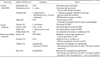
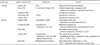
Although it is unclear whether the abnormal composition of the entreric flora contributes to the pathogenesis of IBD, many clinical trials have examined the effect of diverse probiotic strains on the modulation of IBD progression including LGG (78) and E. coli strain Nissle 1917 (79,80). In addition, VSL#3, a mixture of 4 lactobacilli strains (Lactobacillus plantarum, Lactobacillus casei, Lactobacillus acidophilus, Lactobacillus delbrueckii ssp. bulgaricus), 3 bifidobacteria strains (Bifidobacterium infantis, Bifidobacterium breve, Bifidobacterium longum), and 1 strain of Streptococcus (Streptococcus salivarius ssp. thermophilus), has been examined in ulcerative colitis (UC), Crohn's disease (CD) (81,82). The therapeutic effects of several probiotic strains in animal studies and clinical trials were summarized in Table I and Table II, respectively.
Several studies have investigated the influence of probiotic consumption on colitis in animal trials. In particular, IL-10 knockout mouse that develops IBD spontaneously has been largely employed as a IBD model. Interestingly, IL-10 knockout mice spontaneously develop colitis when colonized with a conventional flora but remain disease free when maintained under germ-free conditions (83-87). These studies suggest the role of commensal flora in the development of inflammatory bowel disease. The role of probiotic strains in animal model of IBD was summarized in Table II.
Collectively these studies indicate that although the probiotic administration has potential therapeutic efficacy in IBD, larger controlled trials are necessary before the use of probiotics as a routine medical treatment.
Summary and future perspectives
Gastrointestinal microbes act as an environmental factor that affects the host's physiology, particularly in the context of obesity and its related metabolic disorder as well as allergic disorders (88,89). The significant involvement of the gut microbiota in human health and disease suggests that probiotics could be a novel therapeutic modality to treat diverse diseases by manipulating the composition of commensal microbiota (9). In fact, many reports suggest that enough consumption of probiotics is beneficial to suppress the development of atopic dermatitis by modifying the intestinal microflora and immune responses. Further studies are needed to evaluate the clinical and immunological effects of different strains of probiotics in well-designed clinical trials.
 XML Download