

 PDF
PDF ePub
ePub Citation
 Print
Print


INTRODUCTION
TIM-3 is a transmembrane protein containing immunoglobulin V like domain, mucin domain, transmembrane domain and cytoplasmic domain. TIM-3 plays important roles in immune responses and the pathogenesis of several diseases (1). With its blocking antibody or its soluble fusion protein TIM-3-Ig, Th1 immune response as well as anti-tumor immunity is enhanced (2-4). Also, induction of tolerance in an experimental transplantation model is abrogated by injection of TIM-3 antibody (5). Furthermore, dysregulation of T cell TIM-3 expression is observed in several autoimmune diseases including multiple sclerosis, rheumatoid arthritis and Crohn's disease (6-8).
Galectin-9 and phosphatidylserine have been identified as TIM-3 ligands. Galectin-9 was originally identified as a potent eosinophil chemoattractant produced and released by antigen-stimulated T cells (9). Galectin-9 induces intracellular calcium flux, aggregation and death of in vitro cultured Th1 cells in a manner that is dependent on Tim-3, and administration of galectin-9 in vivo results in selective loss of interferon-gamma-producing cells and suppression of Th1 autoimmunity (10). In the other hand, phosphatidylserine, which is normally localized to the inner leaflet of the plasma membrane, is exposed on the cell surface when the cell is activated or undergoes apoptosis. TIM-3 on monocytes and dendritic cells binds phosphatidylserine on apoptotic cells and enhances presentation of antigen expressed in the apoptotic cells through the upregulation of endocytosis (11).
In this study, we wanted to identify the genes that were differentially expressed in T cells by TIM3 expression. Using TIM3 overexpressing Jurkat T cell lines and panels of degenerate primer sets we found three genes (TIMP-1, IFITM1 and CCL1) that were up-regulated in TIM3 overexpressing cells, and one gene (PAR3) that was down-regulated in TIM3 overexpressing cells. However, only the CCL1 transcript level was significantly increased in Jurkat T cells transiently transfected with TIM3 expression vector as well as in human primary CD4+ T cells expressing abundantly TIM-3.
MATERIALS AND METHODS
Cells and stimulation
The human Jurkat T cell line was cultured in RPMI1640 medium (Gibco BRL, Paisley, Scotland) supplemented with 10% fetal bovine serum (FBS; Gibco BRL), 100 U/ml penicillin and 100µg/ml streptomycin (Gibco BRL) at 37℃ in 5% CO2 incubator.
Construction of plasmids for expression of flag tagged TIM3
The human TIM-3 gene was cloned from peripheral blood mononuclear cells (PBMCs) stimulated with concanavalin A (ConA; 1µg/ml) for 2 days. Total RNA was subjected to reverse transcription-polymerase chain reaction (RT-PCR) using the human TIM-3 specific primers [HTIM3F(KpnI) 5'-GGGGTACCGTTAAAACTGTGCCTAACAG-3', HTIM3R(Hind III): 5'-CCCAAGCTTCAAAAATAAGGTGGTTGG-3'] and then cloned into a pGEM-T vector (Promega, Madison, WI, USA). The TIM3 gene amplified with specific primers [TIM3(Xho I): 5'-CCGCTCGAGTACGAAGTGGAATACAGAGCG GAGG-3' and TIM3(Xma I): 5'-TCCCCCCGGGCTATGGCATTGCAAAGCGACAAC-3'] was inserted into a pIRES2-EGFP vector (Clontech Laboratories, Mountain View, CA, USA). The DNA fragment for leader and flag peptide was inserted upstream of the TIM-3 gene at Nhe I and Xho I sites. The nucleotide sequences were verified.
Establishment of stable cells expressing TIM-3 protein
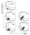
The pIRES2-EGFP-TIM3 vector or empty vector was transfected into Jurkat T cells using the Microporator (Digital Bio Technology, Seoul, Korea) with settings of 1200 V, pulse width 20 and pulse number 1. Among the clones resistant to neomycin, two independent clones (T5 and T7) that expressed TIM-3 on the surface and green fluorescence protein (GFP) in the cytoplasm were selected by flow cytometry. As controls, two independent clones expressing GFP alone were selected (G2 and G6).
Flow cytometry
For TIM-3 detection, cells were labeled with anti-human TIM-3-PE (R&D Systems, Minneapolis, MN, USA) and analyzed using a FACSCanto II apparatus (Becton Dickinson, San Diego, CA, USA).
Screening of differentially expressed genes using annealing control primer (ACP) system
Total RNAs of T5 and G2 were extracted using a RNA isolation kit RNAiso (TaKaRa Bio, Shiga, Japan). cDNA synthesis and RT-PCR were performed following the procedures recommended by the manufacturer using an annealing control primer system (Seegene, Seoul, Korea). PCR products were analyzed by agarose gel electrophoresis. Differentially amplified PCR products eluted from gel were cloned into a pCR2.1-TOPO vector (Invitrogen, Carlsbad, CA, USA) and then sequenced. The identical DNA sequence to the insert was searched using BLASTN.
Semiquantitive RT-PCR and real-time RT-PCR
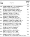
To validate the transcription level of the selected genes, semiquantitive RT-PCR was performed using a specific primer set for each gene (Table I). CCL1 transcription level was assessed by real-time RT-PCR using Sybr Premix Ex Taq (TaKaRa, Shiga, Japan). The CCL1 primers (Bioneer, Daejeon, Korea) were 5'-AATACCAGCTCCATCTGCTCCAA-3' and 5'-GAACCCATCCAACTGTGTCCAAG-3'. CCL-1 transcription was normalized to glyceraldehyde 3-phosphate dehydrogenase (GAPDH) transcription.
Isolation of primary CD4+ T cells and TIM-3high T cells
Primary human CD4+ T cells were isolated from peripheral blood mononuclear cells of healthy donors using magnetic beads (Miltenyi Biotec, Bergisch Gladbach, Germany). The cells were stimulated with antibodies against CD3 (1µg/ml) and CD28 (1µg/ml) for 7 days and then sorted into TIM-3 high and TIM-3 low cells by fluorescence activated cell sorting using a FACS Aria II cell sorter (BD, Franklin Lakes, NJ) after labeling with allophycocyanin (APC)-conjugated anti-TIM-3 antibody (eBioscience, San Diego, CA). All blood samples were obtained in compliance with the Ajou University Institutional Review Board protocol.
RESULTS
Jurkat T cell clones over-expressing TIM-3
To identify the genes that were differentially expressed in T cells by TIM-3 expression, we established cell lines overexpressing TIM-3 by stably transfecting Jurkat T cells with an expression vector containing TIM-3 gene under the control of the cytomegalovirus promoter as well as a gene for GFP under the internal ribosome entry site. Cells expressing GFP were sorted twice using fluorescence activated cell sorting (FACS) and then cloned. Compared to parental Jurkat T cells and GFP expressing clones (G2 and G6), TIM-3 expression in the T5 and T7 stable cell clones was clearly demonstrated (Fig. 1).
The differentially expressed gene candidates by TIM-3 expression
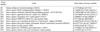
To screen for differentially expressed genes in TIM-3 expressing cells, RNA was isolated from Jurkat T cells, T5 cells stimulated with phorbol 12-myristate 13-acetate (PMA) and A23187, and then RT-PCR was performed using an ACP system. Agarose gel electrophoresis selected 76 PCR products showing up- or down-regulated expression in T5 compared to Jurkat T cells for sequencing (Fig. 2). Sequence analysis of these genes showed that several PCR products with different ACP sets overlapped. Eleven differentially expressed gene candidates were identified by homology search using the NCBI dbEST database (Table II).
Validation of difference in transcription of the candidate genes
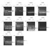
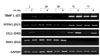
To validate the candidate gene expression levels, semi-quantitative RT-PCR was conducted using sequence specific primers and RNA isolated from Jurkat T cells, G2, G6, T5 and T7 (Fig. 3). The transcription of TIMP-1, IFITM1 and CCL1, respectively, was increased in T5 and T7 cells, compared to control cells. CCL1 expression was induced in all cells when stimulated; the induction level was greater in T5 and T7 cells than in control cells. In contrast, the transcription of PAR3 was reduced in T5 and T7 cells compared to control cells. Next, the transcript levels of these genes were assessed in cells transiently transfected with TIM3 expression vector (Fig. 4). Only the CCL1 transcript level was significantly increased in cells transiently transfected with TIM3 expression vector compared with control cells (p<0.05).
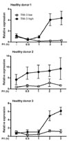
The increased transcription of CCL1 in human primary CD4+ T cells highly expressing TIM-3
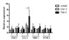
Finally we examined the transcription of CCL1 in primary CD4+ T cells highly expressing TIM-3 (Fig. 5). CD4+ T cells isolated from human subjects were stimulated for 7 days and then sorted into TIM-3 high cells and TIM-3 low cells. The cells were restimulated with PMA and A23187 and then CCL-1 transcription was determined by real-time RT-PCR. Although the induction pattern of CCL-1 showed difference among subjects, CCL-1 transcription was increased in TIM-3high cells after stimulation in all subjects but not in TIM-3 low cells.
Taken together, these data suggest that CCL1 transcription may be enhanced in CD4+ T cells highly expressing TIM-3.
DISCUSSION
Presently, CCL1 was identified as a differentially expressed gene in CD4+ T cells highly expressing TIM-3 compared to cells with TIM-3 low expression. CCL1 is produced by both Th1 and Th2 cells in response to T cell receptor triggering (12). However, interleukin (IL)-12 and interferon (IFN)-alpha, cytokines that promote the differentiation of human Th1 cells selectively inhibit transcription and secretion of CCL1 by Th1 cells (12). In interferon-gamma knockout mice, CCL1 expression is increased along with the elevation of the Th2-associated chemokines CCL11 and CCL17, and the Th17-associated chemokines CCL22 and CXCL2 (13). Given that TIM-3 is preferentially expressed in fully differentiated Th1 cells, the increased CCL1 transcription by CD4+ T cells highly expressing TIM-3 was an unexpected result.
CCL1, originally identified as a gene expressed in activated T cell lines, is secreted by various cells including lymphocytes, Langerhans cells, monocytes, mast cells, endothelial cells and epithelial cells (14,15). Its sole receptor, CC chemokine receptor (CCR)-8, is expressed in polarized Th2 cells, IL-10 producing regulatory T cells and macrophages (16,17). Its chemotactic role for Th2 cells (16), and high serum levels in atopic dermatitis suggest that CCL1 plays a key role in the progression of Th2 type hypersensitivity (18,19). CCL1 together with CCL22 participates in the migration of regulatory T cells, leading to a reduced alloproliferative response (20,21). In mice, CCL1 interaction with CCR8 on macrophages leads to the development of postoperative and postinflammatory peritoneal adhesions (22). Further studies are required to investigate CCL1 production by TIM-3-expressing CD4+ T cells in normal and pathologic immune responses.
Currently, TIM-3 is believed to play a negative role in cytokine production by Th1 and Th17 cells based on the observations that the secretion of IFN-γ, IL-2, IL-17 and IL-6 by human CD4+ T cells is increased in the presence of anti-TIM-3 antibody (23), and that the cytokine production of Th1 cells is enhanced by the injection of TIM-3-Ig into mice (2). It will be interesting to examine whether the expression of other chemokines is affected by TIM3 expression and whether CCL1 expression is changed in the presence of anti-TIM-3 antibody.
In summary, TIM-3 expression in CD4+ T cells enhances transcription of CCL1, a chemotactic factor for CCR8+ polarized Th2 cells, regulatory T cells and macrophages.
 XML Download