Journal List > Immune Netw > v.10(3) > 1033279

Ko, Jang, and Lee: GSK3β Inhibitor Peptide Protects Mice from LPS-induced Endotoxin Shock

Abstract

Background

Glycogen synthase kinase 3β (GSK3β) is a ubiquitous serine/threonine kinase that is regulated by serine phosphorylation at 9. Recent studies have reported the beneficial effects of a number of the pharmacological GSK3β inhibitors in rodent models of septic shock. Since most of the GSK3β inhibitors are targeted at the ATP-binding site, which is highly conserved among diverse protein kinases, the development of novel non-ATP competitive GSK3β inhibitors is needed.

Methods

Based on the unique phosphorylation motif of GSK3β, we designed and generated a novel class of GSK3β inhibitor (GSK3i) peptides. In addition, we investigated the effects of a GSK3i peptide on lipopolysaccharide (LPS)-stimulated cytokine production and septic shock. Mice were intraperitoneally injected with GSK3i peptide and monitored over a 7-day period for survival.

Results

We first demonstrate its effects on LPS-stimulated pro-inflammatory cytokine production including interleukin (IL)-6 and IL-12p40. LPS-induced IL-6 and IL-12p40 production in macrophages was suppressed when macrophages were treated with the GSKi peptide. Administration of the GSK3i peptide potently suppressed LPS-mediated endotoxin shock.

Conclusion

Collectively, we present a rational strategy for the development of a therapeutic GSK3i peptide. This peptide may serve as a novel template for the design of non-ATP competitive GSK3 inhibitors.

INTRODUCTION

Glycogen synthase kinase 3 (GSK3) is a ubiquitous serine/threonine kinase that is regulated by serine phosphorylation at 21 in GSK3α and 9 in GSK3β (1-3). GSK3 phosphorylates a broad range of substrates such as glycogen synthase (4), nuclear factor of activated T-cells (NFATc) (5), cAMP response element binding (CREB) (6), c-Jun (7) and c-Myc (8), and also inactivates many of these substrates. Through this activity, GSK3 regulates many cellular functions, including glycogen metabolism, cell-cycle control and cell proliferation (3,9). Among the GSK3 isoforms, α and β, GSK3β gained prominence as a potential drug target in various disease areas including type 2 diabetes (10,11), Alzheimer's disease (12), mood disorders (13) and cancer (14). Recently, GSK3β was also identified as a regulator of the immune system, suggesting it might be an attractive therapeutic target in inflammatory and autoimmune diseases (15-18).
Due to the growing evidence of GSK3β as a potential therapeutic target in multiple diseases (19,20), several approaches such as high-throughput screening, virtual computer simulations, and structure-based drug design have been used to develop GSK3β kinase inhibitors (21). Most of these inhibitors are a class of ATP-competitive inhibitors. However, a major drawback to the use of the inhibitors is their limited specificity, and therefore there is a concern that such inhibitors exert undesired side effects (22-25).
To overcome these issues, we developed a cell-permeable peptide, the GSK3β inhibitor (GSKi) peptide. We hypothesized that small unique peptides derived from the N-terminal phosphorylation motif of GSK3β containing serine 9 may serve as a pseudo-substrate of GSK3β in cells (26-28).
In this study, we have investigated the effects of a novel GSK3i peptide in a LPS-induced septic shock model. We found that inhibition of GSK3β by the inhibitor peptide decreased LPS-mediated pro-inflammatory cytokine production. In addition, administration of the GSK3i peptide protected mice from LPS-induced endotoxin shock. Therefore, the GSK3i peptide may be useful in the development of selective therapeutic agents for the treatment of septic shock and other related inflammatory diseases.

MATERIALS AND METHODS

Cell culture

Murine bone marrow-derived macrophages (BMDMs) were obtained from the femur of 6~8 week-old C57BL/6 male mice. Bone marrow cells were flushed out from the bone marrow cavity, suspended in DMEM (Hyclone) that was supplemented with 20% heat-inactivated FBS, 100 units/ml penicillin, 100 µg/ml streptomycin. After 1 day, non-adherent cells were cultured in the presence of 10 ng/ml recombinant human M-CSF (R&D Systems). After 7 days, a homogeneous population of adherent macrophages was obtained.

Peptide synthesis

Cell-permeable peptides were synthesized by Peptron (Daejeon, Korea). Peptides were purified by preparative reverse-phase HPLC and were more than 95% pure with the expected amino acid composition and mass spectra. Immediately before use, the peptides were dissolved in phosphate buffered saline (PBS) to prepare stock solutions that were between 5 and 10 mM.

Measurement of cytokines

The level of mouse interleukin (IL)-6 and IL-12p40 in culture supernatants and sera were measured using enzyme-linked immunosorbent assay (ELISA) kits from BD biosciences (San Jose, CA, USA) according to the manufacturer's instructions.

Endotoxin shock model

All animal study protocols were approved by the Animal Care Committee of Ewha Laboratory Animal Genomics Center. The endotoxin shock model used in this study has been described previously (15). In brief, male C57BL/6 mice (6~8 weeks of age; 18~20 g body weight) were injected intraperitoneally with an LD100 (10 µg/ml) of E. coli K235 LPS (Sigma) in 200 µl of PBS containing 0.1% DMSO. Mice survival was monitored over a 7-day period.

RESULTS

Inhibitory effect of the GSKi peptide on LPS-induced pro-inflammatory cytokine production

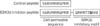
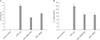
We designed a cell-permeable GSKi peptide spanning the serine 9 phosphorylation motif of GSK3β that was fused with recently characterized cell-permeable sequences derived from the human transcription factor Hph-1 (Fig. 1) (29,30). Since GSK3β is known key regulator of pro-inflammatory cytokine production (16), we examined the ability of the GSKi peptide to regulate cytokine production in response to LPS stimulation. BMDMs from male 6~8 week-old mice were pre-incubated for 2 hours with either 5 µM GSKi peptide or 10 µM SB216763 as a positive control, and then the cells were stimulated with 1 µg/ml LPS for 20 hours. The control peptide containing the cell-permeable sequences only did not affect cytokine production stimulated by LPS (data not shown). As shown in Fig. 2, the presence of the GSK3i peptide was shown to attenuate pro-inflammatory cytokine production; IL-6 and IL-12p40. These inhibitory effects were comparable to that of SB216763 which is a well-characterized pharmacological inhibitor of GSK3. These results demonstrate that the GSKi peptide can regulate LPS-mediated pro-inflammatory cytokine production.

The GSKi peptide protects mice from endotoxin shock

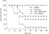
To test the therapeutic potential of the GSKi peptide on septic shock, the effects of the peptide on an experimental LPS-induced endotoxin shock model were investigated. Mice that were given 30 mg/kg of the GSKi peptide before receiving a 100% lethal dose (LD100) of LPS showed significantly improved survival, compared with the control group given LPS (Fig. 3). This protective effect was comparable to that of SB216763. The control peptide did not affect LPS-induced septic shock (data not shown).
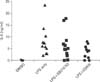
Next, we examined whether GSK3β inhibition regulated the pro-inflammatory cytokine production in mice given an LD100 of LPS. As shown in Fig. 4, we found that the serum level of the pro-inflammatory cytokine IL-6 was significantly reduced in LPS-challenged mice given the GSK3i peptide relative to that of the control mice given LPS. Thus, these results demonstrate that a cell-permeable inhibitor peptide that targets GSK3β may be useful in the treatment of septic shock.

DISCUSSION

In this study, we present a new strategy for developing a novel inhibitor peptide that targets GSK3β. Our study has shown that the peptide motif spanning serine 9 of GSK3β blocks LPS-induced cytokine production and septic shock. The central role of the GSK3β pathway in innate immune responses has been well documented (31). Specifically, it has been shown that GSK3β is involved in the PI3K/Akt pathway and mediates cytokine production in TLR signaling (15). Moreover, many reports have elucidated important roles for the GSK3β in immune diseases such as arthritis, colitis, multiple sclerosis and sepsis (32,33) suggesting that GSK3 might be an attractive therapeutic target in inflammatory diseases. In this regard, our studies suggest that the GSKi peptide may be used as a novel tool for studying GSK3 inhibition.
The exact mechanism of how the GSK3i peptide regulates inflammatory response such as cytokine production is not fully understood. In resting cells, GSK3 is highly active, but its enzyme activity can be inhibited by the PI3-kinase-dependent pathway in response to various ligands stimulation (34-36). It has been well established that PKB/Akt is responsible for the direct phosphorylation of GSK3β on the N-terminal serine 9 residue (37,38). After serine 9 phosphorylation, the phosphorylated N-terminal residues of GSK3β inhibit the enzyme by binding to the active site as a pseudo-substrate (26-28). Since the GSKi peptide contains a serine 9 residue that can be phosphorylated by PKB/Akt in cells, it may act as a pseudo-substrate that binds in the active site of GSK3β and inhibits its enzyme activity. Consistent with our data, a phosphopeptide corresponding to residue 7-14 of GSK3β inhibited GSK3 activity in vitro, whereas the nonphsphorylated peptide did not (27,34).
The development of bioactive peptides as therapeutic alternatives offers novel exciting approaches for target-selective pharmacotherapy (39,40). The in vivo pharmacodynamics of the GSKi peptide was not detemined in this study. However, our data demonstrated that the GSKi peptide acted in vivo to protect mice against LPS-induced shock. Such phenomenon indicates that a more detailed assessment of the in vivo delivery and pharmacokinetic profiles of the GSKi peptide may lead to the design of effective therapeutic reagents directed against septic shock.

Figures and Tables

Figure 1
Structure of the GSKi peptide conjugated with the Hph-1 protein transduction domain. Amino acid sequences corresponding to residues 3~12 of GSK3β were chosen for the design of a GSKi peptide. The serine 9 residue, which can be phosphorylated by PKB/Akt, is highlighted in bold. The control peptide contains 11-mer of the protein transduction domain.
in-10-99-g001
Figure 2
The GSKi peptide decreased pro-inflammatory cytokines production after LPS stimulation. BMDMs were pre-incubated for 2 hours with medium only and either 10 µM SB216763 or 5 µM GSK3i peptide, and then stimulated with 1 µg/ml LPS for 20 hours. Cell-free supernatants were analyzed by ELISA for production of pro-inflammatory cytokines; IL-6 (A) or IL-12p40 (B). Data represent mean±s.d. and are representative of at least three experiments.
in-10-99-g002
Figure 3
The GSK3i peptide protected mice from LPS-induced endotoxin shock. 25 mg/kg of the GSK3 inhibitor SB216763 (n=15) or 30 mg/kg of the GSK3i peptide (n=15) were administered intraperitoneally to mice before injection of 15 mg/kg of the E.coli K235 LPS insult, which is the lethal dose (LD100). Sham-immunized mice were given only PBS containing 0.1% DMSO (n=4). LPS control (n=15). Administration of the GSK3 inhibitor SB216763 or GSK3i peptide protected mice from an LD100 of LPS given therapeutically. Survival of mice following LPS challenge was monitored for 7 days.
in-10-99-g003
Figure 4
In vivo IL-6 in plasma, measured by ELISA 12 hours after mice were given an LD100 of LPS. Sham-immunized mice were given only PBS containing 0.1% DMSO (n=3). n=12 for LPS only, SB216763, and GSKi peptide, respectively.
in-10-99-g004

ACKNOWLEDGEMENTS

This work was supported by the Korea Research Foundation Grant funded by the Korean Government (MOEHRD, Basic Research Promotion Fund) (KRF-2007-C00538).

Notes

The authors have no financial conflict of interest.

References

1. Cohen P, Frame S. The renaissance of GSK3. Nat Rev Mol Cell Biol. 2001. 2:769–776.
crossref
2. Frame S, Cohen P. GSK3 takes centre stage more than 20 years after its discovery. Biochem J. 2001. 359:1–16.
crossref
3. Woodgett JR. Judging a protein by more than its name: GSK-3. Sci STKE. 2001. 2001:re12.
crossref
4. Embi N, Rylatt DB, Cohen P. Glycogen synthase kinase-3 from rabbit skeletal muscle. Separation from cyclic-AMP-dependent protein kinase and phosphorylase kinase. Eur J Biochem. 1980. 107:519–527.
5. Beals CR, Sheridan CM, Turck CW, Gardner P, Crabtree GR. Nuclear export of NF-ATc enhanced by glycogen synthase kinase-3. Science. 1997. 275:1930–1934.
crossref
6. El Jamali A, Freund C, Rechner C, Scheidereit C, Dietz R, Bergmann MW. Reoxygenation after severe hypoxia induces cardiomyocyte hypertrophy in vitro: activation of CREB downstream of GSK3 beta. FASEB J. 2004. 18:1096–1098.
7. Wei W, Jin J, Schlisio S, Harper JW, Kaelin WG Jr. The v-Jun point mutation allows c-Jun to escape GSK3-dependent recognition and destruction by the Fbw7 ubiquitin ligase. Cancer Cell. 2005. 8:25–33.
crossref
8. Gregory MA, Qi Y, Hann SR. Phosphorylation by glycogen synthase kinase-3 controls c-myc proteolysis and subnuclear localization. J Biol Chem. 2003. 278:51606–51612.
crossref
9. Liang J, Slingerland JM. Multiple roles of the PI3K/PKB (Akt) pathway in cell cycle progression. Cell Cycle. 2003. 2:339–345.
crossref
10. Gum RJ, Gaede LL, Koterski SL, Heindel M, Clampit JE, Zinker BA, Trevillyan JM, Ulich RG, Jirousek MR, Rondinone CM. Reduction of protein tyrosine phosphatase 1B increases insulin-dependent signaling in ob/ob mice. Diabetes. 2003. 52:21–28.
crossref
11. Ring DB, Johnson KW, Henriksen EJ, Nuss JM, Goff D, Kinnick TR, Ma ST, Reeder JW, Samuels I, Slabiak T, Wagman AS, Hammond ME, Harrison SD. Selective glycogen synthase kinase 3 inhibitors potentiate insulin activation of glucose transport and utilization in vitro and in vivo. Diabetes. 2003. 52:588–595.
crossref
12. Martinez A, Perez DI. GSK-3 inhibitors: a ray of hope for the treatment of Alzheimer's disease? J Alzheimers Dis. 2008. 15:181–191.
crossref
13. Beaulieu JM, Gainetdinov RR, Caron MG. Akt/GSK3 signaling in the action of psychotropic drugs. Annu Rev Pharmacol Toxicol. 2009. 49:327–347.
crossref
14. Inoki K, Ouyang H, Zhu T, Lindvall C, Wang Y, Zhang X, Yang Q, Bennett C, Harada Y, Stankunas K, Wang CY, He X, MacDougald OA, You M, Williams BO, Guan KL. TSC2 integrates Wnt and energy signals via a coordinated phosphorylation by AMPK and GSK3 to regulate cell growth. Cell. 2006. 126:955–968.
crossref
15. Martin M, Rehani K, Jope RS, Michalek SM. Toll-like receptor-mediated cytokine production is differentially regulated by glycogen synthase kinase 3. Nat Immunol. 2005. 6:777–784.
crossref
16. Hu X, Paik PK, Chen J, Yarilina A, Kockeritz L, Lu TT, Woodgett JR, Ivashkiv LB. IFN-gamma suppresses IL-10 production and synergizes with TLR2 by regulating GSK3 and CREB/AP-1 proteins. Immunity. 2006. 24:563–574.
crossref
17. Rehani K, Wang H, Garcia CA, Kinane DF, Martin M. Toll-like receptor-mediated production of IL-1Ra is negatively regulated by GSK3 via the MAPK ERK1/2. J Immunol. 2009. 182:547–553.
crossref
18. Beurel E, Jope RS. Glycogen synthase kinase-3 promotes the synergistic action of interferon-gamma on lipopolysaccharide-induced IL-6 production in RAW264.7 cells. Cell Signal. 2009. 21:978–985.
crossref
19. Cohen P, Goedert M. GSK3 inhibitors: development and therapeutic potential. Nat Rev Drug Discov. 2004. 3:479–487.
crossref
20. Wada A. GSK-3 inhibitors and insulin receptor signaling in health, disease, and therapeutics. Front Biosci. 2009. 14:1558–1570.
crossref
21. Phukan S, Babu VS, Kannoji A, Hariharan R, Balaji VN. GSK3beta: role in therapeutic landscape and development of modulators. Br J Phamacol. 2010. 160:1–19.
22. Plotkin B, Kaidanovich O, Talior I, Eldar-Finkelman H. Insulin mimetic action of synthetic phosphorylated peptide inhibitors of glycogen synthase kinase-3. J Pharmacol Exp Ther. 2003. 305:974–980.
crossref
23. Fischer PM. CDK versus GSK-3 inhibition: a purple haze no longer? Chem Biol. 2003. 10:1144–1146.
24. Phiel CJ, Klein PS. Molecular targets of lithium action. Annu Rev Pharmacol Toxicol. 2001. 41:789–813.
crossref
25. Sawa M. Strategies for the design of selective protein kinase inhibitors. Mini Rev Med Chem. 2008. 8:1291–1297.
crossref
26. Dajani R, Fraser E, Roe SM, Young N, Good V, Dale TC, Pearl LH. Crystal structure of glycogen synthase kinase 3 beta: structural basis for phosphate-primed substrate specificity and autoinhibition. Cell. 2001. 105:721–732.
27. Frame S, Cohen P, Biondi RM. A common phosphate binding site explains the unique substrate specificity of GSK3 and its inactivation by phosphorylation. Mol Cell. 2001. 7:1321–1327.
crossref
28. ter Haar E, Coll JT, Austen DA, Hsiao HM, Swenson L, Jain J. Structure of GSK3beta reveals a primed phosphorylation mechanism. Nat Struct Biol. 2001. 8:593–596.
29. Choi JM, Ahn MH, Chae WJ, Jung YG, Park JC, Song HM, Kim YE, Shin JA, Park CS, Park JW, Park TK, Lee JH, Seo BF, Kim KD, Kim ES, Lee DH, Lee SK, Lee SK. Intranasal delivery of the cytoplasmic domain of CTLA-4 using a novel protein transduction domain prevents allergic inflammation. Nat Med. 2006. 12:574–579.
crossref
30. Kim H, Choi HK, Shin JH, Kim KH, Huh JY, Lee SA, Ko CY, Kim HS, Shin HI, Lee HJ, Jeong D, Kim N, Choi Y, Lee SY. Selective inhibition of RANK blocks osteoclast maturation and function and prevents bone loss in mice. J Clin Invest. 2009. 119:813–825.
crossref
31. Beurel E, Michalek SM, Jope RS. Innate and adaptive immune responses regulated by glycogen synthase kinase-3 (GSK3). Trends Immunol. 2010. 31:24–31.
crossref
32. Jope RS, Yuskaitis CJ, Beurel E. Glycogen synthase kinase-3 (GSK3): inflammation, diseases, and therapeutics. Neurochem Res. 2007. 32:577–595.
crossref
33. Rayasam GV, Tulasi VK, Sodhi R, Davis JA, Ray A. Glycogen synthase kinase 3: more than a namesake. Br J Pharmacol. 2009. 156:885–898.
crossref
34. Bax B, Carter PS, Lewis C, Guy AR, Bridges A, Tanner R, Pettman G, Mannix C, Culbert AA, Brown MJ, Smith DG, Reith AD. The structure of phosphorylated GSK-3beta complexed with a peptide, FRATtide, that inhibits beta-catenin phosphorylation. Structure. 2001. 9:1143–1152.
crossref
35. Rommel C, Bodine SC, Clarke BA, Rossman R, Nunez L, Stitt TN, Yancopoulos GD, Glass DJ. Mediation of IGF-1-induced skeletal myotube hypertrophy by PI(3)K/Akt/mTOR and PI(3)K/Akt/GSK3 pathways. Nat Cell Biol. 2001. 3:1009–1013.
crossref
36. Mottet D, Dumont V, Deccache Y, Demazy C, Ninane N, Raes M, Michiels C. Regulation of hypoxia-inducible factor-1alpha protein level during hypoxic conditions by the phosphatidylinositol 3-kinase/Akt/glycogen synthase kinase 3beta pathway in HepG2 cells. J Biol Chem. 2003. 278:31277–31285.
crossref
37. Cross DA, Alessi DR, Cohen P, Andjelkovich M, Hemmings BA. Inhibition of glycogen synthase kinase-3 by insulin mediated by protein kinase B. Nature. 1995. 378:785–789.
crossref
38. Doble BW, Woodgett JR. GSK-3: tricks of the trade for a multi-tasking kinase. J Cell Sci. 2003. 116:1175–1186.
crossref
39. Plotkin B, Kaidanovich O, Talior I, Eldar-Finkelman H. Insulin mimetic action of synthetic phosphorylated peptide inhibitors of glycogen synthase kinase-3. J Pharmacol Exp Ther. 2003. 305:974–980.
crossref
40. Sato AK, Viswanathan M, Kent RB, Wood CR. Therapeutic peptides: technological advances driving peptides into development. Curr Opin Biotechnol. 2006. 17:638–642.
crossref
TOOLS
Similar articles