

 PDF
PDF ePub
ePub Citation
 Print
Print


INTRODUCTION
Nonsteroidal anti-inflammatory drugs (NSAIDs) inhibit cyclooxygenase (COX), the rate-limiting enzyme for the synthesis of prostaglandins (1,2). Two COX isoforms have been identified in eukaryotic cells, COX-1 and COX-2. COX-1 is constitutively expressed in most cells, while COX-2 is inducibly expressed in a more limited array of cells at inflammatory sites (3,4). Thus, the ability of NSAIDs to inhibit COX-2 activity may explain their therapeutic effects as anti-inflammatory drugs (2-5). Most of the NSAIDs that are currently in clinical use inhibit both COX-1 and COX-2, although some inhibit one isosome to a greater extent than the other (6).
There is growing evidence showing that NSAIDs may have immunomodulatory activities not apparently related to the inhibition of prostaglandin synthesis. NSAIDs were shown to inhibit T cell proliferation, expression of activation-related molecules such as CD25 and CD71, and the production of cytokines such as IL-2, IFN-γ and TNF-α (7-9). Aspirin and salicylates have been shown to inhibit NF-κB activation, which is indispensible in the transcription of pro-inflammatory cytokines (10,11). Ibuprofen, indomethacin and fenoprofen have also been shown to inhibit PMA-induced cytokine synthesis in human peripheral blood lymphocytes (12). It has also been demonstrated that aspirin, ibuprofen, tylenol and naproxen suppress antibody production in human peripheral blood mononuclear cells (13). In support of the immunomodulatory activity of NSAIDs on lymphocytes, it has been shown that COX-1 is constitutively expressed on T cells, whereas the expression of COX-2 is inducibly up-regulated in T cells upon stimulation (9,14).
In the present study, we examined the effects of aspirin and ibuprofen on the MHC-restricted presentation of the exogenous antigen, ovalbumin (OVA), in DCs, which are the most important accessory cells for the activation of naïve T cells and the generation of primary T cell responses (15). We found that the drugs inhibit MHC-restricted exogenous antigen presentation, especially when the DCs are exposed to the drugs during early the stage of differentiation. Since T cells can only recognize antigens presented on MHC molecules, the inhibition of MHC-restricted antigen processing pathways by the drugs may have profound implications in explaining the immunomodulatory effects of the drugs such as the inhibition of immune activation and the production of pro-inflammatory cytokines by T cells.
MATERIALS AND METHODS
Cells and cell lines
T cell hybridomas, B3Z86/90.14 (B3Z) and DOBW, were kindly provided by Dr. Nilabh Shastri (University of California, Berkeley, CA) and by Dr. Clifford V. Harding (Case Western Reserve University, Cleveland, OH), respectively (16,17). The DC cell line (DC2.4) was obtained from the Dana-Farber Cancer Institute, Boston, MA, USA (18).
Generation of bone marrow-derived DCs (BM-DCs)
DCs were generated from total BM cells as described previously (19). Briefly, BM cells obtained from femurs of BALB/c mmice were cultured in a 6-well plates (5×10⁶/well) in a culture medium supplemented with 200 U/ml rmGM-CSF. At days 3 and 4 from the initiation of the culture, nonadherent cells were discarded by replacing the culture medium with fresh medium containing the cytokines after gentle shaking. DCs were harvested by gentle pipetting at day 6.
Preparation of OVA-microspheres
Microspheres containing OVA were prepared using a solvent-evaporation method, as described previously (20), using OVA dissolved in 3% polyvinyl alcohol (4 mg/ml) and poly (DL-lactide-co-glycolide) (PLGA; lactide:glycolide=50:50; Sigma-Aldrich, St. Louis, MO, USA) dissolved in a mixture of acetone and ethanol (9:1) (5%). The concentration of OVA was determined by micro-bicinchoninic acid assay kit (Pierce, Rockford, IL) according to the manufacturer's instructions after lysing the microspheres in a lysis buffer containing 0.1% SDS and 0.1 N NaOH. For phagocytosis assays, micropheres containing both OVA and fluorescein isothiocyanate (FITC) were prepared by adding FITC (final, 5 mg/ml) to a mixture of acetone and ethanol (9:1) together with PLGA (final, 5%).
MHC class I-restricted presentation assay
LacZ T cell activation assays were used to assess the amounts of cross-presented OVA peptides, as previously described (20). Briefly, DCs were cultured in the presence of different concentrations of aspirin (Sigma-Aldich) or ibuprofen (Sigma-Aldich) for 18 h in 96-well plates (1×10⁵/well), and then added with OVA-microspheres (50µg/ml as OVA). After 2 h incubation at 37℃, the plate was washed twice with 300µl/well of pre-warmed PBS, and then fixed with 100µl/well of ice-cold 1.0% paraformaldehyde for 5 min at room temperature. The plate was washed 3 times with 300µl/well of PBS, and B3Z cells were added (2×10⁵/well). After incubating for 4 h at 37℃, lacZ activity was measured either by colorimetric analysis after incubating freeze-thaw lysed cells with β-galactosidase substrate, chlorophenol red β-D-galactopyranoside (Calbiochem, Darmstadt, Germany), as described previously (20).
MHC class II-restricted presentation assay
BM-DCs were cultured in the presence of different concentrations of aspirin or ibuprofen for 18 h in 96-well plates (1×10⁵/well), and then added with OVA-microspheres (50µg/ml as OVA). After 2 h incubation at 37℃, unphagocytized OVA-microspheres were removed by suction, and then fixed with ice-cold 1.0% paraformaldehyde for 5 min at room temperature. The plate was then washed twice with 300 µl/well of pre-warmed media, and added with DOBW cells (1×10⁵/well). After 24 h incubation at 37℃, the plate was centrifuged at 1,800 rpm, and the culture supernatant was collected and assayed for IL-2 content using an IL-2 ELISA kit (BD Biosciences, San Jose, CA).
Phagocytosis assay
DCs were cultured in the presence of different concentrations of aspirin or ibuprofen for 18 h in 6-well plates (2×10⁶ cells/well), and then added with microspheres (average diameter, 300 nm) containing both ovalbumin (OVA) and fluorescein isothiocyanate (FITC). After 2 h, unphagocytozed microspheres were removed by washing with pre-warmed PBS. The plate was chilled on ice for 20 min, then the cells were harvested by treating with Cell stripper solution (Cellgro Mediatech, Herndon, VA) as suggested in the manufacturer's instruction, fixed in 1% paraformaldehyde in PBS, and flow cytometric analysis was performed on a FACS Calibur flow cytometer (Becton Dickinson).
Phenotypic analysis
DCs were cultured in the presence of different concentrations of aspirin or ibuprofen for 18 h in 6-well plates (2×10⁶ cells/well). The plate was then chilled on ice for 20 min, and the cells were harvested by treating with Cell stripper solution (Cellgro Mediatech). The cells were stained with monoclonal antibodies recognizing murine cell surface molecules after blocking of FcR-binding anti-CD16/CD32 monoclonal antibody (clone 2.4G2), and flow cytometric analysis was performed on a FACS Caliver (Becton-Dickinson). The monoclonal antibodies, anti-H-2Kb (clone AF6-88.5), anti-I-Ab (clone AF6-120.1), anti-CD40 (clone 3/23), anti-CD54 (clone 3E2), anti-B7-1 (clone 16-10A1), anti-B7-2 (clone GL1), and isotype-matched control antibodies were purchased from BD Biosciences.
RESULTS
Aspirin and ibuprofen block the MHC-restricted presentation of exogenous OVA
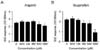
To examine the effects of the aspirin and ibuprofen on the cross-presentation, DC2.4 cells were cultured in the presence of the drugs for 18 h, and then allowed to phagocytose OVA-microspheres for 2 h. The DC2.4 cells were then washed, fixed with paraformaldehyde, and the amount of OVA peptide-class I MHC complexes was measured using a T cell hybridoma, B3Z, which recognizes OVA peptide (SIINFEKL)-H-2Kb complex and expresses β-galactosidase (16). As shown in Fig. 1, aspirin inhibited MHC class I-restricted OVA presentation at concentrations of 500µM or above. The inhibitory activity of ibuprofen on MHC class I-restricted OVA presentation was observed at concentrations of 250µM or above.
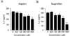
The effects of the drugs on the MHC class II-restricted presentation of exogenous OVA were examined in DCs generated from bone marrow cells (BM-DCs) with GM-CSF. BM-DCs were treated with different concentrations of the drugs for 18 h, and then allowed to phagocytize OVA-microspheres for 2 h. The DCs were then washed, fixed with paraformaldehyde, and then the amount of OVA peptide-class II MHC complexes was measured using OVA-specific CD4 T cell hybridoma, DOBW cells. As shown in Fig. 2, aspirin inhibited MHC class II-restricted OVA presentation at concentration of 250µM or above. The inhibitory activity of ibuprofen on MHC class II-restricted OVA presentation was observed at concentrations of 125µM or above.
DCs developed in the presence of aspirin or ibuprofen are suppressed in the antigen presenting function
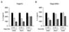
To examine the effects of aspirin and ibuprofen on the acquisition of antigen presenting function, the drugs were added to DC differentiation-inducing cultures at different time points, days 0, 3 and 5 after the initiation of the culture. BM-DCs were harvested on day 6 from the initiation of the culture, and the ability to present exogenous antigen in association with MHC class II molecules was examined as in Fig. 2. As shown in Fig. 3A, DCs generated from mouse bone marrow cells in the presence of aspirin from the initiation of the culture (Day0) or 3 days after the initiation of the culture (Day3) were profoundly suppressed in MHC class II-restricted OVA presenting capability. This inhibitory effect of aspirin appeared to be dose dependent. When developing DCs were exposed to aspirin at a later time point (Day5, 5 days after the initiation of culture), the suppressive activity of aspirin on antigen presenting capability was weaker compared to the earlier exposures (Day0 and Day3). Likewise, DCs generated in the presence of ibuprofen from earlier time points of differentiation (Day0 and Day5) were significantly suppressed in MHC class II-restricted antigen presenting capability. We also found that DCs generated in the presence of aspirin or ibuprofen were suppressed in the MHC class I-restricted exogenous antigen presentation, and the suppressive activities of the drugs were more prominent when developing DCs were exposed to the drugs at earlier time points (data not shown).
Aspirin and ibuprofen do not inhibit the phagocytic activity of DCs
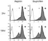
To examine whether the suppressed capacity of aspirin or ibuprofen-treated DCs to present OVA peptides in association with MHC molecules was due to decreased phagocytic activity, DC2.4 cells were cultured with the drugs (1 mg/ml) for 18 h, and then added with microspheres containing both OVA and FITC. After 2 h incubation, unphagocytized microspheres were washed, and the cells were harvested by gentle pipetting after cooling on ice. Flow cytometric analysis of the harvested cells showed that neither of the drugs suppressed the phagocytic activity of DC2.4 cells (Fig. 4).
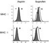
Ibuprofen, but not aspirin, slightly increases the expression of MHC molecules and co-stimulatory molecules
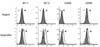
To examine whether the suppressed capacity of aspirin or ibuprofen-treated DCs to present OVA peptides in association with MHC molecules was due to the inhibition of expression of MHC molecules on the cell surface, DC2.4 cells were cultured with the drugs (1 mg/ml) 18 h, and then the expression levels of MHC class I and class II molecules were determined by anti-H-2Kb and anti-I-Ab monoclonal antibodies. As shown in Fig. 5, aspirin did not affect the expression of MHC class I or class II molecules. Ibuprofen, which inhibited MHC-restricted exogenous OVA presentation, even slightly increased the expression of MHC class I and class II molecules. Flow cytometric analysis for major co-stimulatory molecules such as B7-1, B7-2, and CD40 also showed that aspirin did not affect the expression of these co-stimulatory molecules (Fig. 6). Again, ibuprofen, which inhibited MHC-restricted exogenous OVA presentation, even slightly increased the expression of co-stimulatory molecules such as B7-1, B7-2, and CD40 (Fig. 6).
DISCUSSION
The present study demonstrates that DCs exposed to aspirin or ibuprofen for 18 h are suppressed in MHC-restricted exogenous antigen presenting capability. Aspirin and ibuprofen significantly inhibited MHC class I-restricted exogenous antigen presenting capability of DCs at doses of 500µM or above and of 250µM or above, respectively. The MHC class II-restricted exogenous antigen presenting capability of DCs was inhibited at lower concentration of the drugs-250µM or above for aspirin and 125µM or above for ibuprofen. When aspirin is used as an anti-inflammatory drug in doses of 1,200 mg/day, the in vivo therapeutic levels are in between 100~2,000µM (21). Administration of ibuprofen in doses of 400 mg three times/day translates to up to 120µM in the plasma (22). Taken together, our results suggest that aspirin and ibuprofen suppress antigen presenting function of DCs at clinically relevant concentrations.
Because NSAIDs are usually used for prolonged time periods in the treatment of chronic inflammatory diseases, we were curious to investigate whether the DCs exposed to aspirin or ibuprofen during development could acquire normal function in terms of antigen processing and presentation. Our results show that DCs generated from mouse bone marrow cells in the presence of aspirin (100µM) or ibuprofen (100µM) from the early stage of differentiation (Day 0 or 3 after the differentiation initiation culture) were profoundly suppressed in MHC-restricted exogenous antigen presenting capability. These results suggest that aspirin and ibuprofen, when used in prolonged and higher doses, exert suppressive activity on adaptive immunity in addition to the well-known mechanism of COX inhibition.
DCs play a key role in the initiation of primary immune responses (23-25). DCs can acquire and process antigens in the periphery, and migrate to secondary lymphoid tissues where they prime the primary T cell responses. The capability of DCs to activate even naïve T cells in a primary response has been explained by their ability to express high levels of MHC class II molecules and co-stimulatory molecules. The present study shows that aspirin and ibuprofen suppress MHC class II-restricted antigen presentation more strongly than MHC class I-restricted antigen presentation in DCs. The effects of the expression of co-stimulatory molecules appeared to be different between the two drugs. Aspirin did not affect the expression of co-stimulatory molecules in DC2.4 cells, whereas ibuprofen slightly increased the expression of co-stimulatory molecules. Ibuprofen also slightly increased the expression of MHC class I and class II molecules in DC2.4 cells. Although ibuprofen slightly increased the expression of co-stimulatory molecules as well as MHC molecules, the DCs treated with ibuprofen were suppressed in activating OVA-specific CD4 T cells compared to normal DCs. Because T cells can only recognize antigens presented in the form of short peptide-MHC complexes presented by professional antigen presenting cells (22-25), inhibition of the MHC-restricted antigen presenting function of DCs by ibuprofen might have had significant effects in activating T cells.
In all of the experiments described in the present study, DCs were exposed to the drugs for 18 h, and the cells were allowed to phagocytose OVA-microspheres for 2 h. The DCs were washed to remove unphagocytosed OVA-microspheres, fixed with paraformaldehyde, and then washed thoroughly again to remove paraformaldehyde before functional assays with OVA-specific CD4 or CD8 T cells. Thus, the activation of OVA-specific T cells must be due to enhanced expression of OVA peptides on DCs, and not due to the carryover of the drugs to T cell cultures.
In summary, we have demonstrated that aspirin and ibuprofen at high concentrations inhibit both MHC class I and class II-restricted exogenous antigen presentation. In addition, we showed that the DCs generated in the presence of these drugs are profoundly suppressed in MHC-restricted antigen presentation capability. These results suggest that aspirin and ibuprofen may exert their anti-inflammatory activity by the inhibition of the antigen presenting function of DCs in addition to the well-known mechanism of COX inhibition, especially when these drugs are used in prolonged durations.
 XML Download