

 PDF
PDF ePub
ePub Citation
 Print
Print


INTRODUCTION
Recombinant antibodies composed of antigen-binding fragments of whole antibodies such as Fv, scFv or Fab have been acknowledged to have many applications in biological research. They also offer several advantages including the production of functional forms in bacteria and easy genetic manipulation to confer desired features to the antigen-binding fragments (1,2). The smallest functional unit of antibodies required for proper antigen binding is an Fv fragment, a heterodimer of the heavy chain variable domain (VH) and the light chain variable domain (VL). Unlike whole IgG or Fab molecules, however, the dissociation constant between the VH and VL domains of an Fv fragment ranges only from 10-5~10-8 M, which is not sufficient to keep the domains associated with each other since there are no interchain disulfide interactions (3-5).
As an alternative, VH and VL domains are connected via a short peptide linker so that stable Fv fragments can be produced as a single polypeptide in Eschericha coli. Such single-chain variable fragments (scFv) are generally known to retain antigen-binding specificity and affinity of the parent antibody (6-9). The scFv format of an antibody molecule has a distinctive advantage over its Fab counterpart in that high yield can be obtained in E. coli because of a single polypeptide structure in nature. In some cases, however, scFv fragments are unstable and have a much lower affinity against the antigens than that of Fab or whole Ig counterpart, especially when they are fused to toxins for constructing recombinant immunotoxins, probably due to the interference of a peptide linker with antigen binding or in sufficient stabilization of the Fv molecules (10,11). To circumvent this drawback, it had been attempted to uitlize a disulfide-stabilized Fv (dsFv) fragment that can be generated by introducing interchain disulfide bonds artificially at the framework regions of VH and VL domains (12,13). A dsFv fragment is found to be considerably more stable and shows better antigen-binding capacity than the corresponding scFv counterpart, suggesting that dsFv format is more useful than scFv as therapeutic and diagnostic reagents (14,15). Unfortunately, generation of a dsFv is not trivial because appropriate amino residues in conserved framework regions have to be identified for the disulfide bridge by molecular modeling technology, followed by cumbersome point-directed mutagenesis.
Another problem in producing functional antibody fragments, even scFv, in E. coli is that a reduced state of the E. coli cytoplasm acts strongly against the formation of disulfide bonds in proteins (16). Therefore, antibody fragments must be secreted to the periplasmic space, but it frequently leads to a low yield of soluble antibody fragments primarily due to the aggregation and degradation of the fragments in the periplasm, inefficient translocation through the cytoplasmic membrane, and lysis of host cells in many cases (17-19). Expression of antibody fragments in the cytoplasm of E. coli may alleviate cellular toxicity if the mutant trxB E. coli strain that has much oxidizing cytoplasmic environment were utilized so that the formation of disulfide bonds of polypeptides in the cytoplasm is allowed (20). For examples, high yield of functional scFv fragments from the anti-progesterone antibody, DB3, has been obtained by cytoplasmic expression of the antibody fragments using E. coli ADA494 strain (trxB mutant) (21,22), and soluble Fab antibody fragments have also been successfully produced in the cytoplasm of E. coli trxB gor mutant (23,24).
In this study, we accessed the possibility of producing a novel functional Fv fragment, named zipFv, by linking VH and VL fragments via Fos/Jun leucine zipper and its fusion polypeptides with alkaline phosphatase (AP), named zipFv-AP, and demonstrated that the functional zipFv and the zipFv-AP antibody fragments can be expressed in the cytoplasm of E. coli trxB gor mutant using the VH and the VL domains from SP112, the human Fab clone specific for pyruvate dehydrogenase complex-E2 (PDC-E2), as a model system.
MATERIALS AND METHODS
Bacterial strains and oligonucleotides
Escherichia coli strain ElectroTen Blue (Δ(mcrA)183 Δ(mcrCB-hsdSMR-mrr)173 endA1 supE44 thi-1 recA1 gyrA96 relA1 lac KanR [F' proAB lacIqZΔM15 Tn10 (TetR)]) (Stratagene, USA) was used as the bacterial host for the preparation of recombinant vectors and DNA cloning, and Origami(DE3) (Δ(araleu) 7697 ΔlacX74 ΔphoA PvuII phoR araD139 aphC galE galK rpsL F'[lac+ lacIq pro] (DE3) gor522::Tn10 trxB (KanR, StrR, TetR)) (Novagen, USA) for the expression of recombinant antibody fragments. All oligonucleotides used in this study were synthesized from Bioneer Co. (South Korea).
DNA cloning procedure
All DNA cloning experiments were carried out according to the standard procedures (25), and Ex-Taq polymerase and Pfu DNA polymerase (Takara, Japan) were successfully used for the polymerase chain reactions (PCR). The pCzFv, pCzFvHAP and pCzFvLAP vectors were constructed previously as shown in Fig. 1 (IG Therapy, South Korea. unpublished). The VH and the VLκ chain genes were PCR amplified from SP112 Fab clone (26) at the condition of 35 cycles of 94℃ 1 min, 55℃ 1 min, 72℃ 1 min, followed by 72℃ soaking for 10 min using human Ig-specific VH and JH, or VLκ and Jκ primers synthesized according to the previous report with slight modification (VH sense: 5'-GGGGGCCCAGCCGGCCATGGCCGAGGTGCAGCTGGTGGAGTCTGG-3', JH antisense: 5'-GGGGGCCACATTGGCCGATGAGGAGACGGTGACCAKGGTBCCTTGGCCCCA-3', Vκ sense: VLκ forward: 5'-GGGGTCGACATGGAAATTGAGTTGACGCAGTCTCC-3', Jκ antisense: 5'-GGGCCGCGGATACGTTTGATHTCCASYTTGGTCCC-3'; where Sfi I, Sal I and Sac II recognition sites were underlined, and degeneracy is denoted as follows: H=A, C or T; S=G or C; Y=C or T; K=G or T; B=G, T or C) (27).
The VLκ gene fragment of SP112 was recovered from 1.2% agarose gel, treated with Sal I and Sac II (Takara), and cloned into pCzFv, pCzFvHAP and pCzFvLAP at a 2 : 1 molar ratio using T4 DNA ligase (Takara) at 16℃ overnight. The resulting DNA ligated reactions were electroporated into ElectroTen Blue competent cells using Gene Pulser (Biorad, USA) at 2.5 kV, 25µF and 400Ω, and the transformed cells were recovered by growing them in SOC medium for 1 h at 37℃. Then, the cell suspensions were spread onto 2× YT plates containing 50µg/ml ampicillin (2× YT/amp). E. coli colonies carrying pCzFv, pCzFvHAP or pCzFvLAP vectors with correct VLκ insert were identified by cutting the miniprep plasmid with Sal I and Sac II, followed by 1.2% agarose gel electrophoresis. Thereafter, the VH gene obtained from SP112 was treated with Sfi I (Takara), and subsequently cloned into the pCzFv, pCzFvHAP, and pCzFvLAP vectors containing VLκ inserts. The resulting pCzFv, pCzFvHAP and pCzFvLAP vectors having both VH and VLκ genes of SP112 were named as pCzFv-112, pCzFvHAP-112 or pCzFvLAP-112, respectively, and electroporated into Origami(DE3) cells.
The gene encoding a DNA binding domain of AraC (amino acid residues 168~291) (AraCDNA) (28,29) was obtained by PCR amplification from the pBAD/gIII vector (Invitrogen, USA) using PCR primers (sense: 5'-GGGACTAGTAACGAGTCGCTCCATCCACCG-3'; antisense: 5'-GGGGTCGACACTAGTTTATGACAACTTGACGGCTAC ATCA-3', where Spe I recognition site was underlined). The resulting 408 bp PCR products were digested with Spe I restriction enzyme, and cloned into the pCzFvLAP-112 that had been treated with Nhe I. The ligation reaction was electroporated into ElectroTen Blue cells and finally into Origami(DE3) as above. The resulting construct was named pCzFvHADLAP-112.
For the cytopalsmic co-expression of DsbC, the full-length E. coli DsbC gene without signal peptide sequence was obtained from the genomic DNA of BL21(DE3) cells by PCR using following PCR primers (sense: 5'-GGGCATATGGATGACGCGGCAATTCAACAA-3', antisense: 5'-GGGGGTACCTTATTTACCGCTGGTCATTTTTTG-3', Nde I and Kpn I restriction sites were underlined). The resulting PCR products were recovered from 1% agarose gel using Wizard DNA purification kit (Promega, USA). Then the DNA fragments were restricted with Nde I/Kpn I, and cloned into pCDFDuet (Novagen), resulting in the construction of pCDF-DsbC. Origami(DE3) cells were transformed with the construct, and selected with LB medium supplemented with 50µg/ml of spectinomycin (Sigma Co.).
Preparation of antigens
Recombinant inner lipoyl domain of human PDC-E2 (residues 91~227) fused to glutathione S-transferase (GST) was prepared by growing E. coli having pGEX vector with PDC-E2 inner lipoyl domain cDNA in 200 ml of LB containing 50 µg/ml ampicillin (LB/amp) supplemented with 1 mM isopropyl-β-D-1-thiogalactopyranoside (IPTG) until OD600=0.5 at 37℃ (30). The cell culture was centrifuged at 3,000×g for 10 min, and the resulting cell pellet was resuspended in 5 ml of ice-cold phosphate-buffered saline (PBS) supplemented with 1% Triton X-100 (Sigma Co.). The cell suspension was sonicated 5 times for 5 sec each with 2 min intervals on ice in the presence of 100 µg/ml phenylmethylsulfonyl fluoride (PMSF) (Sigma Co.), and microfuged for 5 min at 4℃ to remove cell debris. For the purification of the protein, glutathione-agarose bead (Sigma Co.) in 2 ml PBS were mixed with the supernatant at 4℃ overnight, washed with ice-cold PBS 4 times, and recombinant proteins were eluted with 5 mM reduced glutathione (Sigma Co.) in 50 mM Tris-Cl (pH 8.0). Negative control antigens were prepared as follow. Maltose binding protein (MBP) was prepared by growing E. coli harboring pMAL-c2 (New England Biolab, USA) using amylose-binding resin according to the manufacturer's protocol. Human recombinant IL-15 proteins were produced by using pET expression system and purified with Ni-NTA resin (IG Therapy Co., unpublished). Bovine serum albumin (BSA) was purchased from Sigma Co.
Production of the zipFv and the zipFv-AP fragments
Origami(DE3) cells carrying pCzFv-112, pCzFvHAP-112, pCzFvLAP-112 or pCzFvHADLAP-112 were grown on 5 ml of 2× YT/amp until OD600=approximately 0.5. Then IPTG was added into the culture at 0.1 mM final concentration, and production of the zipFv molecules was induced at 27℃ for 16 h. The cells were harvested by centrifugation (4,000 rpm for 10 min) and resuspended in 2 ml of cold PBS containing 100 µg/ml of PMSF. The cell suspension was sonicated 5 times on ice, and microfuged for 5 min at 4℃ to obtain cytoplasmic extract. The resulting cytoplasmic extracts were used in the enzyme-linked immunosorbent assay (ELISA). For the co-expression of DsbC and the zipFv-AP fusion molecules, electrocompetent cells were prepared with Origami(DE3) cells housing pCDF-DsbC, and pCzFvHADLAP-112 was introduced into the cells by electroporation as described previously. The cells having both pCDF-DsbC and pCzFvHADLAP-112 were selected by growing them in 2× YT/amp medium supplemented with 50µg/ml of spectinomycin.
ELISA
Maxisorp 96-well plates (Nunc, Denmark) were coated with 50 µl of 10 µg/ml PDC-E2, recombinant human IL-15, MBP, or BSA in coating buffer (0.1 M NaHCO3, pH 9.6) at 4℃ overnight and blocked with 3% powdered milk in PBS (MPBS) for 2 h at room temperature. Then, serial dilutions of the cytoplasmic extracts (50 µl) were added into each of the microwells, and incubated for 2 h at 27℃. The plates were washed four times with PBS containing 0.05% Tween 20 (PBST), and mouse anti-myc tag monoclonal antibody (mAb) (IG Therapy, Co.) followed by goat anti-mouse IgG-horse radish peroxidase (HRPO) conjugated (Sigma Co.) was added and incubated for 1 h at 27℃, respectively. The signal was visualized by adding 50 µl of 3.3',5.5'-tetramethyl benzidine (TMB) substrate (Sigma Co.), and the enzymatic reaction was stopped by adding an equal volume of 1 N HCl into each well. Finally, absorbance at 450 nm was measured using ELISA reader (Biorad). In order to determine the alkaline phosphatase activity (AP) of the zipFv-AP fusions bound to antigens, 50 µl of p-nitrophenyl phosphate (pNPP) substrate (Sigma Co.) were directly added into each well instead of using anti-myc tag mAb. The enzymatic reaction was stopped by adding 3 N NaOH into each well, and absorbance at 405 nm was measured using ELISA reader.
Western blot
Origami(DE3) cells carrying pCzFv-112, pCzFvHAP-112 or pCzFvLAP-112 were grown in 2 ml of 2× YT/amp medium at 37℃ until OD600 reached approximately 0.5, and the expression of the zipFv and its AP fusion proteins was induced as above. The cells were harvested by microfugation and resuspended with SDS sample buffer with or without β-2-mercaptoethanol, and total cellular proteins were separated with 12% SDS-PAGE (25). The proteins were transferred to nitrocellulose membrane (Amersham Pharmacia, Sweden), and the membrane was blocked with MPBS. After washing the membrane three times with PBST, anti-myc tag mAb followed by incubation with goat anti-mouse IgG-alkaline phosphatase (AP) conjugated (Sigma Co.), or nitro blue tetrazolium chloride (NBT)/5-bromo-4-chloro-3-indolyl phosphate (BCIP) substrate (Sigma Co.) directly was used to detect the presence of the zipFv or its AP activity in total E. coli lysate. The bands on the blot were analyzed with densitometer (Biorad).
RESULTS
Construction of zipFv expression vectors
It is well accepted that bacterially expressed antibody fragments (Fv, scFv or Fab) provide a simple and cost-effective alternative to the traditional production of diagnostic mAbs by animal cell tissue culture, and numerous recombinant antibody fragments have been successfully produced through periplasmic expression using E. coli. Although scFv format is advantageous in prokaryotic expression because of its single polypeptide structure, disulfide-stabilized Fv (dsFv) had also been recognized to be more useful than scFv as therapeutic and diagnostic agent through its superior stabilization (14). SP112, a human Fab binds to PDC-E2, was chosen in this study because it is available in our laboratory at hand and a scFv version of SP112 does not show any significant antigen-binding reactivity to PDC-E2 (data not shown).
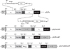
To evaluate whether heterodimerization of VH and VL of SP112 via Fos (40 amino acid residues in size)/Jun (40 amino acid residues in size) leucine zipper could retain the antigen-binding reactivity, a new Fv expression vector, pCzFv, was constructed using the pET22b expression vector as a backbone (Fig. 1). The Fos : Jun leucine zippers were used to link VH and VL fragments since they preferentially form heterodimers (Fos : Jun) at least 1,000 times over homodimers (Fos : Fos or Jun : Jun) due to greater thermodynamic stability (31), and the same amphiphilic helix leucine zipper had already been successfully used in the generation of dimeric and bispecific scFv fragments (2).
The bicistronic expression of VH-Fos-myc and VL-Jun-S1 gene fragments driven by a single p7 promoter ensures the equal transcription of the fragments, and partial mouse IgG3 hinge fragments (10 amino acid residues in size) are introduced between the VH and Fos domains as well as between the VL and Jun domains to confer structural flexibility. Two cysteine residues are also added at the flanking region of each Fos and Jun segment, respectively, so that two interchain disulfide bonds could be formed in the zipper region to reinforce the binding of Fos/Jun dimer (2). In addition, myc tag and S1 tag are placed at the end of Fos or Jun, respectively, to facilitate the detection of each antibody fragments.
As similar to a scFv-AP fusion produced in the periplasm of bacteria (32), fusion of AP to the zipFv might be advantageous in that binding of the antibody fragments to antigen can be analyzed in an ELISA or Western blot without use of additional enzyme-conjugated antibodies. Thereby, the pCzFv vector was further modified to pCzFvHAP or pCzFvLAP vector by linking leaderless phoA gene (1,470 bp in length) encoding alkaline phosphatase (AP) (46.5 kDa in size) at the 3' end of either the VH-Fos-myc or the VL-Jun-S1 gene fragment, respectively, to produce the zipFv-AP fusion proteins [zipFv-112(H-AP) or zipFv-112(L-AP)]. Finally, the VH and the VL genes of SP112 were cloned into the pCzFv, pCzFvHAP and pCzFvLAP vectors, and the resulting vector constructs, named as pCzFv-112, pCzFvHAP-112 and pCzFvLAP-112, respectively, were electroporated into Origami(DE3) trxB gor mutant E. coli cells.
Expression and characterization of the zipFv and the zipFv-AP fusion proteins
The Origami(DE3) trxB gor cells carrying pCzFv-112, pCzFvHAP-112 or pCzFvLAP-112 were grown in 2× YT/amp medium, and the recombinant antibody fragments were expressed by adding 0.1 mM IPTG at 27℃ for 16 h. The zipFv and the zipFv-AP fusion proteins produced by each of the host cells were named as zipFv-112, zipFv-112(H-AP) or zipFv-112(L-AP), respectively, The optimal induction condition had been pre-determined using various concentrations of IPTG (0~1 mM), temperature (27℃ or 37℃) and induction time (2~16 h). Growing cells at 27℃ for 16 h produced at least 1~3 times more functional zipFv and zipFv-AP fusion fragments than at 37℃ for 4 h, and 0.1 mM final concentration of IPTG gave the highest yield of recombinant antibody fragments (data not shown). It was noteworthy that no host cell lysis was observed during IPTG induction of the zipFv or zipFv-AP fusion proteins whereas over-expression of SP112 in the periplasm of E. coli caused host cell lysis (data not shown).
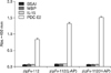
To determine the antigen-binding specificity of the antibody fragments present in the cytoplasmic extracts of the host cells, ELISA was performed using microtiter plates coated with recombinant PDC-E2, recombinant human IL-15, MBP or BSA. Anti-myc tag mAb was used for detecting the antibody fragments bound to antigens. As shown in Fig. 2, the zipFv-112 and its AP fusions, the zipFv-112(H-AP) and the zipFv-112(L-AP), specifically reacted to PDC-E2 only, but not to any other negative control antigens, demonstrating that the recombinant antibody fragments of the zipFv and the zipFv-AP formats retained the antigen-binding specificity of SP112, even if they were produced in the cytoplasm of the host cells. This also implied that the proper disulfide bonds were formed between the VH-Fos-myc fragment and the VL-Jun-S1 fragment in the cytoplasm of the trx gor mutant E. coli.
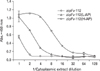
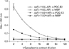
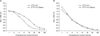
Since Fig. 2 showed that the zipFv-112 fragment exhibited the weakest antigen-binding signal among the three antibody fragments, yield of the functional zipFv-112, the zipFv-112 (H-AP) and the zipFv-112(L-AP) in the cytoplasmic extracts were compared by ELISA (Fig. 3). The cytoplasmic extracts from the cells that grown at the exactly same experimental conditions were prepared and serial dilutions were made in MPBS. Mouse anti-myc tag mAb followed by goat anti-mouse IgG-HRPO conjugated was used to detect the VH-Fos-myc fragments of the zipFv-112 and the zipFv-112(L-AP) or the VH-Fos-myc-AP fragments of the zipFv-112(H-AP) that bound to PDC-E2. The results revealed that the zipFv-112(H-AP) showed about 4 times higher binding signal to PDC-E2 than that of the zipFv-112(L-AP) and 10 times higher than that of the zipFv-112, implying that the AP fusion at the C-terminus of the VH-Fos-myc of the zipFv increases yield of the functional antibody fragments uppermost. To confirm this finding, experiments were performed as same as Fig. 3 using the same cytoplasmic extracts containing the zipFv-112(H-AP) or the zipFv-112(L-AP), except that AP substrate (pNPP) instead of anti-myc tag mAb was directly applied to visualize ELISA signals (Fig. 4). Unexpectedly, the data indicated that PDC-E2-binding signal of the zipFv-112(H-AP) was about 5-fold lower than that of the zipFv-112(L-AP). It implies that that an AP domain of the VL-Jun-S1 would block the myc-tag epitope of the VH-Fos-myc domain that recognized by anti-myc tag mAb in the zipFv-112(L-AP).
Western blot analysis to determine the expression level of the VH and the VL fusion molecules
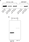
To understand these contradictory results, cell lysates of Origami(DE3) trxB gor cells housing pCzFv-112, pCzFvHAP-112 or pCzFvLAP-112 were prepared, and the same concentration of total proteins were analyzed by western blot (Fig. 5A). Mouse anti-myc tag mAb followed by anti-mouse IgG-AP conjugated was used to determine expression level of the VH-Fos-myc of the zipFv-112 or the zipFv-112(L-AP), and the VH-Fos-myc-AP fragment of the zipFv-112(H-AP) in the cell lysates. In addition, the AP activity of the VH-Fos-myc-AP of the zipFv-112(H-AP) and the VL-Jun-S1-AP of the zipFv-112 (L-AP) was also measured by applying NBT/BCIP substrate directly to the blot. The denstometric analysis of the blot revealed that the VH-Fos-myc (21 kDa) of the zipFv-112 was expressed about 5-fold higher than the VH-Fos-myc-AP (67.5 kDa) of the zipFv-112(H-AP) and 4-fold higher than the VH-Fos-myc of the zipFv-112(L-AP), even though the zipFv-112 showed the weakest antigen-binding signal in ELISA as shown in Fig. 3. To resolve this contradiction we carried out western blot analysis of the cell lysate expressing the zipFv-112 at reducing and non-reducing conditions of 12% SDS-PAGE using anti-myc mAb, and the results revealed that only 7% of total VH-Fos-myc fragments in the cytoplasm of Origami(DE3) cells formed the functional zipFv molecules according to densitometry analysis, indicating that majority of the VH-Fos-myc and the VL-Jun-S1 fragments were not properly dimerized each other in the cytoplasm of Origami(DE3) cells (Fig. 5B). On the other hand, the fusion of an AP moiety at the C-terminus of either the VH-Fos-myc or the VL-Jun-S1 resulted in lower expression of the VH-Fos-myc of both zipFv-112(H-AP) and zipFv-112(L-AP). Difference in expression level between the VH-Fos-myc-AP of the zipFv-112(H-AP) and the VH-Fos-myc of the zipFv-112(L-AP) was meager, although the zipFv-112(H-AP) showed a 4-fold higher binding signal to PDC-E2 than the zipFv-112(L-AP) as shown in Fig. 3.
Interestingly, blot data by using AP substrate (NBT/BCIP) directly showed that the VL-Jun-S1-AP of the zipFv-112(L-AP) seemed to be produced 50-fold higher than the VH-Fos-myc-AP of the zipFv-112(H-AP) in the cytoplasm of the cells. This may explain a 5-fold higher antigen-binding signal of the zipFv-112(L-AP) than that of the zipFv-112(H-AP) as shown in Fig. 4. However, additional SDS-PAGE analysis followed by Coomassie blue staining revealed that the higher AP activity of the VL-Jun-S1-AP of the zipFv-112(L-AP) didn't come from the higher expression level of the fragment than the VH-Fos-myc-AP of the zipFv-112(H-AP) (data not shown). Taken together, the data imply that the positioning of the AP moiety at the C-terminus of the VH-Fos-myc somehow hinders AP activity. Unfortunately, we were not able to present western blot data showing VL-Jun-S1 and VL-Jun-S1-AP expression level because commercial anti-S1 tag mAb produced too much background on the blot. Our unpublished observation indicated that VH domain usually has a higher tendency to aggregate each other compared to VL domain when it is expressed in E. coli probably due to the hydrophobic residues exposed outward. Therefore, we assume that presence of AP seemed to reduce this self-aggregation probably by concealing hydrophobic residues in the variable regions (33), resulting in the increase of functional zipFv-AP molecules compared with a zipFv format.
Effect of an irrelevant fusion moiety at the C-terminus of the VH-Fos-myc on the functionality of the zipFv-112(L-AP) construct
We further investigated to see whether incorporation of an irrelevant protein, such as DNA-binding domain of AraC (AraCDNA), at the C-terminus of the VH-Fos-myc of the zipFv-112(L-AP) might improve detection of the antigen-binding zipFv-112(L-AP) by anti-myc tag mAb while keeping its high AP activity. AraCDNA was chosen because the gene fragment encoding the protein with appropriate restriction enzyme recognition sites was readily available in our laboratory, and it had been acknowledged that the domain could be produced in soluble form in the cytoplasm of E. coli (34). The gene fragment of the AraCDNA was cloned at the 3' end of myc tag of the pCzFvLAP-112 to construct the pCzFvHADLAP as shown in Fig. 1, and the resulting zipFv-112(H-AD/L-AP) fragments were produced in Origami(DE3) cells. The antigenbinding intensity of the zipFv-112(L-AP) and the zipFv-112 (H-AP) was compared by ELISA using anti-myc tag mAb as Fig. 3 (Fig. 6). The ELISA results showed that the fusion of AraCDNA domain at the C-terminus of the VH-Fos-myc of the zipFv-112(L-AP) restored the antigen-binding signal of the zipFv-112(H-AD/L-AP) to the same degree as that of the zipFv-112(H-AP), indicating that the presence of a irrelevant protein moiety at the C-terminus of the VH-Fos-myc exposes myc epitope properly. In terms of AP activity, the zipFv-112(H-AD/L-AP) showed the similar degree of antigen-binding intensity as the zipFv-112(L-AP) when AP substrate was used directly in ELISA (data not shown).
Effect of DsbC on the functional expression of the zipFv-AP fragments
Co-expression of chaperone proteins such as the Skp chaperone protein with antibody fragments had been reported to reduce toxicity and to enhance the proper folding of scFv or Fab fragments expressed in E. coli (35), and Levy et al demonstrated that co-expression of Skp or DsbC chaperone protein increased the cytoplasmic production of functional Fab fragments up to 5-6-fold (23). Additionally, DsbC co-expression increases the yield of tissue plasminogen activator (tPA) in the cytoplasm of E. coli by more than 20-fold (36). Since only minor fraction (<10%) of total VH-Fos-myc dimerized with the VL-Jun-S1 fragments to become functional zipFv or zipFv-AP, we assumed that the same strategy may work for the improved expression of the functional zipFv fragments.
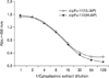
To test whether co-expression of DsbC further improves a production of the functional zipFv-112(H-AD/L-AP), DsbC gene without signal peptide was cloned into pCDFDuet vector. The resulting pCDF-DsbC construct was electroporated into Origami(DE3) cells, and the cytoplasmic expression of a leaderless version of DsbC (217 amino acids, approximately 24 kDa in size) upon IPTG induction was confirmed by 12% SDS-PAGE (data not shown). Electrocompetent Origami(DE3) cells housing pCDF-DsbC were transformed with pCzFvHADLAP-112, and ELISA was performed to compare PDC-E2-binding intensity of the zipFv-112(H-AD/L-AP) expressed with or without DsbC (Fig. 7). The data revealed that the zipFv-112(H-AD/L-AP) with DsbC gave about 1.5-fold higher ELISA signal to the antigen than that of zipFv-112 (H-AD/L-AP) without DsbC when anti-myc tag mAb was used for detecting signals (Fig. 7A). However, no beneficial effect was observed on the VL-Jun-S1-AP stabilization since no improvement on antigen-binding signal was found (Fig. 7B), suggesting that cytoplasmic co-expression of DsbC does not improve functional dimerization of the VH-Fos-myc-AD and the VL-Jun-S1-AP significantly.
Considering that cytoplasmic Fab showed substantial antigen-binding reactivity even at 200-300-fold dilution (23,24), it appeared that the zipFv-AP fusions showed much weaker antigen-binding intensity compared to the Fab fragments produced by cytoplasmic expression. We assume that this might come from the different experimental setups in that we prepared 1.5 ml of the cytoplasmic extracts from 3 ml culture, but Venturi et al used 10 ml of the cytoplasmic extracts obtained from 2 liters of culture for ELISA (24), meaning that their Fab preparation should give an 100-fold higher signal than ours. In the case of the zipFv-112(H-AD/L-AP), it gave a good antigen-binding signal at 1/32 dilution factor, and it can be inferred from the results that this construct would produce as much as 10-fold higher yield of functional cytoplasmic antibody fragments than Fab format. It is possible that high expression rate of our zipFv-AP fusions compared to Fab fragments may come from the different IPTG induction protocol for recombinant antibody production that we used 27℃ 16 h, whereas cytoplasmic Fab expression was performed at 37℃ 4 h. Venturi et al also reported that 50~250 times higher yield of Fab fragments could be obtained by cytoplasmic expression than in the periplasmic space, but we were not able to confirm this observation since we didn't have periplasmic expression module for the zipFv or the zipFv-AP fusions.
It is possible that functional zipFv fragments might form more efficiently than Fab fragments in the cytoplasm of the mutant E. coli because the zipFv requires four pairs of disulfides, two pairs of disulfides in the variable regions and another two pairs between Fos and Jun zipper, whereas Fab needs five pairs in total, including one intramolecular disulfide in the CH1, one in CL kappa, and an intermolecular disulfide linking. However, comparison of the yield between the zipFv and Fab above is only circumstantial unless an identical antibody gene is expressed using the same expression module at the same experimental condition. Additionally, (His)6 tag needs to be implemented to zipFv or zipFv-AP format for purification of the molecules to address the issues on the yield of the antibody molecules precisely and quantitatively.
DISCUSSION
In this study, we demonstrated that a functional Fv fragment can be generated by dimerizing VH and VL fragments with a well-known Fos-Jun zipper, and a resulting zipFv and its derivative fused with AP (zipFv-AP) can be produced as a functional form in the cytoplasm of Origami(DE3) trxB gor mutant E. coli strain. Interestingly, fusion of AP moiety to the zipFv-112 improved formation of the functional zipFv-AP molecules in the cytoplasm of host cells regardless of its location, and also influenced the conformation of the zipFv-AP molecules depending on its location as shown by the zipFv112 (H-AP) and the zipFv-112(L-AP). Additionally, incorporation of the AraCDNA domain into the zipFv-112(L-AP) [zipFv-112(H-AD/L-AP)] improved folding of the zipFv-AP molecules. In contrast to previous reports, cytoplasmic co-expression of DsbC seemed not as quite beneficial as expected. Currently, we are exploring the possibility of replacing the AraCDNA domain of the zipFv-112(H-AD/L-AP) with other functional proteins, for example green fluorescent protein (GFP), to make tri-functional antibody fragments. Considering that the production of recombinant antibody fragments fused with functional molecules such as AP is important in the development of molecular diagnostic reagents for biomedical research, we believe that our zipFv-AP constructs may provide an alternative antibody format for producing bi-functional antibody fragments in the cytoplasm of E. coli in a secure and convenient manner, and possibly for the development of diverse antibody contents for proteome analysis.
 XML Download