

 PDF
PDF ePub
ePub Citation
 Print
Print


Abstract
Purpose
Tracheal bronchus is an aberrant bronchus arising from tracheal wall above the carina. Most cases of tracheal bronchus are asymptomatic, so they are diagnosed incidentally. Tracheal bronchus may be associated with other anomalies. The aim of this study is to evaluate the clinical characteristics of tracheal bronchus.
Methods
This study was conducted on 19 children who were diagnosed as tracheal bronchus by the chest computed tomography from January 2000 to December 2011. Based on the medical record, clinical features, such as symptoms, radiologic findings, combined anomalies were evaluated retrospectively.
Results
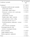
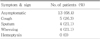
The age at diagnosis was ranged from 2 day to 14 years (mean, 51 months). Among the 19 children, twelve children (63.2%) were boys and seven children (36.8%) were girls. Eighteen children (94.7%) had right-side tracheal bronchus and one child (5.3%) had left-side tracheal bronchus. Displaced type were fourteen children (73.7%), supernumerary type were five children (26.3%). Thirteen children (68.4%) had no respiratory symptoms, but five children (26.3%) had persistent cough and four children (21.1%) had recurrent wheezing. Combined congenital abnormalities were present in seventeen children (89.5%), including congenital cardiovascular anomaly (n=14, 73.7%), trachea-esophageal fistula (n=3, 15.8%), Down syndrome (n=2, 10.5%).
Conclusion
The patients with tracheal bronchus had not severe respiratory symptoms, but had many combined anomalies. So, it is necessary to consider the presence of tracheal bronchus in children with respiratory symptom, like recurrent wheezing, and to evaluate clinical significance, like combined anomaly, in tracheal bronchus patients. The limitation of this study is that the study group includes many cardiovascular disease patients (84.2%).
Figures and Tables
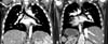 | Fig. 1Tracheal bronchus. (A) Right-side tracheal bronchus. Coronal image in 5-month-old boy shows right upper lobe bronchus arising from carina. (B) Left-side tracheal bronchus. Coronal image in 13-year-old girl shows left upper lobe bronchus arising from the trachea 2 cm proximal to the carina and situs inversus totalis.
|
Table 1
Characteristics of Patients
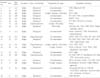
VSD, ventricular septal defect; SVC, superior vena cana; LPA, left pulmonary artery; PA, pulmonary atresia; DORV, double outlet right ventricle; CoA, coarctation of aorta; AS, aortic stenosis; ASD, atrial septal defect; PDA, patent ductus arteriosus; EA, esophageal atresia; TEF, tracheoesophageal fistula; LV, left ventricle; PAPVR, partial anomalous pulmonary venous return; PS, pulmonary stenosis.
![]()
References
1. Ghaye B, Szapiro D, Fanchamps JM, Dondelinger RF. Congenital bronchial abnormalities revisited. Radiographics. 2001. 21:105–119.

2. Siegel MJ, Shackelford GD, Francis RS, McAlister WH. Tracheal bronchus. Radiology. 1979. 130:353–355.
3. Ritsema GH. Ectopic right bronchus: indication for bronchography. AJR Am J Roentgenol. 1983. 140:671–674.
4. McLaughlin FJ, Strieder DJ, Harris GB, Vawter GP, Eraklis AJ. Tracheal bronchus: association with respiratory morbidity in childhood. J Pediatr. 1985. 106:751–755.
5. Venkateswarlu T, Turner CJ, Carter JD, Morrow DH. The tracheal bronchus: an unusual airway probelm. Anesth Analg. 1976. 55:746–747.
6. Vredevoe LA, Brechner T, Moy P. Obstruction of anomalous tracheal bronchus with endotracheal intubation. Anesthesiology. 1981. 55:581–583.
7. Shipley RT, McLoud TC, Dedrick CG, Shepard JA. Computed tomography of the tracheal bronchus. J Comput Assist Tomogr. 1985. 9:53–55.
8. Wong KS, Lien R, Lin TY. Clinical and computed tomographic features of tracheal bronchus in children. J Formos Med Assoc. 1999. 98:646–648.
9. Won JH, Park JY, Kang TK, Park KS, Kim YJ, Kim CH, et al. A clinical experience of tracheal bronchus. Tuberc Respir Dis. 1998. 45:583–586.
10. Kim JC, Kim YJ, Kang BJ, Youn YD, Lee SY, Kwon YL, et al. The clinical evaluation of anomalous bronchi arising from the trachea and main bronchi. Tuberc Respir Dis. 2005. 59:664–669.
11. Choi JA, Chung NG, Lee JS, Whang KT, Cho SH. A case of tracheal bronchus. Pediatr Allergy Respir Dis. 1998. 8:112–118.
12. Kong BG, Lee YK, Jeong EY, Lee WK, Kim KW, Koh JK. A demonhstration of a tracheal bronchus by bronchoscopy and computed tompgraphy. J Korean Pediatr Soc. 2000. 43:1501–1504.
13. Choi AR, Choi SH, Kim SW, Sung DW, Rha YH. A case of recurrent respiratory infection resulting from a congenital anomaly of the bronchial tree tracheal bronchus. Korean J Pediatr. 2008. 51:660–664.
14. Harris JH Jr. The clinical significance of the tracheal bronchus. Am J Roentgenol Radium Ther Nucl Med. 1958. 79:228–234.
15. Foster-Carter AF. Broncho-pulmonary abnormalities. Br J Tuberc Dis Chest. 1946. 40:111–124.
16. Inada K, Kishimoto S. An anomalous tracheal bronchus to the right upper lobe; report of two cases. Dis Chest. 1957. 31:109–112.
17. Vevecka E, De Boeck K, Moerman P, van Raemdonck D, Lerut T. Tracheal bronchus associated with congenital cystic adenomatoid malformation. Pediatr Pulmonol. 1995. 20:413–416.
18. Alescio T, Cassini A. Induction in vitro of tracheal buds by pulmonary mesenchyme grafted on tracheal epithelium. J Exp Zool. 1962. 150:83–94.
19. Bertrand P, Navarro H, Caussade S, Holmgren N, Sánchez I. Airway anomalies in children with Down syndrome: endoscopic findings. Pediatr Pulmonol. 2003. 36:137–141.
20. Keane MP, Meaney JF, Kazerooni EA, Whyte RI, Flint A, Martinez FJ. Accessory cardiac bronchus presenting with haemoptysis. Thorax. 1997. 52:490–491.
21. Barat M, Konrad HR. Tracheal bronchus. Am J Otolaryngol. 1987. 8:118–122.
22. Gerson CE, Rothstein E. An anomalous tracheal bronchus to the right upper lobe. Am Rev Tuberc. 1951. 64:686–690.
23. Ming Z, Lin Z. Evaluation of tracheal bronchus in Chinese children using multidetector CT. Pediatr Radiol. 2007. 37:1230–1234.
 XML Download