

 PDF
PDF ePub
ePub Citation
 Print
Print


Abstract
Purpose
To evaluate the clinical characteristics and radiologic patterns of adolescents with pulmonary tuberculosis (TB), and to assess whether they are related with primary TB or reactive TB.
Methods
Among the enrolled patients who were diagnosed with pulmonary TB from March 2000 to May 2011, 36 with plain radiography and/or chest computed tomography (CT) were reviewed. We reviewed retrospectively their medical charts to collect clinical data and past history. Among these 36 patients, plain radiography of the 36 patients and chest CT of the 34 patients were retrospectively evaluated.
Results
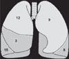
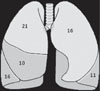
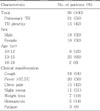
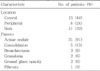
The patients consisted of 18 males and 18 females, and their median age was 14 years old. The most common clinical presentation was cough and fever. Half of them had chronic cough for more than two weeks. Ten patients had history of close contact with adult patients with active pulmonary TB: 7 patients with their parents, 2 patients with friends, 1 patient with their grandmother. The most frequent pattern of plain radiography was pleural effusion (16/36). In the chest CT findings, all cases showed parenchymal lesions and lymphadenopathy. In addition, 91% of the cases showed acinar nodules. The pattern of pleural effusion revealed associated ipsilateral pleural lymph node and subpleural nodule. Rim enhancement and calcification of the lymph node demonstrated 9% (3/34) and 12% (4/34), respectively. Only two of them showed typical hilar lymphadenopathy in chest X ray and CT.
Figures and Tables
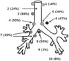 | Fig. 3Location of lymph node involved area in the chest computed tomography scan. 1, highest mediastinal nodes; 2, pre-vascular and retrotracheal nodes; 3, right upper paratracheal nodes; 4 and 5, right and left lower paratracheal nodes; 6, subaortic nodes; 7, right hilar nodes; 8, subcarinal nodes; 9, paraoesophageal nodes; 10, pulmonary ligament nodes.
|
Table 1
Demographic and Clinical Characteristics of the Adolescents with Pulmonary Tuberculosis (TB)
![]()
References
1. Eun BW. Characteristics of tuberculosis in children and adolescents. Korean J Pediatr. 2009. 52:513–518.

2. Korea Center for Disease Control and Prevention, Korean Institute of Tuberculosis, Korean National Tuberculosis Association. 2010 Annual report on the notified tuberculosis patients in Korea. 2011. Seoul: Korea Center for Disease Control and Prevention.
3. American Thoracic Society. Targeted tuberculin testing and treatment of latent tuberculosis infection. MMWR Recomm Rep. 2000. 49(RR-6):1–51.
4. Lobue P, Menzies D. Treatment of latent tuberculosis infection: An update. Respirology. 2010. 15:603–622.
5. de Pontual L, Balu L, Ovetchkine P, Maury-Tisseron B, Lachassinne E, Cruaud P, et al. Tuberculosis in adolescents: A French retrospective study of 52 cases. Pediatr Infect Dis J. 2006. 25:930–932.
6. Inselman LS. Tuberculosis in children: an update. Pediatr Pulmonol. 1996. 21:101–120.
7. Starke JR. Tuberculosis in children. Curr Opin Pediatr. 1995. 7:268–277.
8. Koh WJ, Jeong YJ, Kwon OJ, Kim HJ, Cho EH, Lew WJ, et al. Chest radiographic findings in primary pulmonary tuberculosis: observations from high school outbreaks. Korean J Radiol. 2010. 11:612–617.
9. Im BC, Kim Y, Kim KS, Kim YW, Kim EY, You EJ, et al. Clinical review of pulmonary tuberculosis in teenagers according to the involved lung. Korean J Pediatr Infect Dis. 2010. 17:148–155.
10. Byeon JH, Lee Y, Lee JC, Yoo Y, Lee KH, Lee KC, et al. Three cases of pulmonary and/or intestinal tuberculosis in adolescents. Korean J Pediatr. 2007. 50:1134–1138.
11. Andreu J, Cáceres J, Pallisa E, Martinez-Rodriguez M. Radiological manifestations of pulmonary tuberculosis. Eur J Radiol. 2004. 51:139–149.
12. Kim WS, Moon WK, Kim IO, Lee HJ, Im JG, Yeon KM, et al. Pulmonary tuberculosis in children: evaluation with CT. AJR Am J Roentgenol. 1997. 168:1005–1009.
13. Harisinghani MG, McLoud TC, Shepard JA, Ko JP, Shroff MM, Mueller PR. Tuberculosis from head to toe. Radiographics. 2000. 20:449–470.
14. Kim HJ, Lee HJ, Kwon SY, Yoon HI, Chung HS, Lee CT, et al. The prevalence of pulmonary parenchymal tuberculosis in patients with tuberculous pleuritis. Chest. 2006. 129:1253–1258.
15. Joint Committee for the Development of Korean Guidelines for Tuberculosis Korea Centers for Disease Control and Prevention. Korean guidelines for tuberculosis. 2011. Seoul: Korea Center for Disease Control and Prevention.
16. Miller WT, Miller WT Jr. Tuberculosis in the normal host: radiological findings. Semin Roentgenol. 1993. 28:109–118.
17. Jeong YJ, Lee KS. Pulmonary tuberculosis: up-to-date imaging and management. AJR Am J Roentgenol. 2008. 191:834–844.
18. Andronikou S, Joseph E, Lucas S, Brachmeyer S, Du Toit G, Zar H, et al. CT scanning for the detection of tuberculous mediastinal and hilar lymphadenopathy in children. Pediatr Radiol. 2004. 34:232–236.
19. Leung AN. Pulmonary tuberculosis: the essentials. Radiology. 1999. 210:307–322.
20. Dosanjh DP, Hinks TS, Innes JA, Deeks JJ, Pasvol G, Hackforth S, et al. Improved diagnostic evaluation of suspected tuberculosis. Ann Intern Med. 2008. 148:325–336.
21. de Charnace G, Delacourt C. Diagnostic techniques in paediatric tuberculosis. Paediatr Respir Rev. 2001. 2:120–126.
22. Lee JY, Choi HJ, Park IN, Hong SB, Oh YM, Lim CM, et al. Comparison of two commercial interferon-gamma assays for diagnosing Mycobacterium tuberculosis infection. Eur Respir J. 2006. 28:24–30.
23. Rigouts L. Clinical practice: diagnosis of childhood tuberculosis. Eur J Pediatr. 2009. 168:1285–1290.
24. Horsburgh CR Jr, Goldberg S, Bethel J, Chen S, Colson PW, Hirsch-Moverman Y, et al. Latent TB infection treatment acceptance and completion in the United States and Canada. Chest. 2010. 137:401–409.
 XML Download