

 PDF
PDF ePub
ePub Citation
 Print
Print


Abstract
Purpose
Mycoplasma pneumoniae pneumonia is rarely found in children under 3 years old. Nevertheless, infants have been recently and frequently diagnosed with M. pneumoniae pneumonia. Therefore, the clinical characteristics of such children were investigated in this study.
Methods
Subjects were 232 infants (group A: 0 to 11 months, group B: 12 to 23 months, group C: 24 to 35 months) who were diagnosed between January 2004 and December 2009 with M. pneumoniae pneumonia infection at Myongji Hospital. We reviewed their medical records, the early and monthly incidence of infection, clinical manifestations, and laboratory findings.
Results
Group A consisted of 28 patients (12.1%), group B 78 (33.6%), and group C 126 (54.3 %). The younger patient group in the peak season, 2006, demonstrated a high incidence rate. Wheezing was more often auscultated in group A than in the other groups. Negative findings on chest X-rays were more often observed in group A. Serologically, high titers of mycoplasma-specific antibody (>1:1,280) were observed in lobar pneumonia and correlated with the severity of clinical manifestations.
Conclusion
The age at which M. pneumoniae infection has been diagnosed has recently decreased and has been found with a particularly high frequency in infants. Despite shorter fever duration before hospitalization and uncertain radiological findings, including M. pneumoniae in the differential diagnosis of pneumonia is recommended for children under 1 year.
Go to : 
References
1. Kim JW, Seo HK, Yoo EG, Park SJ, Yoon SH, Jung HY, et al. Mycoplasma pneumoniae pneumonia in Korean children, from 1979 to 2006-a metaanalysis. Korean J Pediatr. 2009; 52:315–23.

2. Hong JY, Nah SY, Nam SG, Choi EH, Park JY, Lee HJ. Occurrence of Mycoplasma pneumoniae pneumonia in Seoul, Korea, from 1986 to 1995. J Korean Pediatr Soc. 1997; 40:607–13.
3. Pyun BY, Kim HH, Chung JT, Lee JS. A Study as epidemiologic and clinical aspect of Mycoplasma pneumoniae pneumonia during the last 5 years. Pediatr Allergy Respir Dis. 1998; 8:240–7.
4. Lee SS, Youn KL, Kang HH, Cho BS, Cha SH. Clinical findings of Mycoplasma pneumoniae pneumonia under 3 year-old children. Korean J Pediatr Infect Dis. 1999; 6:78–84.
5. Stevens D, Swift PG, Johnston PG, Kearney PJ, Corner BD, Burman D. Mycoplasma pneumoniae infections in children. Arch Dis Child. 1978; 53:38–42.
6. Sabato AR, Martin AJ, Marmion BP, Kok TW, Cooper DM. Mycoplasma pneumoniae: acute illness, antibiotics, and subsequent pulmonary function. Arch Dis Child. 1984; 59:1034–7.
7. Glezen P, Denny FW. Epidemiology of acute lower respiratory disease in children. N Engl J Med. 1973; 288:498–505.
8. Lee SH, Noh SM, Lee KY, Lee HS, Hong JH, Lee MH, et al. Clinico-epidemiologic study of Mycoplasma pneumoniae pneumonia (1993 through 2003). Korean J Pediatr. 2005; 48:154–7.
9. Broughton RA. Infections due to Mycoplasma pneumoniae in childhood. Pediatr Infect Dis. 1986; 5:71–85.
10. Hammerschlag MR. Mycoplasma pneumoniae infections. Curr Opin Infect Dis. 2001; 14:181–6.
11. Lee HS, Choi KM. Recent trends in the prevalence of Mycoplasma pneumoniae pneumonia according to age. Korean J Pediatr Infect Dis. 2008; 15:162–6.
12. Kim KW, Kim KE. Mycoplasma and chlamydia infection in Korea. Korean J Pediatr. 2009; 52:277–82.
13. Denny FW. Acute respiratory tract infections. Taussing LM, Landau LI, editors. editors.Pediatric Respiratory Medicine. St. Louis: Mosby;1999. p. 556–71.
14. Layani-Milon MP, Gras I, Valette M, Luciani J, Stagnara J, Aymard M, et al. Incidence of upper respiratory tract Mycoplasma pneumoniae infections among outpatients in Rhone-Alpes, France, during five successive winter periods. J Clin Microbiol. 1999; 37:1721–6.
15. Kang KS, Woo HO. Pattern of occurrence of Mycoplasma pneumoniae pneumonia in admitted children: southern central Korea, from 1989 to 2002. J Korean Pediatr Soc. 2003; 46:474–9.
16. Park IS, Lee HB. A clinical study of mycoplasma pneumonia in children during recent 5 years. J Korean Pediatr Soc. 1992; 35:1082–8.
17. Korea Centers for Disease Control & Prevention. Laboratory Diagnosis for Infection Disease: Methods for Individual Disease. 3rd ed.Reston: Inomax;2005. 188–201,. p. 267–78.
18. Clyde WA Jr, Denny FW. Mycoplasma infections in childhood. Pediatrics. 1967; 40:669–84.
19. Choi SK, Jung JA, Kim KH, Kim GH. Study of seroprevalence of antimycoplasma antibody in healthy children and its diagnostic value. J Korean Pediatr Soc. 1998; 41:489–97.
20. Lieberman D, Lieberman D, Printz S, Ben-Yaa-kov M, Lazarovich Z, Ohana B, et al. Atypical pathogen infection in adults with acute exacerbation of bronchial asthma. Am J Respir Crit Care Med. 2003; 167:406–10.
21. Esposito S, Blasi F, Arosio C, Fioravanti L, Fagetti L, Droghetti R, et al. Importance of acute Mycoplasma pneumoniae and Chlamydia pneumoniae infections in children with wheezing. Eur Respir J. 2000; 16:1142–6.
22. Kim KW, Lee BC, Lee KE, Kim ES, Song TW, Park MY, et al. Mycoplasma pneumoniae-induced production of proasthmatic mediators in airway epithelium. Korean J Pediatr. 2006; 49:977–82.
23. Waites KB. New concepts of Mycoplasma pneumoniae infections in children. Pediatr Pulmonol. 2003; 36:267–78.
24. Rytel MW. Primary atypical pneumonia: current concepts. Am J Med Sci. 1964; 247:84–104.
25. Kim JH, Chae SA, Lee DK. Clinical findings of Mycoplasma pneumonia in children, from 1998 to 2003. Korean J Pediatr. 2005; 48:969–75.
26. Arav-Boger R, Assia A, Spirer Z, Bujanover Y, Reif S. Cholestatic hepatitis as a main manifestation of Mycoplasma pneumoniae infection. J Pediatr Gastroenterol Nutr. 1995; 21:459–60.
27. Jacobs E. Serological diagnosis of Mycoplasma pneumoniae infections: a critical review of current procedures. Clin Infect Dis. 1993; 17(Suppl 1):S79–82.
28. Carlisle LK. Bilateral spontaneous pneumothorax associated with Mycoplasma pneumoniae infection. Clin Pediatr (Phila). 1999; 38:301–4.
29. McCormick DP, Wenzel RP, Senterfit LB, Beam WE Jr. Relationship of pre-existing antibody to subsequent infection by Mycoplasma pneumoniae in adults. Infect Immun. 1974; 9:53–9.
30. Ahn YH, Park SH. Clinical considerations about Mycoplasma pneumoniae pneumonia in the young, between 2003 and 2006. Pediatr Allergy Respir Dis. 2007; 17:249–59.
Go to :
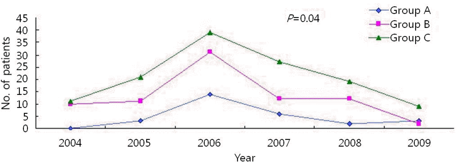
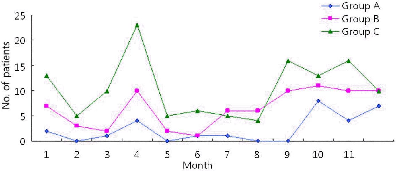
Table 1.
Pulmonary Symptoms of Patients with Mycoplasma pneumoniae Pneumoniae
Table 2.
Extrapulmonary Symptoms of Patients with Mycoplasma pneumoniae Pneumonia
Table 3.
Physical Findings of Patients with Mycoplasma pneumoniae Pneumoniae
| Physical findings | No. of patients (%) | ||
|---|---|---|---|
| Group A | Group B | Group C | |
| Auscultation finding | |||
| Rale | 20 (71.4) | 57 (73.1) | 91 (72.2) |
| Decreased breathing sound | 2 (7.1) | 19 (24.4) | 17 (13.5) |
| Wheezing∗ | 10 (35.7) | 2 (2.6) | 2 (1.6) |
| Others | |||
| Pharyngeal injection∗ | 6 (21.4) | 32 (41.0) | 70 (55.6) |
| Rash | 1 (3.6) | 3 (3.8) | 3 (2.4) |
| Otitis media | 2 (7.1) | 4 (5.1) | 11 (8.7) |
| Conjuntiva injection | 1 (3.6) | 0 (0.0) | 0 (0.0) |
Table 4.
Radiologic Findings of Patients with Mycoplasma pneumoniae Pneumoniae
| Radiologic findings | No. of patients (%) | ||
|---|---|---|---|
| Group A | Group B | Group C | |
| Pneumonic infiltration | 10 (35.7) | 40 (51.3) | 83 (65.9) |
| Lobar pneumonia | |||
| Single lung | |||
| Multilobar | 1 (3.57) | 0 (0.0) | 0 (0.0) |
| Right lower lobe | 0 (0.0) | 6 (7.7) | 9 (7.1) |
| Right middle lobe | 0 (0.0) | 6 (7.7) | 4 (3.2) |
| Left lower lobe | 1 (3.57) | 5 (6.4) | 2 (1.6) |
| Left upper lobe | 0 (0.00) | 1 (1.3) | 1 (0.8) |
| Right upper lobe | 3 (10.71) | 6 (7.7) | 4 (3.2) |
| Both lungs | 1 (3.57) | 0 (0.0) | 0 (0.0) |
| Pleural effusions | 1 (3.6) | 1 (1.3) | 2 (1.6) |
| Atelectasis | 0 (0.0) | 1 (1.3) | 1 (0.8) |
| Negative finding∗ | 12 (42.86) | 14 (17.9) | 23 (18.3) |
Table 5.
Laboratory Findings of Patients with Mycoplasma pneumoniae Pneumoniae
| Laboratory findings | No. of patients (%) | ||
|---|---|---|---|
| Group A | Group B | Group C | |
| WBC counts (/mm3) | |||
| <5,000 | 1 (3.6) | 4 (5.1) | 8 (6.3) |
| 5,000–10,000 | 6 (21.4) | 34 (43.6) | 53 (42.1) |
| 10,000–15,000 | 11 (39.3) | 31 (39.7) | 41 (32.5) |
| >15,000 | 10 (35.7) | 9 (11.5) | 24 (19.0) |
| ESR∗ | |||
| >20 mm/hr | 4 (14.3) | 16 (20.5) | 67 (53.2) |
| CRP | |||
| >0.8 mg/dL | 12 (42.9) | 29 (37.2) | 76 (60.3) |
| AST∗/ALT | |||
| AST>45 U/L | 23 (82.1) | 43 (55.1) | 74 (58.7) |
| ALT>55 U/L | 5 (17.9) | 11 (14.1) | 7 (5.6) |
 XML Download