Abstract
Purpose
Bronchiolitis obliterans (BO), an uncommon chronic obstructive lung disease in children, is most often seen following a severe lower respiratory tract infection (LRTI). We investigated the clinical characteristics, etiology, possible risk factors, radiological findings, and response to treatment in children diagnosed with post-infectious BO.
Methods
A retrospective study was performed on 62 patients diagnosed with post-infectious BO based on clinical and high-resolution computed tomography (HRCT) findings from 2005 to 2010. Forty-eight age-matched children who were admitted with the first episode of LRTI and did not subsequently develop BO were also studied as control subjects.
Results
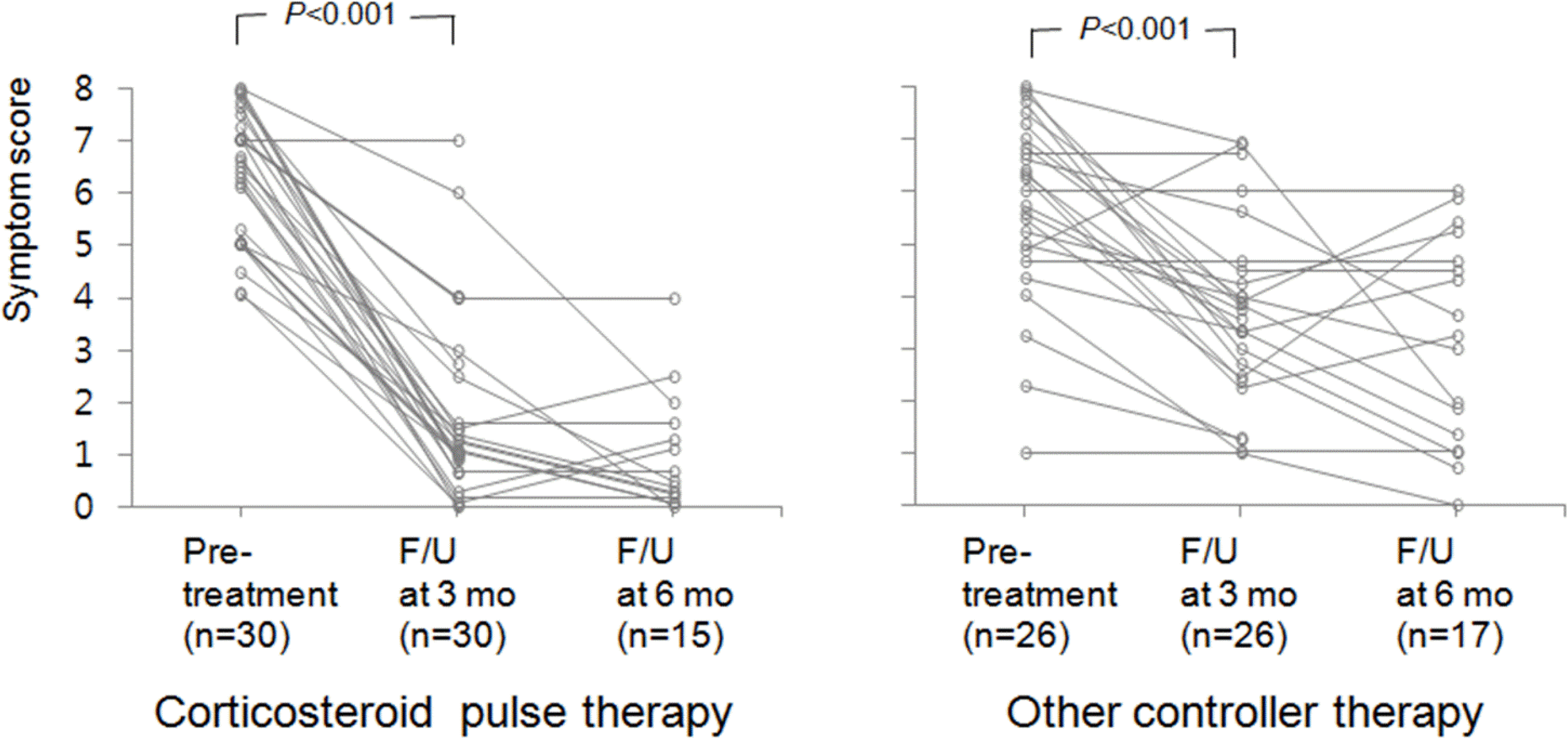
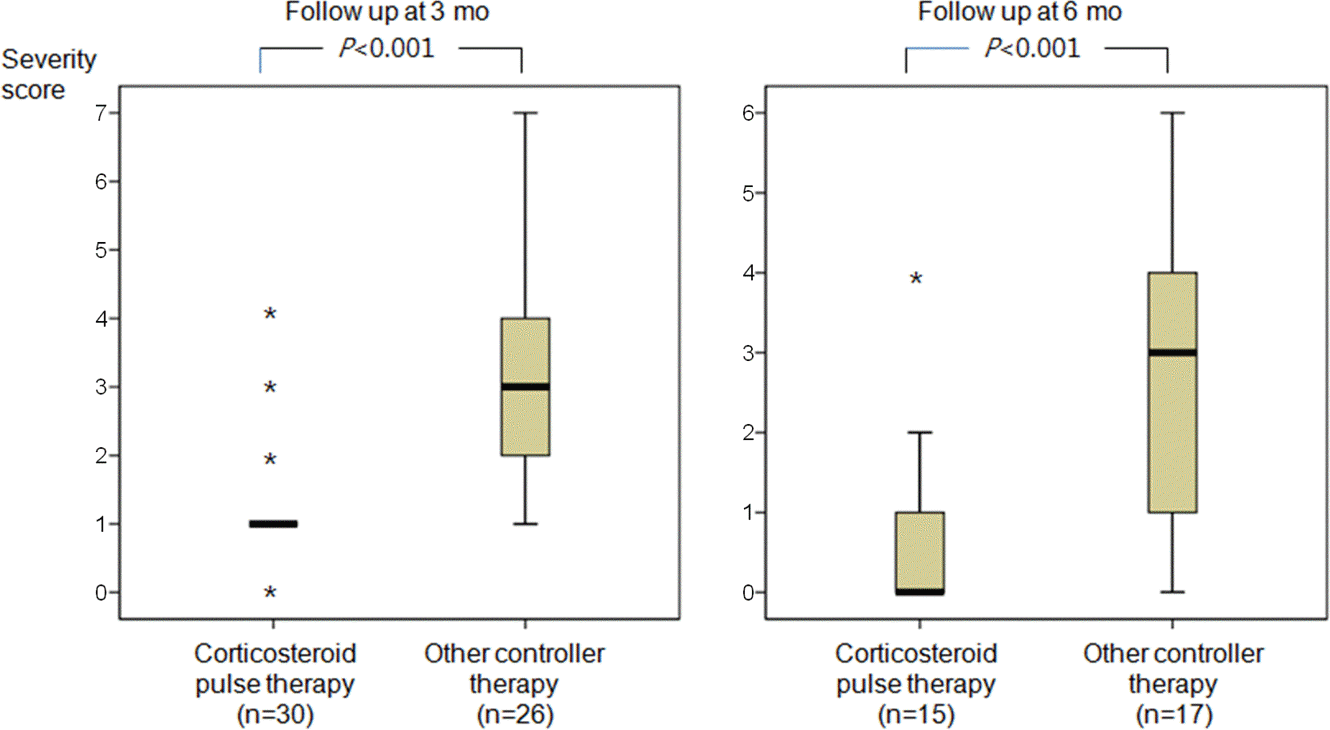
Median ages at diagnosis and initial insult were 28 and 17 months, respectively. The median duration from initial LRTI until diagnosis was 5 months. Children who developed BO showed more respiratory compromise during their acute episodes of LRTI than those who did not. Symptom severity score decreased significantly after adequate treatment, which was significantly greater in patients treated with pulse steroid therapy than those treated with other controllers.
Conclusion
The results suggest that the development of post-infectious BO should be suspected in the children showing persistent respiratory symptoms after severe LRTIs. They also suggest that adequate treatment including pulse steroid therapy may improve clinical status and the prognosis of these patients.
Go to : 
References
1. Schlesinger C, Veeraraghavan S, Koss MN. Constructive (obliterative) bronchiolitis. Curr Opin Pulm Med. 1998; 4:288–93.
2. Shaw RJ, Djukanovic R, Tashkin DP, Millar AB, du Bois RM, Orr PA. The role of small airways in lung disease. Respir Med. 2002; 96:67–80.

3. Kurland G, Michelson P. Bronchiolitis obliterans in children. Pediatr Pulmonol. 2005; 39:193–208.

4. Fischer GB, Sarria EE, Mattiello R, Mocelin HT, Castro-Rodriguez JA. Post infectious bronchiolitis obliterans in children. Paediatr Respir Rev. 2010; 11:233–9.

5. Castro-Rodriguez JA, Daszenies C, Garcia M, Meyer R, Gonzales R. Adenovirus pneumonia in infants and factors for developing bronchiolitis obliterans: a 5-year follow-up. Pediatr Pulmonol. 2006; 41:947–53.

6. Kim CK, Chung CY, Kim JS, Kim WS, Park Y, Koh YY. Late abnormal findings on high-resolution computed tomography after Mycoplasma pneumonia. Pediatrics. 2000; 105:372–8.

7. Chiu CY, Wong KS, Huang YC, Lin TY. Bronchiolitis obliterans in children: clinical presentation, therapy and longterm follow-up. J Paediatr Child Health. 2008; 44:129–33.

8. Kim CK, Kim SW, Kim JS, Koh YY, Cohen AH, Deterding RR, et al. Bronchiolitis obliterans in the 1990s in Korea and the United States. Chest. 2001; 120:1101–6.

9. Colom AJ, Teper AM, Vollmer WM, Diette GB. Risk factors for the development of bronchiolitis obliterans in children with bronchiolitis. Thorax. 2006; 61:503–6.

10. Moonnumakal SP, Fan LL. Bronchiolitis obliterans in children. Curr Opin Pediatr. 2008; 20:272–8.

11. Colom AJ, Teper AM. Clinical prediction rule to diagnose postinfectious bronchiolitis obliterans in children. Pediatr Pulmonol. 2009; 44:1065–9.

12. Hong SJ, Kim BS, Ahn KM, Lee SI, Kim KE, Lee KY, et al. Multicenter Study of Bronchiolitis Obliterans in Korean Children. Pediatr Allergy Respir Dis(Korea). 2002; 12:136–45.
13. Mauad T, Dolhnikoff M. Sao Paulo Bronchiolitis Obliterans Study Group. Histology of childhood bronchiolitis obliterans. Pediatr Pulmonol. 2002; 33:466–74.

14. McLoud TC, Epler GR, Colby TV, Gaensler EA, Carrington CB. Bronchiolitis obliterans. Radiology. 1986; 159:1–8.

15. Panitch HB, Callahan CW Jr, Schidlow DV. Bronchiolitis in children. Clin Chest Med. 1993; 14:715–31.

16. Laraya-Cuasay LR, DeForest A, Huff D, Lisch-ner H, Huang NN. Chronic pulmonary complications of early influenza virus infection in children. Am Rev Respir Dis. 1977; 116:617–25.
17. Krasinski K. Severe respiratory syncytial virus infection: clinical features, nosocomial acquisition and outcome. Pediatr Infect Dis. 1985; 4:250–7.
18. Massie R, Armstrong D. Bronchiectasis and bronchiolitis obliterans post respiratory syncytial virus infection: think again. J Paediatr Child Health. 1999; 35:497–8.

19. Chan PW, Muridan R, Debruyne JA. Bronchiolitis obliterans in children: clinical profile and diagnosis. Respirology. 2000; 5:369–75.

20. Yoo Y, Yu J, Kim DK, Choi SH, Kim CK, Koh YY. Methacholine and adenosine 5'-monophosphate challenges in children with postinfectious bronchiolitis obliterans. Eur Respir J. 2006; 27:36–41.
Go to : 
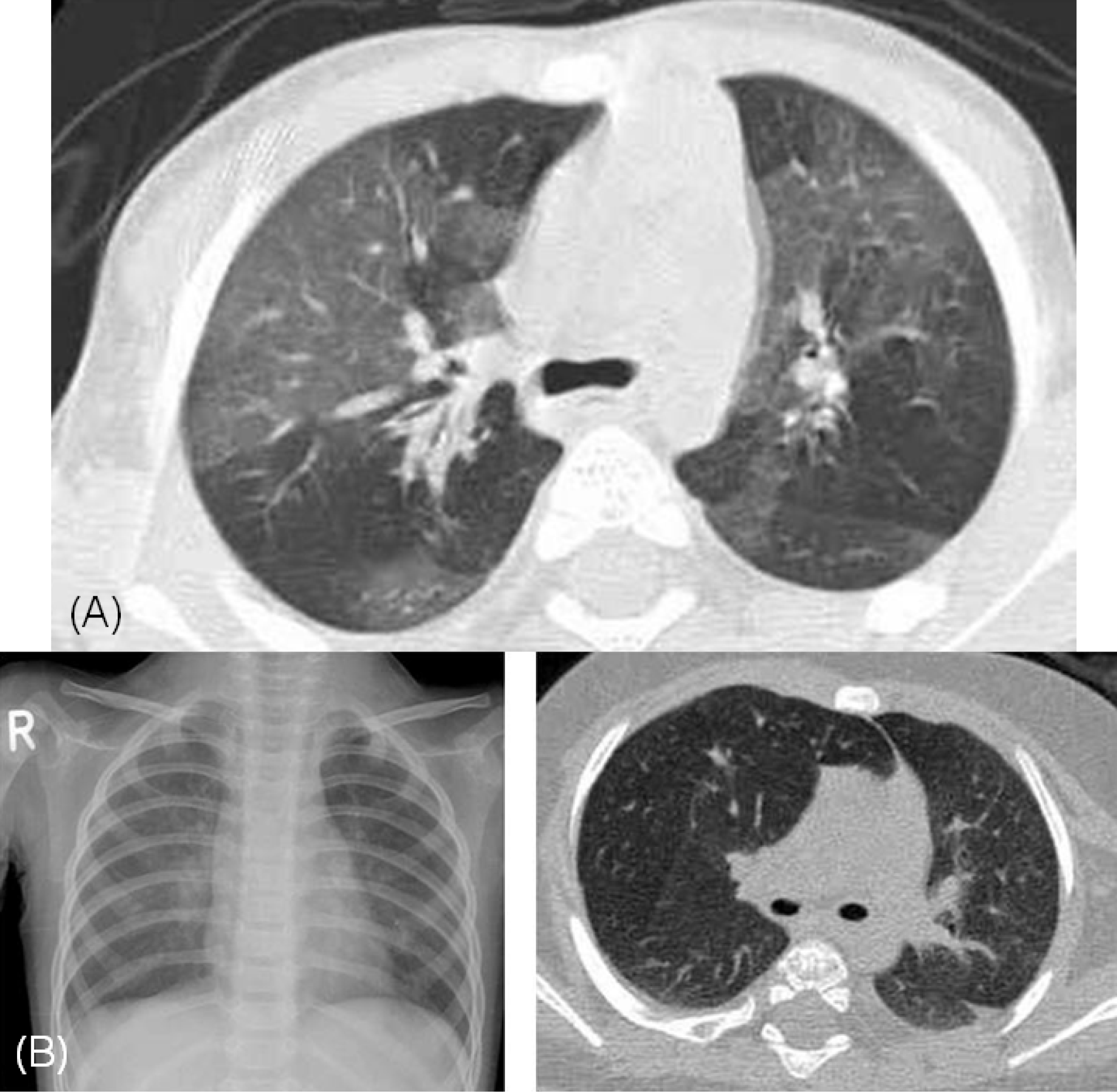
 | Fig. 1.(A) High resolution computed tomography (HRCT) shows mosaic perfusion in both lung fields. (B) Severe air-trapping is shown on plain chest X-ray and HRCT. |
 | Fig. 2.Symptom severity score was reduced significantly after treatment in both patient groups: the patients treated with corticosteroid pulse therapy and other controller therapy. F/U, follow-up. |
 | Fig. 3.Symptom severity score was significantly lower in the patients treated with corticosteroid pulse therapy than in the patients treated with other controller therapy at 3 and 6 month-follow up after treatment, respectively. |
Table 1.
Age Distribution of the Patients at Diagnosis and Initial Insults
| Age (mo) | At diagnosis (n=62) | At initial insult (n=62) |
|---|---|---|
| <6 | 11 (18) | |
| 6–12 | 6 (10) | 6 (10) |
| 12–36 | 6 (10) | 30 (49) |
| 36–72 | 25 (40) | 8 (12) |
| >72 | 17 (27) | 4 (6) |
| Unknown | 8 (13) | 3 (5) |
Table 2.
Duration Between Initial Insults and Diagnosis of Bronchiolitis Obliterans (n=62)
| Duration (mo) | No. (%) |
|---|---|
| <3 | 21 (34) |
| 3–6 | 13 (21) |
| 6–12 | 15 (24) |
| >12 | 10 (16) |
| Unknown | 3 (5) |
Table 3.
Clinical Characteristics of the Patients Diagnosed on Admission and at Out Patient Department
Table 4.
High Resolution Computed Tomography Findings of the Patients at Diagnosis (n=62)
| Findings | No. (%) |
|---|---|
| Mosaic perfusion | 55 (89) |
| Air trapping | 51 (82) |
| Atelectasis | 16 (26) |
| Bronchiectasis | 1 (2) |
| Effusion | 1 (2) |
Table 5.
Clinical Features During Acute Episodes in Children With vs. Without Bronchiolitis Obliterans (BO)
| BO group (n=44) | Non-BO group (n=48) | |
|---|---|---|
| Age at onset (mo), median (range) | 19 (2–46) | 15 (1–36) |
| Sex, M/F | 31/13 | 29/19 |
| Season, SS/FW | 23/21 | 14/34 |
| Fever, n (%)∗ | 31 (70) | 22 (46) |
| Wheezing, n (%) | 43 (98) | 39 (81) |
| Crackles, n (%) | 43 (98) | 37(77) |
| Tachypnea, n (%)† | 25 (57) | 9 (19) |
| Accessory muscle use, n (%)† | 19 (43) | 7 (15) |
| O2 saturation <92%, n (%) | 14 (32) | 10 (21) |
| Systemic corticosteroid use, n (%) | 37 (84) | 21 (42) |
| Systemic corticosteroid, mean, day† | 10.1±5.9 | 4.5±2.1 |
| IV theophylline use, n (%) | 17 (39) | 5 (10) |
| Duration of admission, mean, day† | 11.9±6.7 | 6.2±2.3 |
| Re-admission within 6 mo, n (%) | 12 (27) | 13 (27) |
| Re-admission within 6–12 mo, n (%)‡ | 12 (27) | 5 (10) |
| Severity score≥3, n (%)† | 23 (52) | 7 (15) |


 PDF
PDF ePub
ePub Citation
 Print
Print


 XML Download