

 PDF
PDF ePub
ePub Citation
 Print
Print


Abstract
The seminiferous tubules merge and connect with the tubuli recti that form the spaces known as the rete testis. Cystic ectasia of the rete testis is a rare benign testicular lesion. We report the cystic ectasia of the rete testis in a 66-year-old man.
Go to : 
REFERENCES
1). Burrus JK, Lockhart ME, Kenney PJ, Kolettis PN. Cystic ectasia of the rete testis: clinical and radiographic features. J Urol. 2002; 168:1436–8.

2). Colangelo SM, Fried K, Hyacinthe LM, Fracchia JA. Tubular ectasia of the rete testis: an ultrasound diagnosis. Urology. 1995; 45:532–4.
3). Nistal M, Mate A, Paniagua R. Cystic transformation of the rete testis. Am J Surg Pathol. 1996; 20:1231–9.
4). Jequier AM, Phillips N. Cystic dilatation of the rete testis: a hidden diagnosis among infertile men. Reprod Biomed Online. 2009; 18:190–4.
5). Jimenez-López M, Ramírez-Garrido F, López-González Garrido JD, Mantas-Avila JA, Nogueras-Ocaña M, Jimenez-Verdejo A, et al. Dilatation of the rete testis: ultrasound study. Eur Radiol. 1999; 9:1327–9.
6). Brown DL, Benson CB, Doherty FJ, Doubilet PM, DiSalvo DN, Van Alstyne GA, et al. Cystic testicular mass caused by dilated rete testis: sonographic findings in 31 cases. AJR Am J Roentgenol. 1992; 158:1257–9.
7). Fawzy MA, Kelly BE, Johnston SR. Cystic dilatation of the rete testis. Ulster Med J. 2001; 70:59–60.
8). Gooding GA, Leonhardt W, Stein R. Testicular cysts: US findings. Radiology. 1987; 163:537–8.
Go to :
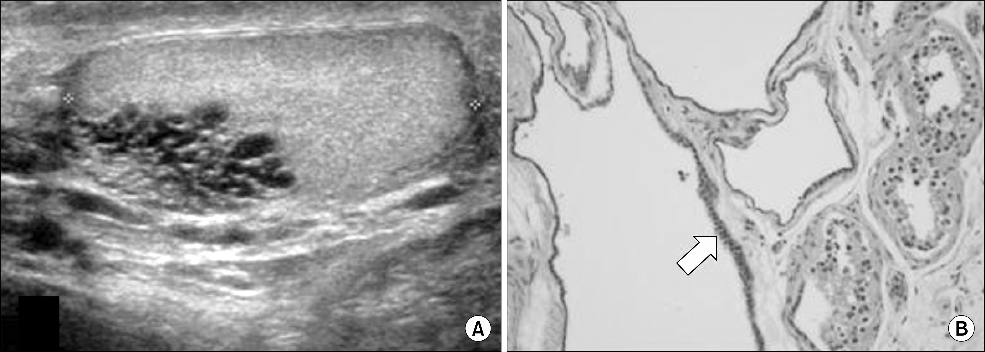 | Fig. 1.(A) Ultrasound of the testis showed clustered anechoic tubular structures within the rete testis of left testicle (1.63×1.19× 2.20 cm). (B) Microscopic examination in the rete testis region showed multilocular cysts, lined by simple cuboidal to columnar epithelial cells and consistent with a cystic ectasia of the rete testis and consistent with a cystic ectasia of the rete testis (arrow). H&E, ×200. |
 XML Download