

 PDF
PDF ePub
ePub Citation
 Print
Print


Abstract
Cystic lymphangiomas are a benign tumor caused by lymphatic malformation. They are normally seen in the head and neck region and very rarely occur in the scrotum. We report a rare case of a 20-year-old man who presented with a gradually enlarging, painful scrotal mass which was identified ultrasonographically and histologically as a scrotal cystic lymphangioma and treated by surgical excision.
REFERENCES
1). Mosca RC, Pereira GA, Mantesso A. Cystic hygroma: characterization by computerized tomography. Oral Surg Oral Med Oral Pathol Oral Radiol Endod. 2008; 105:e65–9.

2). Park JS, Ahn BC, Kim YS, Joo HJ, Kim SJ. Retroperitoneal cystic lymphangioma simultaneously diagnosed with bladder cancer. Korean J Urol. 2002; 43:535–7.
3). Singla SL, Rattan KN, Singh S. Cystic hygroma of the gluteal region. Indian J Pediatr. 2000; 67:779–80.
4). Weiss SW, Goldblum JR. Tumors of lymph vessels. Weiss SW, Goldblum JR, editors. Enzinger and Weiss's soft tissue tumors. 4th ed.St. Louis: Mosby;2001. p. 956–67.
5). Grossgold ET, Kusuda L. Scrotal lymphangioma in an adult. Urology. 2007; 70:590e. 1–2.
6). Hurwitz RS, Shapiro E, Hulbert WC, Diamond DA, Casale AJ, Rink RC. Scrotal cystic lymphangioma: the misdiagnosed scrotal mass. J Urol. 1997; 158:1182–5.
7). Vikicevic J, Milobratovic D, Vukadinovic V, Golubovic Z, Krstic Z. Lymphangioma scroti. Pediatr Dermatol. 2007; 24:654–6.
8). Weidman ER, Cendron M, Schned AR, Harris RD. Scrotal lymphangioma: an uncommon cause for a scrotal mass. J Ultrasound Med. 2002; 21:669–72.
9). Demir Y, Latifoğlu O, Yenidünya S, Atabay K. Extensive lymphatic malformation of penis and scrotum. Urology. 2001; 58:106.
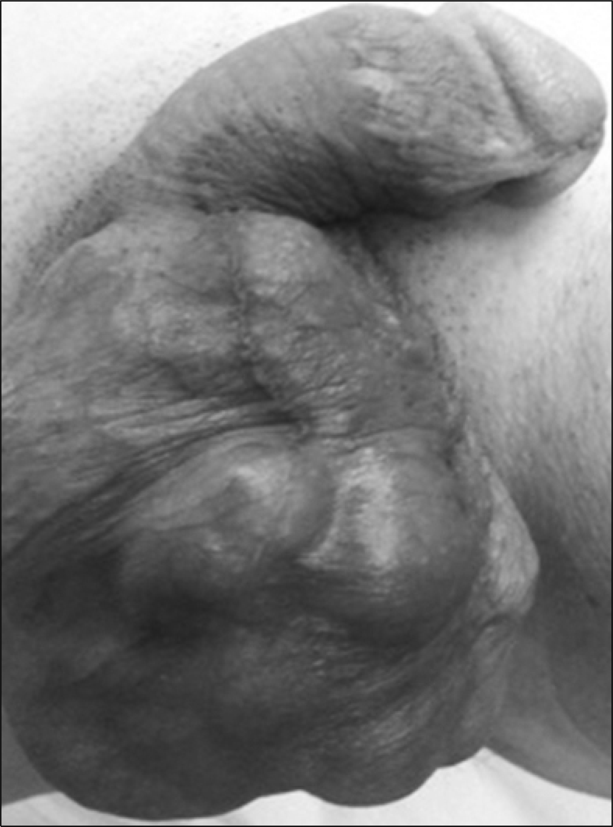
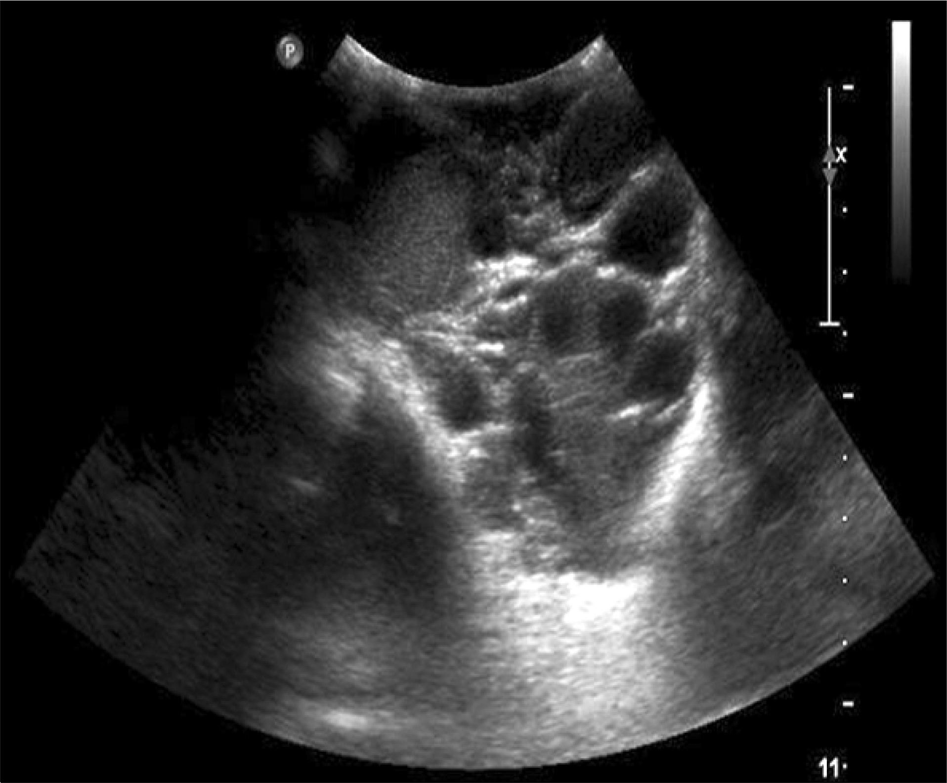
Fig. 2.
Ultrasonography shows ill-defined multiloculated cystic lesion with various echogenecity in scrotum.

 XML Download