

 PDF
PDF ePub
ePub Citation
 Print
Print


Abstract
Xanthogranulomatous prostatitis is a rare inflammatory condition of the prostate. We report a case of xanthogranulomatous prostatitis. An 82-year-old man presented to our emergency department complaining of gross hematuria. A hard, enlarged, non-tender prostate was palpated on digital rectal examination. Urinalysis showed hematuria and pyuria, and the complete blood count (CBC) revealed leukocytosis. The prostate-specific antigen (PSA) level was elevated at 86.8 ng/ml. Computed tomography (CT) showed an enlarged prostate (volume 100 ml) that protruded into the bladder base. In the left lobe of the prostate, a 1.2×1.1-cm abscess was seen. Transurethral resection of the prostate was done. The pathological diagnosis was xanthogranulomatous prostatitis. The natural progression of the disease is unknown because of the paucity of cases and longterm follow-up reports. To evaluate the pathogenesis and longterm features of progression of this disease, more clinical cases should be collected.
REFERENCES
1). Hayashi N, Wada T, Kiyota H, Ueda M, Oishi Y. Xanthogranulomatous cystitis. Int J Urol. 2003; 10:498–500.

2). Epstein JI, Hutchins GM. Granulomatous prostatitis: distinction among allergic, nonspecific, and post-transurethral resection lesions. Hum Pathol. 1984; 15:818–25.
3). O'Dea MJ, Hunting DB, Greene LF. Non-specific granulomatous prostatitis. J Urol. 1977; 118:58–60.
4). Tanner FH, McDonald JR. Granulomatous prostatits: a histologic study of a group of granulomatous lesions collected from prostate glands. Arch Pathol. 1943; 36:358–70.
5). Epstein JI. The prostate and seminal vesicles. Diagnostic surgical pathology. 2nd ed.New York: Raven;1994. p. 1807–48.
6). Stillwell TJ, Engen DE, Farrow GM. The clinical spectrum of granulomatous prostatitis: a report of 200 cases. J Urol. 1987; 138:320–3.
7). Rafique M, Yaqoob N. Xanthogranulomatous prostatitis: a mimic of carcinoma of prostate. World J Surg Oncol. 2006; 4:30.
8). Srigley JR. Benign mimickers of prostatic adenocarcinoma. Mod Pathol. 2004; 17:328–48.
9). Miekoś E, Włodarczyk W, Szram S. Xanthogranulomatous prostatitis. Int Urol Nephrol. 1986; 18:433–7.
Fig. 1.
Computed tomography (CT) of the abdomen and pelvis. (A) The coronal view shows an enlargedprostate. (B) The axial view shows focal abscess formation (white arrow) in the left lobe of the prostate.
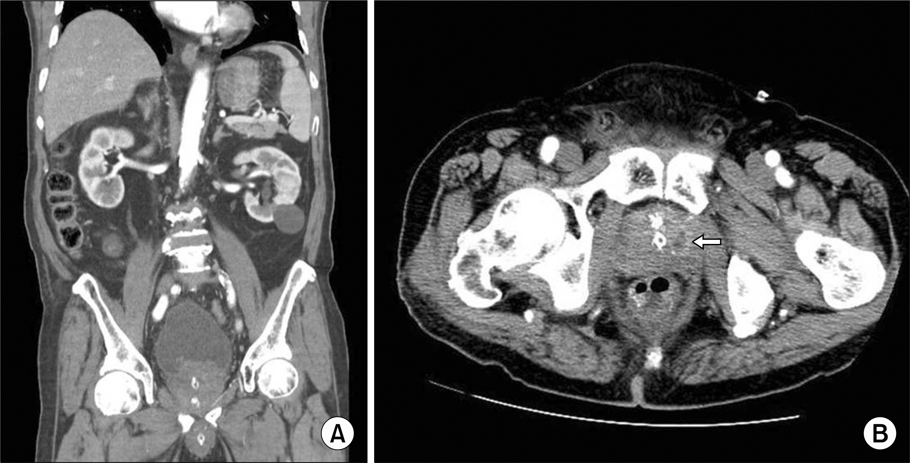
Fig. 2.
(A) Microscopically, the TURP specimen reveals foci of granulomatous inflammation, which consists ofdistended glandular lumens surrounded by macrophages and lymphocytes. The granulomas are composed offoam cells, which are macrophages with lipid components (H&E, ×200). (B) The aggregated macrophagesare positive for CD68 immunostaining, which appears brown (×200).

 XML Download