Journal List > Korean J Androl > v.29(2) > 1033069

Yang, Kim, Lee, Chung, Kim, Ryu, Won, Park, and Lee: Differential Expression of Proteins Related with Penile Apoptosis in a Rat after Cavernous Nerve Resection

Abstract

Purpose

The mechanism including changes of proteome within cavernosal tissue after cavernous nerve injury were not evaluated. We performed proteomics and functional analysis to identify proteins of penile corpus cavernosum whose expression was or was not altered by cavernous nerve resection (CNR).

Materials and Methods

Using 8-week-old male WKY rats, sham and CNR operation under microscope were performed. After 8 weeks, penile tissues of sham and CNR group were harvested. We used 2-DE and MALDI-TOF/TOF (AB 4700) to identify of differently expressed penile proteins. 2-DE gels were stained with silver nitrate and the gels were analyzed with PDQuest.

Results

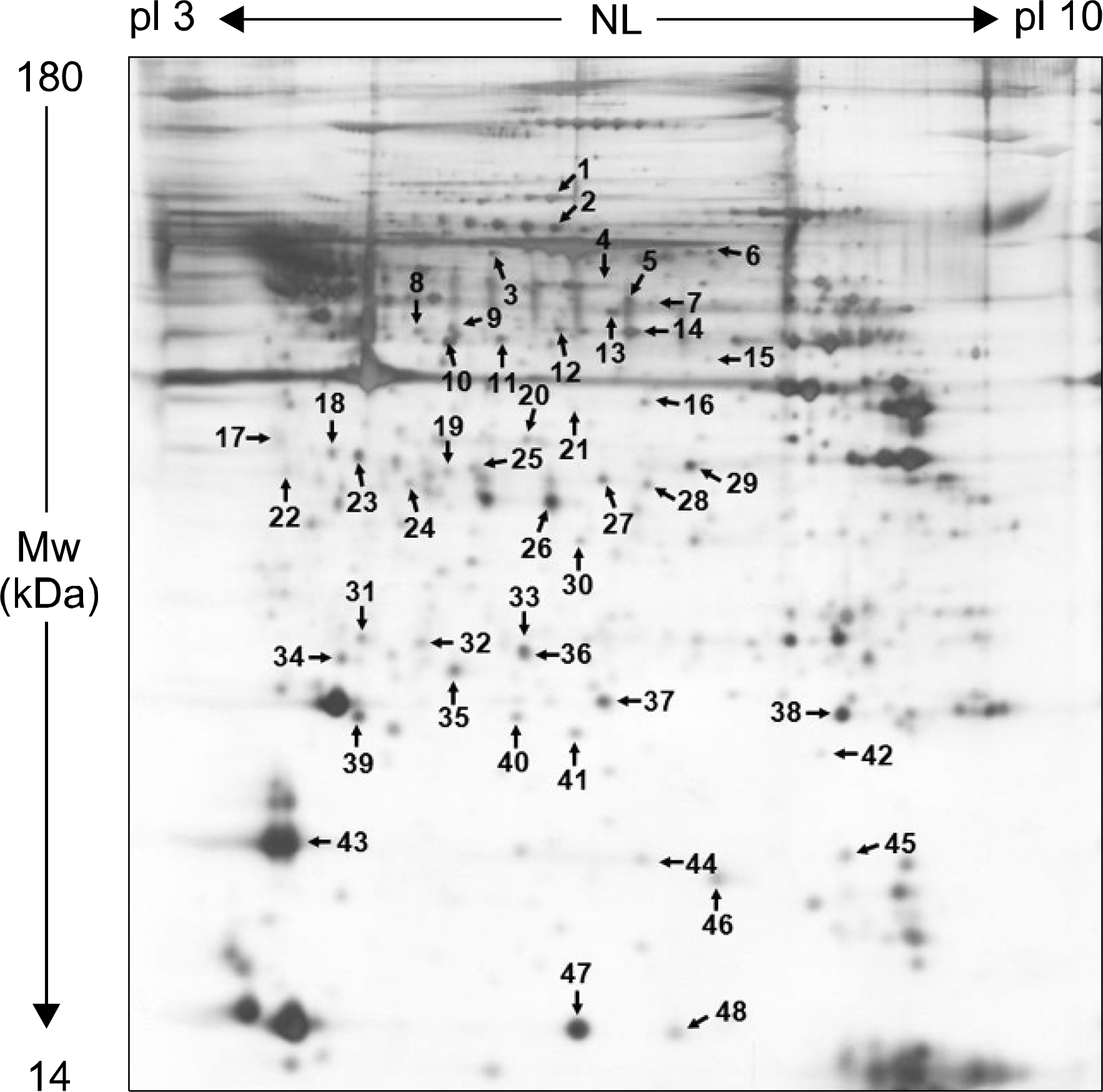
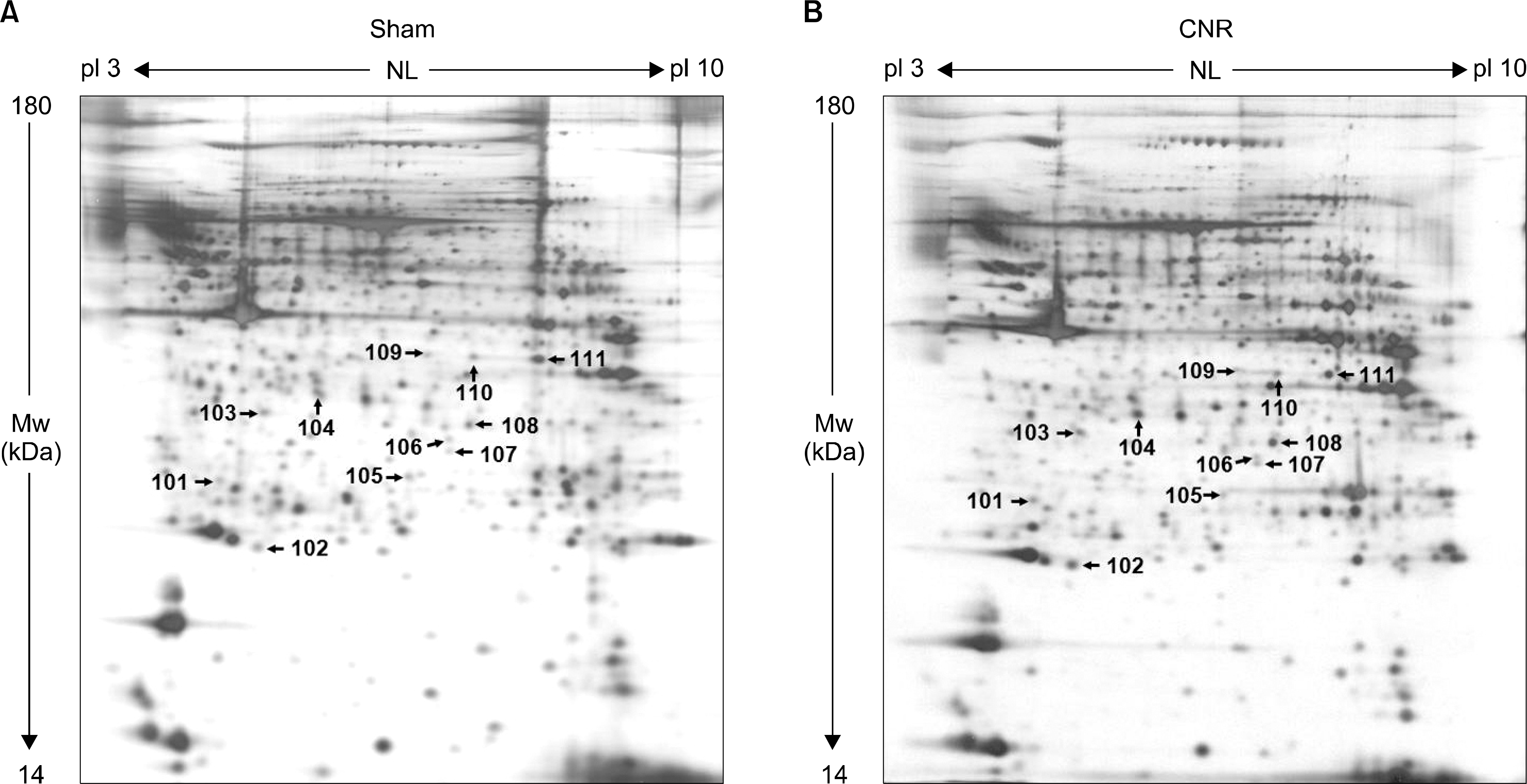
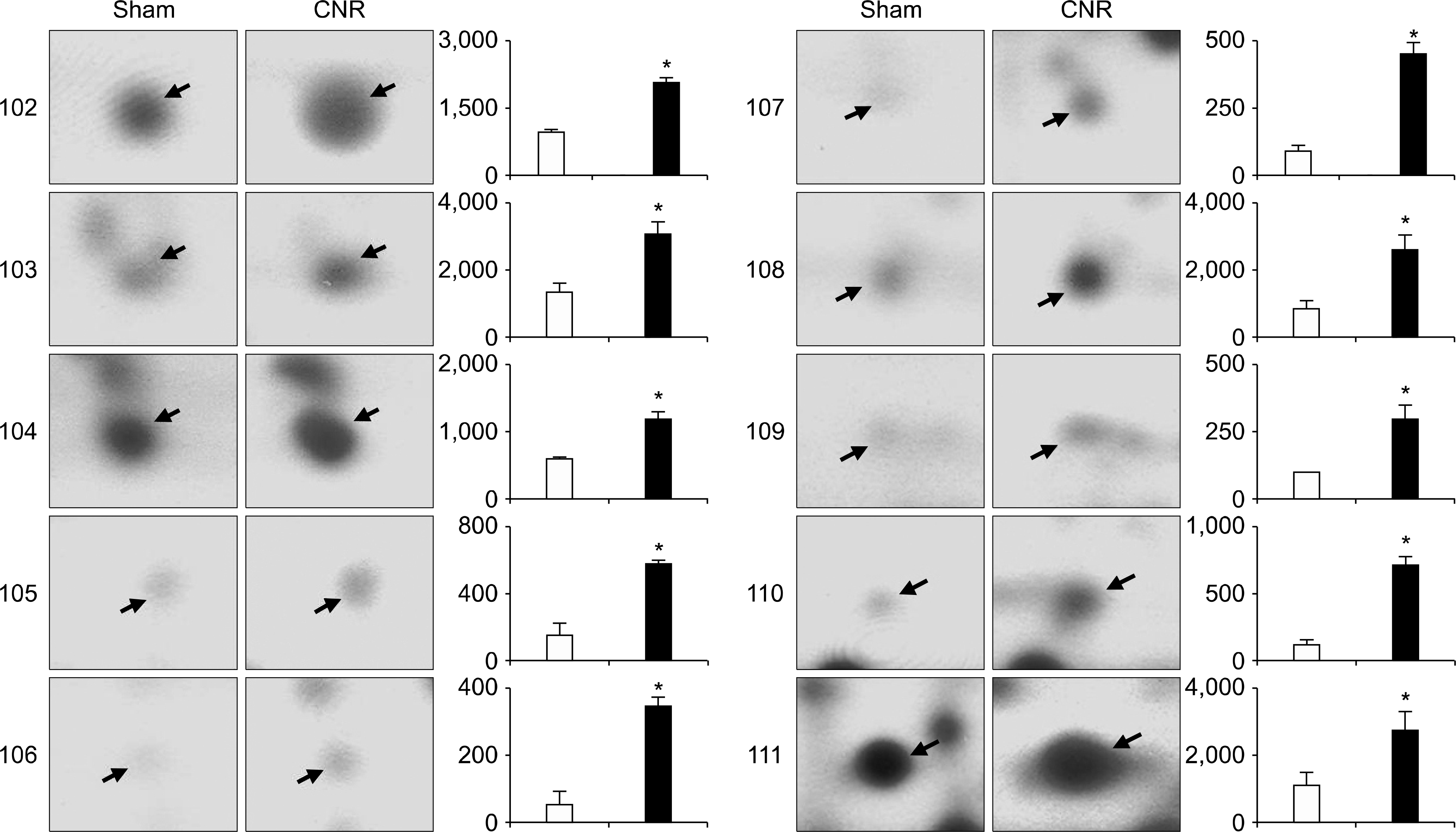
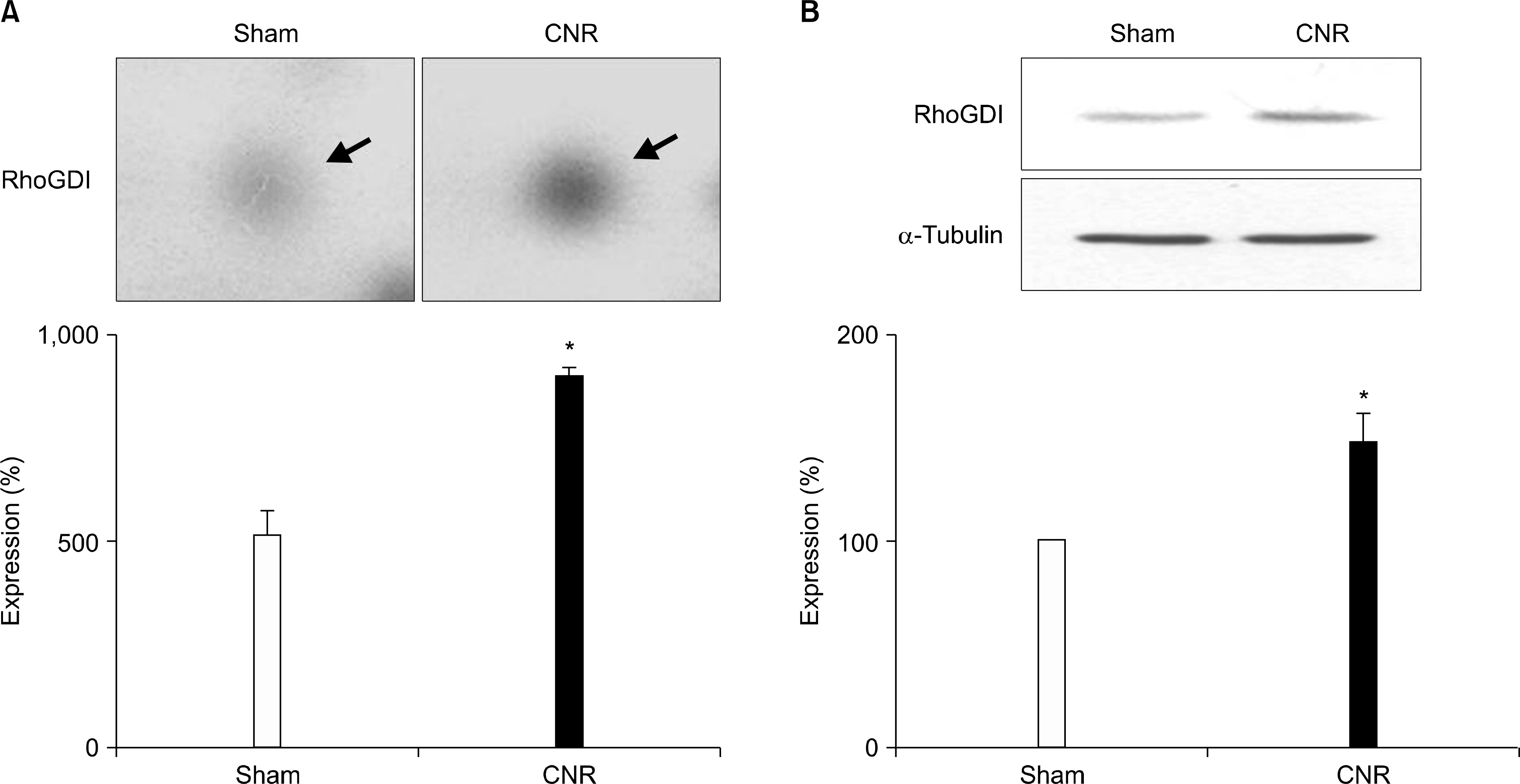
We isolated more than 950 proteins on silver-stained gels of whole protein extracts from normal rat penile corpus cavernous. Of these proteins, 48 prominent proteins were identified using MALDI-TOF/TOF. Protein characterization revealed that the most prominent penile corpus cavernous proteins were those with antioxidant, chaperone, or cytoskeletal structure. Moreover, 11 proteins having levels elevated by CNR were annexin proteins, endoplasmic reticulum protein 29, glutathione s-transferase w-1, and others. In addition, Rho-GDP dissociation inhibitor (RhoGDI), a regulator of Rho proteins, was also increased in CNR rats compared with sham-operated control rats.

Conclusions

The apoptotic signals observed in penile tissues was greatly increased in CNR rats than in sham-operated rats. These results suggest that RhoGDI is one of the proteins regulated by CNR in penile smooth muscle strips, and has a crucial role in the early stage of penile apoptosis.

REFERENCES

1). Burnett AL. Strategies to promote recovery of cavernous nerve function after radical prostatectomy. World J Urol. 2003; 20:337–42.
crossref
2). Olsson CA, Goluboff ET. Detection and treatment of prostate cancer: perspective of the urologist. J Urol. 1994; 152:1695–9.
crossref
3). Walsh PC, Donker PJ. Impotence following radical prostatectomy: insight into etiology and prevention. J Urol. 1982; 128:492–7.
crossref
4). Takenaka A, Murakami G, Matsubara A, Han SH, Fujisawa M. Variation in course of cavernous nerve with special reference to details of topographic relationships near prostatic apex: histologic study using male cadavers. Urology. 2005; 65:136–42.
crossref
5). Lepor H, Gregerman M, Crosby R, Mostofi FK, Walsh PC. Precise localization of the autonomic nerves from the pelvic plexus to the corpora cavernosa: a detailed anatomical study of the adult male pelvis. J Urol. 1985; 133:207–12.
crossref
6). Montorsi F, Briganti A, Salonia A, Rigatti P, Burnett AL. Current and future strategies for preventing and managing erectile dysfunction following radical prostatectomy. Eur Urol. 2004; 45:123–33.
crossref
7). Costello AJ, Brooks M, Cole OJ. Anatomical studies of the neurovascular bundle and cavernosal nerves. BJU Int. 2004; 94:1071–6.
crossref
8). Sezen SF, Burnett AL. Intracavernosal pressure monitoring in mice: responses to electrical stimulation of the cavernous nerve and to intracavernosal drug administration. J Androl. 2000; 21:311–5.
9). Lysiak JJ, Yang SK, Klausner AP, Son H, Tuttle JB, Steers WD. Tadalafil increases Akt and extracellular signal-regulated kinase 1/2 activation, and prevents apoptotic cell death in the penis following denervation. J Urol. 2008; 179:779–85.
crossref
10). Mullerad M, Donohue JF, Li PS, Scardino PT, Mulhall JP. Functional sequelae of cavernous nerve injury in the rat: is there model dependency. J Sex Med. 2006; 3:77–83.
crossref
11). Klein LT, Miller MI, Buttyan R, Raffo AJ, Burchard M, Devris G, et al. Apoptosis in the rat penis after penile denervation. J Urol. 1997; 158:626–30.
crossref
12). User HM, Hairston JH, Zelner DJ, McKenna KE, McVary KT. Penile weight and cell subtype specific changes in a post-radical prostatectomy model of erectile dysfunction. J Urol. 2003; 169:1175–9.
crossref
13). Hu WL, Hu LQ, Song J, Li SW, Zheng XM, Cheng B, et al. Fibrosis of corpus cavernosum in animals following cavernous nerve ablation. Asian J Androl. 2004; 6:111–6.
14). Carrier S, Zvara P, Nunes L, Kour NW, Rehman J, Lue TF. Regeneration of nitric oxide synthase-containing nerves after cavernous nerve neurotomy in the rat. J Urol. 1995; 153:1722–7.
crossref
15). Iacono F, Giannella R, Somma P, Manno G, Fusco F, Mirone V. Histological alterations in cavernous tissue after radical prostatectomy. J Urol. 2005; 173:1673–6.
crossref
16). Domes T, De Young L, O'Gorman DB, Gan BS, Bella AJ, Brock G. Is there a role for proteomics in Peyronie's disease? J Sex Med. 2007; 4:867–77.
crossref
17). Liu X, Gao X, Pang J, Zhang Y, Wang K, Fang Y, et al. Proteomic analysis of rat penile tissue in a model of erectile dysfunction after radical prostatectomy. BJU Int. 2007; 99:1500–5.
crossref
18). User HM, Zelner DJ, McKenna KE, McVary KT. Microarray analysis and description of SMR1 gene in rat penis in a post-radical prostatectomy model of erectile dysfunction. J Urol. 2003; 170:298–301.
crossref
19). Lee CK, Park HJ, So HH, Kim HJ, Lee KS, Choi WS, et al. Proteomic profiling and identification of cofilin responding to oxidative stress in vascular smooth muscle. Proteomics. 2006; 6:6455–75.
crossref
20). Lee CK, Han JS, Won KJ, Jung SH, Park HJ, Lee HM, et al. Diminished expression of dihydropteridine reductase is a potent biomarker for hypertensive vessels. Proteomics. 2009; 9:4851–8.
crossref
21). Tostes RC, Carneiro FS, Lee AJ, Giachini FR, Leite R, Osawa Y, et al. Cigarette smoking and erectile dysfunction: focus on NO bioavailability and ROS generation. J Sex Med. 2008; 5:1284–95.
crossref
22). Wolf BB, Green DR. Suicidal tendencies: apoptotic cell death by caspase family proteinases. J Biol Chem. 1999; 274:20049–52.
crossref
23). Sasaki T, Takai Y. The Rho small G protein family-Rho GDI system as a temporal and spatial determinant for cytoskeletal control. Biochem Biophys Res Commun. 1998; 245:641–5.
crossref
24). Hoffman GR, Nassar N, Cerione RA. Structure of the Rho family GTP-binding protein Cdc42 in complex with the multifunctional regulator RhoGDI. Cell. 2000; 100:345–56.
crossref
25). Miñambres R, Guasch RM, Perez-Aragó A, Guerri C. The RhoA/ROCK-I/MLC pathway is involved in the ethanol-induced apoptosis by anoikis in astrocytes. J Cell Sci. 2006; 119:271–82.
26). Harenberg A, Girkontaite I, Giehl K, Fischer KD. The Lsc RhoGEF mediates signaling from thromboxane A2 to actin polymerization and apoptosis in thymocytes. Eur J Immunol. 2005; 35:1977–86.
crossref
27). He H, Baldwin GS. Rho GTPases and p21-activated kinase in the regulation of proliferation and apoptosis by gastrins. Int J Biochem Cell Biol. 2008; 40:2018–22.
crossref
28). Alici B, Gümüstas MK, Ozkara H, Akkus E, Demirel G, Yencilek F, et al. Apoptosis in the erectile tissues of diabetic and healthy rats. BJU Int. 2000; 85:326–9.
crossref
29). Jin L, Liu T, Lagoda GA, Champion HC, Bivalacqua TJ, Burnett AL. Elevated RhoA/Rho-kinase activity in the aged rat penis: mechanism for age-associated erectile dysfunction. FASEB J. 2006; 20:536–8.
crossref
30). Wang H, Eto M, Steers WD, Somlyo AP, Somlyo AV. RhoA-mediated Ca2+ sensitization in erectile function. J Biol Chem. 2002; 277:30614–21.
31). Sakamoto T, Repasky WT, Uchida K, Hirata A, Hirata F. Modulation of cell death pathways to apoptosis and necrosis of H2O2-treated rat thymocytes by lipocortin I. Biochem Biophys Res Commun. 1996; 220:643–7.
crossref
32). Parente L, Solito E. Annexin 1: more than an anti-phospholipase protein. Inflamm Res. 2004; 53:125–32.
crossref
33). Townsend DM, Tew KD. The role of glutathione-S-transferase in anti-cancer drug resistance. Oncogene. 2003; 22:7369–75.
crossref
34). Bambang IF, Xu S, Zhou J, Salto-Tellez M, Sethi SK, Zhang D. Overexpression of endoplasmic reticulum protein 29 regulates mesenchymal-epithelial transition and suppresses xenograft tumor growth of invasive breast cancer cells. Lab Invest. 2009; 89:1229–42.
crossref

Fig. 1.
Increment in apoptosis of penile corpus cavernosum from CNR rats. Immunohistochemical staining shows higher immunoreactivity in endothelial cells (arrows) and in smooth muscle cells (arrowheads) of CNR group than those of control group. The results are typical examples in 5 independent experiments. 2nd Ab: secondary antibody control. Magnification ×100 or ×400.
kja-29-111f1.tif
Fig. 2.
Images of a silver-stained 2-DE showing whole proteins identified in rat penile corpus cavernosum strips. Proteins were subjected to IEF on IPG strips (pH 3∼10, nonlinear, 17 cm) and then separated by 12% SDS-PAGE. The protein spots were visualized with silver stain. The arrows indicate proteins detected with MALDI-TOF/TOF. The numbers indicated in the gels correspond to the numbers in Tables 1. The results are typical examples in 4 independent experiments.
kja-29-111f2.tif
Fig. 3.
Silver-stained 2-DE gels in whole proteins of penile corpus cavernosum strips from sham (A) and CNR (B) rats. Proteins were subjected to IEF on IPG pH 3∼10 nonlinear (NL) strips and then separated by 12% (w/v) polyacrylamide SDS-PAGE. Arrows show proteins subsequently examined by MALDI-TOF/TOF. The numbers indicated in the gels correspond to the numbers in Table 2. The images are typical of those obtained in gels from 4 independent experiments.
kja-29-111f3.tif
Fig. 4.
Enlargement of a silver-stained 2-DE image showing rat penile corpus cavernosum proteins changed by CN resection. The arrowheads in both the left (sham control) and right (CNR) panels indicate proteins up-regulated compared with controls. The numbers indicated in the gels correspond to the numbers in Fig. 3 and Table 2. Regions containing proteins with statistically significant changed levels following the CNR were cropped and enlarged for this figure (∗p<0.05). Statistical analysis was performed on gels from 4 independent experiments using PDQuest. White bars, sham control; black bars, CNR.
kja-29-111f4.tif
Fig. 5.
Change of RhoGDI in penile corpus cavernosum strips from CNR rats. (A) Enlargement of a silver-stained 2-DE image showing RhoGDI changed by CNR as described in Fig. 4. (B) Western blot analysis using 1-DE gels. The separated proteins were immunoblotted with anti-RhoGDI or α-tubulin antibodies. Right panels; statistical analysis from left panels. The bands (n=5) of the RhoGDI in the sham control strips were defined as 100%. Denotes significant differences between the sham and CNR rats (∗p<0.05). The arrow indicates RhoGDI.
kja-29-111f5.tif
Table 1.
Summary of proteins prominently expressed in penile tissue from WKY rats as identified by MALDI–TOF/TO
No Protein name Peptide sequences Score/ SC (%) Protein pI/Mr (kDa) (Experimental) Accession No /Database Known function
1 Gelsolin VSETRPSTMVVEHPEFLK 476/15 5.76/86.0 51854227/NC Actin-modulating protein
EVQGFESSTFQGYFK (5.76/101.5) P13020/SP
HVVPNEVVVQR
VSNSGGSMSVSLVADENPFAQSALR
SEDCFILDHGR
TPSAAYLWVGTGASDAEK
AQHVQVEEGSEPDGFWEALGGK
2 Hemopexin protein GEFVWR 457/15 7.58/51.3 60688311/NC Heme binding protein
LFQEEFPGIPYPPDAAVECHR (5.76/88.9) P20059/SP
GECQSEGVLFFQGNR
FNPVTGEVPPR
DYFISCPGR
GATYAFSGSHYWR
SGAQATWAELSWPHEK
3 Dihydrolipoamide S-acetyltransferase VPLPSLSPTMQAGTIAR 142/7 5.70/58.7 11580/NC Pyruvate dehydrogenase activity
ILVPEGTR (5.53/80.9) P08461/SP
VPEANSSWMDTVIR
4 Serum albumin precursor FPNAEFAEITK 342/13 6.09/68.7 55391508/NC Body fluid osmorregulation
LGEYGFQNAILVR (5.83/74.2) P02770/SP
APQVSTPTLVEAAR
CCTLPEAQRR
PCFSALTVDETYVPK
AADKDNCFATEGPNLVAR
5 Chaperonin containing TCP1, subunit 2 (beta) KIHPQTIIAGWR 162/6 6.01/57.4 53733839/NC Molecular chaperone
IHPQTIIAGWR (5.85/65.8) P80314/SP
HGINCFINR
GATQQILDEAER
6 WD repeat1 protein VFASLPQVER 133/13 6.21/66.1 20269306/NC Disassembly of actin filaments
LATGSDDNCAAFFEGPPFK (6.21/83.5) O88342/SP
MTVDESGQLVSCSMDDTVR
7 Selenium binding protein 1 EEIVYLPCIYR 100/6 6.10/52.5 18266692/NC Selenium binding protein
SPHYSQVIHR (5.87/66.9)
IYVVDVGSEPR
8 Serine (or cysteine) proteinase inhibitor, clade F, member 1 LAAAVSNFGYDLY 376/18 6.04/46.4 29293811/N Neuronal differentiation
SSFVAPLE (5.21/59.2) P97298/SP
TTLQDFHLDED
YGLDSDLNC
AAFEWNEEGAGTSSNPDLQPV
DTDTGALLFIGR
9 Serine (or cysteine) proteinase inhibitor, clade F, member 1 LAAAVSNFGYDLY 534/19 6.04/46.4 29293811/N Neuronal differentiatio
SSFVAPLE (5.36/62.6) P97298/SP
KTTLQDFHLDED
TTLQDFHLDED
YGLDSDLNC
AAFEWNEEGAGTSSNPDLQPV
DTDTGALLFIGR
10 Guanine deaminase TPQLALIF 433/17 5.48/50.9 7533042/N Guanine deaminase activity
IVFLEESSQQE (5.33/57.8) Q9WTT6/SP
EWCFKPCEI
FQSTDVAEEVYT
THDLYIQSHISEN
LATLGGSQALGLD
FLYLGDDR
11 Serine (or cysteine) proteinase inhibitor, clade F, member 1 LAAAVSNFGYDLY 394/16 6.04/46.4 29293811/N Neuronal differentiatio
SSFVAPLE (5.57/57.6) P97298/SP
KTTLQDFHLDED
TTLQDFHLDED
AAFEWNEEGAGTSSNPDLQPV
DTDTGALLFIGR
12 Alpha enolase EIFDS 347/13 6.16/47.1 P17182/SP Glycolysis
IGAEVYHNL (5.77/60.8)
YITPDQLADLY
SCNCLLL
VNQIGSVTESLQAC
YNQILR
13 Aldehyde dehydrogenase, mitochondrial AAFQLGSPW 446/16 5.83/54.3 3121992/N Carbohydrate and alcohol metabolism
TIPIDGDFFSYT (5.83/64.4) P81178/SP
TFVQEDVYDEFVE
VVGNPFDS
GYFIQPTVFGDV
TIEEVVG
ELGEYGLQAYTEVK
14 Alpha enolase EIFDS 367/13 6.16/47.0 P04764/SP Glycolysis
AAVPSGASTGIYEALEL (5.88/60.5)
YITPDQLADLY
VNQIGSVTESLQAC
YNQILR
15 Tu translation elongation factor, mitochondrial KYEEIDNAPEE 215/6 7.23/49.5 27370092/NC Protein biosynthesis
YEEIDNAPEE (6.20/53.9)
GITINAAHVEYSTAAR
16 Actin-capping protein gCap39 QAALQVADGFIS 146/7 6.73/39.2 109521/N Cytoskeleton organization
YSPNTQVEILPQGR (5.87/48.0) P24452/SP
17 Tropomyosin 2 CKQLEEEQQALQ 465/17 4.66/32.8 11875203/N Smooth muscle contraction
IQLVEEELD (4.41/41.2) P58774/SP
IQLVEEELDRAQE
KLVILEGELE
TIDDLEDEVYAQK
18 Preprohaptoglobin GSFPWQA 175/10 7.16/30.0 204657/N Hemoglobin binding protein
MGYVSGWG (4.82/39.5) P06866/SP
SCAVAEYGVYVR
19 Preprohaptoglobin VMPICLPSKDYVAPG 151/13 7.16/30.0 204657/N Hemoglobin binding protein
MGYVSGWG (5.34/37.1) Q60574/SP
SCAVAEYGVYVR
20 Alpha-actin VAPEEHPTLLTEAPLNP 263/23 5.45/37.8 49864/N Cytoskeleton organization
GYSFVTTAE (5.67/41.7) P68134/SP
SYELPDGQVITIGNE
DLYANNVMSGGTTMYPGIAD
QEYDEAGPSIVHR
21 Alpha 2-actin AVFPSIVGRP 177/13 5.23/42.0 6671507/N Cytoskeleton organization
VAPEEHPTLLTEAPLNP (5.79/46.0) P62737/SP
GYSFVTTAE
QEYDEAGPSIVHR
22 Tropomyosin 1 RIQLVEEELD 332/16 4.69/32.7 P58771/SP Muscle contraction
IQLVEEELD (4.61/37.1)
IQLVEEELDRAQE
AEFAE
SIDDLEDELYAQK
23 Preprohaptoglobin GSFPWQA 263/16 7.16/30.0 204657/N Hemoglobin binding protein
VMPICLPSKDYVAPG (4.95/39.1) P06866/SP
MGYVSGWG
MGYVSGWG
SCAVAEYGVYVR
24 Pyruvate dehydrogenase (lipoamide) beta EAINQGMDEELERDE 180/10 6.41/38.9 18152793/N Acetyl-Co A biosynthesis
EGIECEVINL (5.17/36.3) Q9D051/SP
VTGADVPMPYAK
25 Capping protein (actin filament) muscle Z-line, alpha 2 LLLNNDNLL 164/17 5.57/32.9 38322760/N Cytoskeleton organization
EATDPRPYEAENAIESW (5.44/37.3) P47754/SP
NFWNG
IQVHYYEDGNVQLVSHK
26 Similar to osteoglycin precursor ESAYLYA 265/16 5.85/34.1 27683465/N Bone formation
DFADMPNL (5.76/35.5) Q62000/SP
MEEIRLEGNPIALG
HPNSFICL
RLPTGSYF
27 Malate dehydrogenase, cytosolic ENFSCLT 175/11 6.16/36.5 319837/N Dihydropyri midinase activity
NVIIWGNHSSTQYPDVNHA (5.83/36.3) P14152/SP
GEFITTVQQR
28 Glycerol 3-phosphate dehydrogenase VCIVGSGNWGSAIA 50/10 6.32/37.4 2317252/N Glycerol synthesis
IVGSNASQLAHFDPR (5.87/36.5) O35077/SP
29 Aldehyde reductase 1 VAIDMGYR 567/30 6.26/35.8 6978491/N Carbohydrate metabolism
HIDCAQVYQNEK (6.03/39.3) P07943/SP
HIDCAQVYQNEKEVGVALQEK
RQDLFIVSK
AIGVSNFNPLQIER
YKPAVNQIECHPYLTQEK
LIEYCHCK
TTAQVLIR
HKDYPFHAEV
30 3-Mercaptopyruvate sulfurtransferase HIPGAAFFDID 382/25 5.88/32.9 55824737/NC Sulfate transport
AFGHHSVSLLDGGF (5.79/33.3) P97532/SP
SPSEPAEFCAQLDPSFI
THEDILENLDA
FQGTQPEP
AQPEHVISQGR
31 Heat shock protein 27 LFDQAFGVP 107/13 6.12/22.9 204665/N Protein folding
AVTQSAEITIPVTFEAR (4.95/28.2) P42930/SP
32 Heat shock protein 1 RVPFSLL 252/20 6.45/22.9 7305173/N Protein folding
VPFSLL (5.22/28.0) P14602/SP
DWYPAHS
LFDQAFGVP
AVTQSAEITIPVTFEAR
33 Heat shock protein 27 RVPFSLL 444/32 6.12/22.9 204665/N Protein folding
VPFSLL (5.66/27.2) P14602/SP
SPSWEPFRDWYPAHS
DWYPAHS
LFDQAFGVP
VSLDVNHFAPEELTV
AVTQSAEITIPVTFEAR
34 Myosin light polypeptide 3 AAPAPAAAPAAAPEPERP 395/36 5.03/22.1 6981240/N Regulatory light chain of myosin
EAFQLFD (4.87/26.9) P16409/SP
ITYGQCGDVL
ALGQNPTQAEVL
DTGTYEDFVEGL
HVLATLGER
35 Preproapolipoprotein A-I DSGRDYVSQFESSTLG 342/24 5.52/30.1 55747/N Cholesterol transport
MQPHLDEFQE (5.37/26.3) P04639/SP
WNEEVEAY
VVAEEF
FGLYSDQM
NHPTLIEYHTK
36 Peroxiredoxin 6 DFTPVCTTELG 204/17 5.72/24.7 O08709/SP Antioxdant activity
VVFIFGPDK (5.66/27.1)
LSILYPATTG
NFDEILR
37 DJ-1 protein GAEEMETVIPVDIM 162/15 6.32/20.0 16924002/N Redox-sensitive chaperon
DVVICPDTSLEEAK (5.82/25.6) O88767/SP
38 Adenylate kinase 1 YGYTHLSTGDLL 180/12 7.71/21.6 13242235/N Cell growth
ATEPVISFYDKR (7.67/24.8) P39069/SP
39 Peroxiredoxin 2 SLSQNYGVL 250/18 5.20/21.8 885932/N Antioxident activity
NDEGIAY (4.94/24.8) Q61171/SP
QITVNDLPVG
SVDEALR
40 Adenine phosphoribosyltransferase DISPLLKDPDSF 49/12 6.31/19.7 P08030/SP AMP biosynthesis
IDYIAGLDSR (5.63/24.9)
41 Subunit d of mitochondrial H-ATP synthase SWNETFHT 257/31 5.78/18.8 220904/N ATP synthesis coupled proton transport
LASLSEKPPAIDWAYY (5.79/24.3) P31399/SP
NCAQFVTGSQA
KYPYWPHQPIENL
42 Alpha B-crystallin RPFFPFHSPS 193/18 6.84/19.9 57580/N Protein folding
APSWIDTGLSEM (7.44/23.5) P23928/SP
QDEHGFISR
43 Fast skeletal muscle troponin C SYLSEEMIAEF 333/34 4.07/18.1 6678371/N Muscle contraction
AAFDMFDADGGGDISV (4.87/20.7) P20801/SP
SEEELAECF
NADGYIDAEELAEIFR
44 Cofilin, muscle isoform LLPLNDC 48/11 7.66/18.7 P45591/SP Actin polymerization
YALYDATYETK (5.65/20.0)
45 Adenylate kinase 1 YGYTHLSTGDLL 111/12 7.71/21.6 13242235/N Cell growth
ATEPVISFYDKR (7.71/20.0) P39069/SP
46 Destrin HFVGMLPEKDX 89/15 8.20/18.4 Q9R0P5/SP Actin polymerization
HEYQANGPEDLNR (6.36/19.3)
47 Chain D, rat transthyretin TADGSWEPFASG 355/48 6.04/13.1 3212535/N Thyroid hormone-binding protein
TAESGELHGLTTDEKFTEGVY (5.80/15.4) P02767/SP
FTEGVY
ALGISPFHEYAEVVFTANDSGHR
48 Similar to protein kinase C inhibitor AQVAQPGGDTIFG 70/17 5.91/13.4 34868506/N Hydrolysis of adenosine 5'-monophosphoramidate substrates
IIFEDDR (5.91/15.3) P70349/SP

The identity of each protein is shown. The names of the proteins, peptides representative of the identified sequences and the sequence coverage (SC), theoretical and experimental pI and Mr values, accession numbers in both the SWISS-PROT (SP) and NCBI (NC) databases, and known functions are presented.

Table 2.
Summary of proteins up-regulated in the penile tissue in CNR rats compared with sham-operated WKY rats. Protein of penile tissue were quantified in CNR and sham-operated WKY rats
No Δ Protein name Peptide sequence Score/ SC (%) Protein pI/Mr (kDa) (Experimental) Accession No /Database Known function
101 +1.7 Rho GDP dissociation inhibitor 1 SIQEIQELDKDDESL 316/33 5.12/23.4 56541074/N Inhibition of Rho family GTPase
QSFVLKEGVEY (4.87/28.8) Q99PT1/SP
YIQHTY
IDKTDYMVGSYGPR
AEEYEFLTPMEEAPK
102 +2.1 Phosphatidyletha nolamine binding protein VDYGGVTVDELG 326/41 5.48/20.7 8393910/N Endoprptidas e inhibitor activity
VLTPTQVMNRPSSISWDGLDPG (5.10/24.5) P31044/SP
LYTLVLTDPDAPS
FREWHHFLVVNM
GNDISSGTVLSEYVGSGPP
FKVESFR
103 +2.0 Annexin A4 AASGFNATEDAQVL 217/17 5.31/35.9 37999910/N Calcium/phospholipid binding protein
INQTYQQQYG SLEEDICSDTSFMFQ (5.15/34.1) P55260/SP
NKPAYFAER
104 +2.3 osteoglycin ESAYLYA 272/24 5.85/34.0 27683465/N Bone formation
precursor DFADMPNL LSFLYLDHNDLESVPPNLPESL (5.50/35.6) Q62000/SP
MEEIRLEGNPIALG
HPNSFICL
RLPTGSYF
105 +3.8 Endoplasmic retuclum protein 29 FDTQYPYGEKQDEFK 204/17 6.23/28.5 16758848/N Intracellular protein transport
DGDFENPVPYSGAVK (5.84/29.1) P52555/SP
ILDQGEDFPASELAR
106 +6.5 Proteasome subunit alpha type 1 NQYDNDVTVWSPQGR 63/9 6.15/29.5 38328483/N Peptidase activity
FVFDRPLPVSR (5.90/32.1) P18420/SP
107 +5.1 Glutathione S-transferase omega 1 GSAPPGPVPEGQIR 71/8 6.54/27.7 50925894/N Glutathion metabolism
FCPFAQR (5.90/31.5) Q9Z339/SP
108 +3.0 Purine-nucleoside phosphorylase TAEWL 346/15 6.46/32.3 34869683/NC Purine nucleoside phopshorylase activity
HRPQVAVICGSGLGGLTA (6.07/33.2)
SCVMMQG
FHMYEGYSLS
DHINLPGFCGQNPL
FPAMSDAYD
FPAMSDAYDRDMR
109 +3.0 NADH dehydrogenase 1 alpha subcomplex 10-like protein LQSWLYASR 51/4 7.14/40.5 30171809/NC Mitochondria l electron transport
IYDSFR (5.86/42.5) Q99LC3/SP
10-like protein
110 +5.5 Annexin A1 TPAQFDADELR 37/3 6.97/38.8 235879/N Lipid metabolism
(6.17/41.7) P07150/SP
111 +2.4 Annexin A1 GGPGSAVSPYPSFNPSSDVAAL HK 329/16 6.97/38.8 235879/N Lipid metabolism
TPAQFDADELR (7.13/41.6) P07150/SP
GTDVNVFNTILTTR
KVFQNYR
VFQNYR

Increases of at least 1.5-fold in 4experiments were considered to be significant (p<0.05). The names of the proteins, peptides representative of the identified sequences and the sequence coverage (SC), theoreticaland experimental pI and Mr values, accession numbers in both the SWISS-PROT (SP) and NCBI (NC) databases, and known functions are presented.

TOOLS
Similar articles