

 PDF
PDF ePub
ePub Citation
 Print
Print


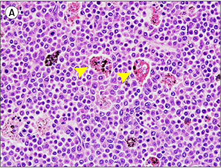
A 72-year-old woman presented with bleeding, swollen gums, and painful cervical lymphadenopathy. A CT scan revealed diffuse lymphadenopathy and hepatosplenomegaly. Initial laboratory tests showed the following: WBC count, 110.5×10⁹/L; hemoglobin level, 10.0 g/dL; platelet count, 218×109/L; LDH level, 528 IU/L; β2 microglobulin level, 11.1 µg/mL; and a differential count with marked leukocytosis with a left shift. Bone marrow biopsy indicated prefibrotic myelofibrosis. There was no evidence of JAK2 or BCR/ABL mutation or Epstein-Barr virus load. Trisomy 8 mosaicism was detected (47, XY, +8[6]/46, XY[24]) on karyotyping. Excisional lymph node biopsy revealed immature myeloid cells admixed with mature myeloid components and occasional megakaryocytes (A: H&E, ×400). Most notably, there were numerous hemophagocytic macrophages (arrowheads). Blasts comprised 40% of the total cellularity and showed a mixture of strongly MPO-positive myeloblasts and MPO-negative, CD68-positive, and CD163-positive monoblastic cells. The patient was diagnosed with primary myelofibrosis and extramedullary blastic transformation (granulocytic sarcoma) with acute myelomonoblastic differentiation accompanied by hemophagocytosis. Therefore, hydroxyurea chemotherapy was initiated. Hemophagocytosis can be seen in leukemic transformation of myelofibrosis.
 XML Download