Journal List > Korean J Hematol > v.46(2) > 1032943

Baek, Lee, Ryu, Lee, Song, Lee, Yun, Kim, and Jo: Clinical features and outcomes of autoimmune hemolytic anemia: a retrospective analysis of 32 cases

Abstract

Background

There has been no report on the clinical features or natural history of autoimmune hemolytic anemia (AIHA) in the Korean adult population. This study retrospectively analyzed the clinical characteristics and long-term outcomes of AIHA in the Korean adults.

Methods

Patients newly diagnosed with AIHA between January 1994 and December 2010 at Chungnam National University Hospital were enrolled. Patient characteristics at diagnosis, response to treatment, and the natural course of the disease were documented.

Results

Thirty-two patients (31 females and 1 male) with a median age of 48 years (range, 17-86) were enrolled. Of these, 21.9% were initially diagnosed with secondary AIHA. Thirteen patients (40.6%) were initially diagnosed with Evans' syndrome. Of the 29 patients who were placed on therapy, 27 (93.1%) showed a partial response or better. Nevertheless, 1 year after initiating treatment, 80% of the patients were still treatment-dependent. During follow-up (median length 14 months; range, 0.5-238), 14 of 25 patients (56.0%) who were initially diagnosed with primary warm antibody AIHA were found to have systemic lupus erythematosus (SLE). Median time to conversion to SLE was 8.0 months (95% CI, 4.3-11.7), and the probabilities of conversion at 12 and 24 months were 63% and 91%, respectively. Younger age (<60 years) and a positive fluorescent anti-nuclear antibody test were associated with a higher probability of SLE conversion (P=0.01 and P<0.001, respectively).

Conclusion

Primary AIHA is rare. Regular, vigilant testing for SLE is required in patients initially diagnosed with AIHA.

INTRODUCTION

Autoimmune hemolytic anemia (AIHA) is defined as the increased destruction of red blood cells (RBCs) in the presence of anti-RBC autoantibodies [1]. AIHA is a relatively uncommon cause of anemia. Recent population-based studies have calculated the incidence of AIHA to be 0.8/100,000/year [2], and its prevalence to be 17/100,000 [3]. AIHA may be primary (idiopathic) or secondary to various diseases, including systemic autoimmune disorders [4-6], malignancies [7], and infections [8, 9]. AIHA can also be induced by certain drugs [10, 11]. This disorder is heterogeneous with respect to the type (warm or cold) of antibodies involved. In spite of a long history of this disorder, management of AIHA is still mainly based on empirical data and on the results of small, retrospective, uncontrolled studies. Therapies for AIHA have been reviewed by several experts [12-15], but treatment guidelines have not yet been established. The current recommendations for the diagnosis and management of this disorder originate from Western Europe and North America, where the epidemiology of hematologic disorders may be different from that in the Orient. Although a few studies have described the clinical characteristics of AIHA in the Asian populations [11, 16-20], information from Asian regions is still limited. Furthermore, there has been no report on the clinical features or natural history of AIHA in the Korean adults. In the present study, we retrospectively analyzed clinical characteristics and outcomes of patients with AIHA in our institute.

MATERIALS AND METHODS

1. Patients

Patients who were consecutively diagnosed with AIHA based on positive results to either Coombs' test or cold agglutinin assay, at Chungnam National University Hospital between January 1994 and December 2010, were enrolled. All patients were Koreans. Patients with drug-induced hemolytic anemia were excluded. All patients underwent the following laboratory investigations: CBC with reticulocyte counts, peripheral blood smear, chemistry (including lactate dehydrogenase [LDH] and direct and indirect bilirubins), urine analysis, serum haptoglobin, plasma hemoglobin, direct and indirect Coombs' tests, and cold agglutinin assay. Screening tests for SLE, including fluorescent anti-nuclear antibody (FANA), complement-3 (C3), and -4 (C4) tests, were also performed. Patients who were positive for FANA underwent additional studies for autoantibodies, such as anti-double strand (ds) DNA antibody and anti-Smith antibody. Lupus anticoagulants (LA) and anti-cardiolipin antibodies (aCL) were examined. Bone marrow studies were performed to rule out lymphoproliferative disorders. SLE was diagnosed according to the American College of Rheumatology revised classification criteria for SLE [21]. Patients fulfilling only 3 of the revised classification criteria for SLE from the American College of Rheumatology were defined as having "incomplete" SLE [22]. Evans' syndrome was diagnosed, if the patient tested positive for hemolytic anemia by the Coomb's test, and for idiopathic thrombocytopenic purpura, in the absence of any known underlying etiology.

2. Treatment and response evaluation

Patients with AIHA who did not present with Evans' syndrome (hereafter described as "AIHA only") received oral prednisolone (Pd) alone (1 mg/kg/d) or intravenous (IV) methylprednisolone (methyl-Pd; 10 mg/kg/d for 3 d) followed by Pd, whereas patients with Evans' syndrome received corticosteroids (oral Pd alone or IV methyl-Pd followed by Pd) or IV immune globulin (IVIg) as first-line therapy. Response criteria [22] were classified as follows: (a) complete response (CR), defined as a stable hemoglobin level of >12 g/dL, no requirement for transfusion, and absence of clinical and laboratory signs of hemolysis (no jaundice, normalization of serum LDH, haptoglobin and indirect bilirubin levels, and normal reticulocyte count), irrespective of Coombs' test result; (b) partial response (PR), defined as a rise in hemoglobin levels of >2 g/dL, no or reduced transfusion requirement, and improvement of clinical and laboratory signs of hemolysis; and (c) no response.

3. Follow-up

Patients were followed up every 1-2 weeks until responses were achieved, and then every 2-3 months. Investigations for SLE were performed annually in most patients. Convertsion to SLE and complications such as renal insufficiency and thrombosis were evaluated.

4. Statistical analyses

Continuous variables were analyzed using a Student's t-test, and binary variables were analyzed using a Chi-square test. The probability of conversion to SLE in patients with primary AIHA was plotted as Kaplan-Meier curves, and probabilities with respect to risk factors were compared using the log-rank test and a Cox proportional hazard model. All analyses were performed using SPSS version 17.0 (SPSS Inc., Chicago, IL). P-value of <0.05 was considered statistically significant.

RESULTS

1. Patient characteristics at diagnosis

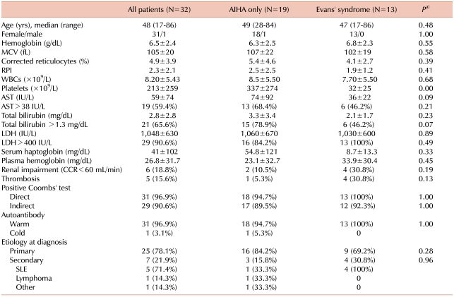
Thirty-two patients with a median age of 48 years (range, 17-86) were enrolled. The majority of the patients (31 of 32) were female, and 13 (40.6%) presented with Evans' syndrome. The occurrence of Evans' syndrome in patients initially diagnosed with primary and secondary AIHA was 36% and 57.1%, respectively. Total bilirubin and LDH levels were above the upper normal limit (UNL) in 65.6% and 90.6% of the patients, respectively. Renal impairment and thrombosis were found in 18.8% and 15.6% of the patients, respectively. Of the 32 patients, 31 (96.9%) were positive for warm antibody, whereas only 1 (3.1%) was positive for cold agglutinin. Underlying disorders were found in 7 patients (21.9%; SLE was found in 5 patients, and lymphoma and unidentified lung neoplasm in 1 patient each). No underlying disorders were detected in 25 patients (78.1%). There were no statistically significant differences between patients with AIHA only and those with Evans' syndrome in terms of demographic features, CBC, chemistry, complications, autoantibody profiles, and etiology, with the exception of lower platelet counts in patients with Evans' syndrome (Table 1).
FANA and anti-dsDNA antibody were positive in 16 of 27 (59.3%) and 12 of 23 (52.2%) patients, respectively. Serum levels of C3 and C4 decreased in 14 of 26 (53.8%) and 21 of 27 (77.8%) patients, respectively. LA and IgG aCL were detected in 7 of 17 (41.2%) and 5 of 15 (33.3%) patients, respectively. The number of positive results to FANA, anti-dsDNA antibody, IgG aCL, IgM aCL, and LA tests, and serum levels of C3 and C4, did not differ between patients with AIHA only and those with Evans' syndrome (Table 2). As anticipated, FANA (100% vs. 52.4%; P=0.03) and anti-dsDNA antibody (100% vs. 38.9%; P=0.03) were more frequently positive in patients with SLE than in patients initially diagnosed with primary AIHA (data not shown). However, there were no statistically significant differences between patients initially diagnosed with AIHA and those with AIHA secondary to SLE, in terms of demographic features, CBC, chemistry, or complications (data not shown).

2. Response to treatment

Twenty-nine patients were placed on therapy, including Pd alone, methyl-Pd followed by Pd, or IVIg followed by Pd. More patients with Evans' syndrome received methyl-Pd or IVIg than did patients with AIHA only. Of the 29 patients, 8 (27.6%) and 19 (65.5%) patients achieved complete and partial responses within 1 month of treatment initiation, respectively. Nevertheless, 2 years after initiating treatment, 80% of the patients still required Pd or other drugs such as azathiopurine or cyclophosphamide to maintain at least a partial response. There were no differences in response rates or Pd dependence between patients with AIHA only and those with Evans' syndrome (Table 3).

3. Natural course of warm antibody AIHA

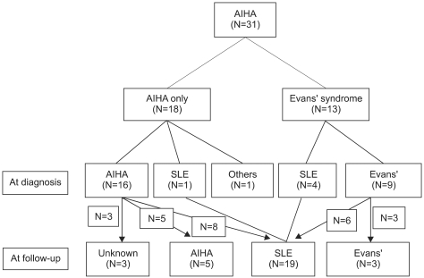
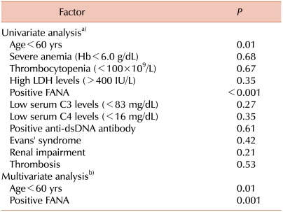
The natural course and complications were analyzed in 25 patients who were initially diagnosed with primary warm antibody AIHA. During a median follow-up of 14 months (range, 0.5-238), 12 patients (48.0%) developed SLE with a median time to conversion of 9.5 months (range, 3-28). Two of these patients (8.0%) were found to have incomplete SLE, with a median time to conversion of 7 months (range, 6-8). Overall, 56% of the patients who were initially diagnosed with primary AIHA were found to have SLE or incomplete SLE during the follow-up. Of these, 8 were AIHA-only patients (out of 16 total; 43.8%), and 6 were Evans' syndrome patients (out of 9 total; 66.7%), revealing no statistical difference in SLE conversion frequencies between the 2 groups. The median time to conversion was shorter, but not significantly so, in patients with Evans' syndrome (6 months; range, 3-28) than in patients with AIHA only (12 months; range, 6-22). Renal impairment and thrombosis developed in 2 (8.0%) and 3 (12.0%) patients, respectively (Table 4, Fig. 1). Kaplan-Meier analysis revealed that the median time to conversion to SLE or incomplete SLE was 8 months (95% CI, 4.3-11.7), and that the probabilities of conversion at 12 and 24 months were 63% and 91%, respectively (Fig. 2). Univariate analysis using the log-rank test revealed that age <60 years (P=0.01) and a positive FANA test (P<0.001) were risk factors for conversion to SLE. Multivariate analysis using a Cox proportional hazard model indicated that these 2 parameters were independent risk factors (P=0.01 and P=0.001, respectively). Severe anemia (hemoglobin <6.0 g/dL), thrombocytopenia, elevated serum LDH levels, low serum C3 or C4 levels, and renal impairment or thrombosis did not increase the risk of conversion to SLE (Table 5). Although patients with Evans' syndrome tended to develop SLE earlier than did those with AIHA only, the difference was not statistically significant (Fig. 2). SLE was found much earlier in most patients with Evans' syndrome than in those with AIHA only, with the exception of a single patient with Evans' syndrome who was diagnosed with SLE long after the initial diagnosis. This patient was not followed up for some time, and thus did not undergo regular testing for SLE. No AIHA-related mortality was found, but 1 patient with lung neoplasm died of respiratory failure 3 weeks after initial presentation.

DISCUSSION

The present study demonstrates that AIHA is an uncommon hematologic disorder in the Korean adult population and that cold agglutinin disease is extremely rare, which may explain why the epidemiology, clinical features, and clinical outcomes of AIHA in the Korean adults have not been previously described. Primary (idiopathic) AIHA is known to be less common than secondary AIHA in the Western populations. However, several reports suggest the presence of ethnic differences. For example, secondary AIHA accounted for 49% and 51% of Coombs'-positive AIHA in the Chinese and French populations, respectively [23, 24]. In contrast, secondary AIHA was found in only 34.3% of cases in the Indian population [19]. In the present study, primary AIHA appeared to be much more frequent at diagnosis, which contradicts previous observations from the Western countries. However, during follow-up, it was revealed that 56% of patients initially diagnosed with primary AIHA actually had secondary AIHA, and that SLE dominated as an underlying disorder. Only 32% of the patients initially diagnosed with primary AIHA were true primary cases. The lower frequency of diagnosis of secondary AIHA in Korea than in other countries may be attributed to the exclusion of a considerable number of patients with secondary AIHA, as most SLE patients are primarily seen and managed by rheumatologists or nephrologists. Chronic lymphocytic leukemia (CLL) is known to be a common underlying disorder in AIHA [25], but is rare in Korea, which may be another reason why secondary AIHA is less frequently diagnosed and why SLE was the predominant underlying disorder in our cases.
Evans' syndrome is an autoimmune disorder defined by the simultaneous or sequential combination of AIHA and immune thrombocytopenia or immune neutropenia [26]. It may reveal underlying conditions such as SLE and common variable immunodeficiency [27]. Although its frequency is unknown, Evans' syndrome is known to be a rare disorder. A review reported that only 6 of 399 adult patients with AIHA (1.5%) were found to have Evans' syndrome [28]. In the present study, Evans' syndrome initially accounted for 28.1% of the AIHA cases; however, 66.7% of these patients were found during follow-up to have secondary AIHA with SLE. In the final count, patients with Evans' syndrome accounted for 9.4% of AIHA cases; this frequency is still higher than that reported in the literature. At present, we have no explanation for this discrepancy. A nationwide study enrolling a large number of patients may reveal whether it can be attributed to ethnic differences or not. There were no statistically significant differences between patients with AIHA only and those with Evans' syndrome in terms of clinical features, with the exception of a higher frequency of IgG aCL antibody positivity. Given that more Evans' syndrome patients than AIHA-only patients had SLE at diagnosis of AIHA, the absence of a statistically significant difference in clinical features between the two groups may be attributed to the small number of patients. Further studies are warranted.
In the present study, we showed that a population of patients who were initially diagnosed with primary AIHA turned out to have SLE during follow-up. The probability of conversion to SLE within 2 years of follow-up was 91%, indicating that the majority of AIHA patients will be diagnosed with SLE during the first 2 years of follow-up, and that true primary AIHA is rare; this is different from the Western, Indian, and Chinese reports [19, 23, 24]. This may be explained, at least in part, by the fact that SLE is 2-4 times more frequent, and more severe, among non-white populations. SLE is a chronic autoimmune disease with a variety of hematological manifestations. Anemia is found in approximately 50% of patients, with the chronic form of anemia being the most common [29]. An impaired erythropoietin response and the presence of antibodies against erythropoietin may contribute the pathogenesis of this type of anemia. Patients with AIHA usually belong to a distinct category, characterized by aCL antibodies, thrombosis, thrombocytopenia, and renal disease, often in the context of secondary anti-phospholipid antibody syndrome [29]. In a previous study of a cohort of 1,251 unrelated females with SLE, 76 patients (6.1%) were found to have hemolytic anemia [30]. The presence of hemolytic anemia was associated with a subset of SLE patients characterized by earlier disease onset, more severe disease, and a higher likelihood of renal involvement, seizures, serositis, and other cytopenias. It has been shown that ethnicity can influence disease manifestations at diagnosis of SLE, even within an Oriental population [31]. In one study of the Korean population [32], AIHA was found in 3.5% of patients with SLE.
In the present study, we found that some patients who were initially diagnosed with primary AIHA developed renal insufficiency or thrombosis during follow-up, indicating that such patients need to be carefully monitored. The present study showed that patients with AIHA aged <60 years and those who were positive for FANA were at an increased risk of conversion to SLE, indicating that patients with these features require regular testing for SLE. There was a tendency for the patients with Evans' syndrome to develop SLE earlier than those with AIHA alone. The fact that the difference was not statistically significant may be attributed to the small number of patients. One patient with Evans' syndrome who did not undergo regular testing for SLE because of poor compliance was diagnosed with SLE very late during the follow-up, and this greatly affected the result for that group.
In conclusion, true primary AIHA is rare. The majority of patients with AIHA are found to have SLE during follow-up. Regular, vigilant testing for SLE is required in patients who are initially diagnosed with primary AIHA.

References

1. Packman CH. Hemolytic anemia due to warm autoantibodies: new and traditional approaches to treatment. Clin Adv Hematol Oncol. 2008; 6:739–741. PMID: 18997664.
2. Klein NP, Ray P, Carpenter D, et al. Rates of autoimmune diseases in Kaiser Permanente for use in vaccine adverse event safety studies. Vaccine. 2010; 28:1062–1068. PMID: 19896453.
crossref
3. Eaton WW, Rose NR, Kalaydjian A, Pedersen MG, Mortensen PB. Epidemiology of autoimmune diseases in Denmark. J Autoimmun. 2007; 29:1–9. PMID: 17582741.
crossref
4. Budman DR, Steinberg AD. Hematologic aspects of systemic lupus erythematosus. Current concepts. Ann Intern Med. 1977; 86:220–229. PMID: 835948.
5. Rosenthal DS, Sack B. Autoimmune hemolytic anemia in scleroderma. JAMA. 1971; 216:2011–2012. PMID: 5108636.
crossref
6. Giannadaki E, Potamianos S, Roussomoustakaki M, Kyriakou D, Fragkiadakis N, Manousos ON. Autoimmune hemolytic anemia and positive Coombs test associated with ulcerative colitis. Am J Gastroenterol. 1997; 92:1872–1874. PMID: 9382055.
7. Ellis LD, Westerman MP. Autoimmune hemolytic anemia and cancer. JAMA. 1965; 193:962–964. PMID: 14343919.
crossref
8. Saif MW. HIV-associated autoimmune hemolytic anemia: an update. AIDS Patient Care STDS. 2001; 15:217–224. PMID: 11359664.
crossref
9. Khan FY, A yassin M. Mycoplasma pneumoniae associated with severe autoimmune hemolytic anemia: case report and literature review. Braz J Infect Dis. 2009; 13:77–79. PMID: 19578637.
crossref
10. Territo MC, Peters RW, Tanaka KR. Autoimmune hemolytic anemia due to levodopa therapy. JAMA. 1973; 226:1347–1348. PMID: 4202390.
crossref
11. Shen Y. Autoimmune hemolytic anemia associated with a formulation of traditional Chinese medicines. Am J Health Syst Pharm. 2009; 66:1701–1703. PMID: 19767374.
crossref
12. Valent P, Lechner K. Diagnosis and treatment of autoimmune haemolytic anaemias in adults: a clinical review. Wien Klin Wochenschr. 2008; 120:136–151. PMID: 18365153.
crossref
13. King KE, Ness PM. Treatment of autoimmune hemolytic anemia. Semin Hematol. 2005; 42:131–136. PMID: 16041662.
crossref
14. Barros MM, Blajchman MA, Bordin JO. Warm autoimmune hemolytic anemia: recent progress in understanding the immunobiology and treatment. Transfus Med Rev. 2010; 24:195–210. PMID: 20656187.
15. Lechner K, Jäger U. How I treat autoimmune hemolytic anemias in adults. Blood. 2010; 116:1831–1838. PMID: 20548093.
crossref
16. Yi Y, Zhang GS, Gong FJ, Yang JJ. Multiple myeloma complicated by Evans syndrome. Intern Med J. 2009; 39:421–422. PMID: 19580625.
crossref
17. Hara A, Wada T, Kitajima S, et al. Combined pure red cell aplasia and autoimmune hemolytic anemia in systemic lupus erythematosus with anti-erythropoietin autoantibodies. Am J Hematol. 2008; 83:750–752. PMID: 18626921.
crossref
18. Lim YA, Kim MK, Hyun BH. Autoimmune hemolytic anemia predominantly associated with IgA anti-E and anti-c. J Korean Med Sci. 2002; 17:708–711. PMID: 12378029.
crossref
19. Naithani R, Agrawal N, Mahapatra M, Pati H, Kumar R, Choudhary VP. Autoimmune hemolytic anemia in India: clinico-hematological spectrum of 79 cases. Hematology. 2006; 11:73–76. PMID: 16522555.
crossref
20. Karasawa M. Autoimmune hemolytic anemia. Nippon Rinsho. 2008; 66:520–523. PMID: 18326320.
21. Hochberg MC. Updating the American College of Rheumatology revised criteria for the classification of systemic lupus erythematosus. Arthritis Rheum. 1997; 40:1725. PMID: 9324032.
crossref
22. Michel M, Chanet V, Dechartres A, et al. The spectrum of Evans syndrome in adults: new insight into the disease based on the analysis of 68 cases. Blood. 2009; 114:3167–3172. PMID: 19638626.
crossref
23. Genty I, Michel M, Hermine O, Schaeffer A, Godeau B, Rochant H. Characteristics of autoimmune hemolytic anemia in adults: retrospective analysis of 83 cases. Rev Med Interne. 2002; 23:901–909. PMID: 12481390.
24. Zhang Y, Chu Y, Shao Z. The clinical implications of IgG subclass in 84 patients with autoimmune hemolytic anemia. Zhonghua Xue Ye Xue Za Zhi. 1999; 20:524–526. PMID: 11721398.
25. Zent CS, Ding W, Reinalda MS, et al. Autoimmune cytopenia in chronic lymphocytic leukemia/small lymphocytic lymphoma: changes in clinical presentation and prognosis. Leuk Lymphoma. 2009; 50:1261–1268. PMID: 19811329.
crossref
26. Evans RS, Takahashi K, Duane RT, Payne R, Liu C. Primary thrombocytopenic purpura and acquired hemolytic anemia: evidence for a common etiology. AMA Arch Intern Med. 1951; 87:48–65. PMID: 14782741.
27. Norton A, Roberts I. Management of Evans syndrome. Br J Haematol. 2006; 132:125–137. PMID: 16398647.
crossref
28. Silverstein MN, Heck FJ. Acquired hemolytic anemia and associated thrombocytopenic purpura: with special reference to Evans' syndrome. Proc Staff Meet Mayo Clin. 1962; 37:122–128. PMID: 13912960.
29. Giannouli S, Voulgarelis M, Ziakas PD, Tzioufas AG. Anaemia in systemic lupus erythematosus: from pathophysiology to clinical assessment. Ann Rheum Dis. 2006; 65:144–148. PMID: 16079164.
crossref
30. Jeffries M, Hamadeh F, Aberle T, et al. Haemolytic anaemia in a multi-ethnic cohort of lupus patients: a clinical and serological perspective. Lupus. 2008; 17:739–743. PMID: 18625652.
31. Thumboo J, Fong KY, Chng HH, et al. The effects of ethnicity on disease patterns in 472 Orientals with systemic lupus erythematosus. J Rheumatol. 1998; 25:1299–1304. PMID: 9676760.
32. Lee SY, Chi HS. Hematologic findings in systemic lupus erythematosus. Korean J Clin Pathol. 1998; 18:14–19.
Fig. 1
Natural course of patients with warm antibody AIHA.
kjh-46-111-g001
Fig. 2
Probability of conversion to SLE in patients initially diagnosed with primary warm antibody AIHA (N=25). (A) Probability of conversion to SLE in all patients. (B) Comparison of SLE conversion probabilities between patients with AIHA only and those with Evans' syndrome.
kjh-46-111-g002
Table 1
Patient characteristics.
kjh-46-111-i001

a)AIHA only vs. Evans' syndrome.

Abbreviations: AIHA, autoimmune hemolytic anemia; MCV, mean corpuscular volume; PRI, red cell production index; WBC, white blood cell; AST, aspartate aminotransferase; LDH, lactate dehydrogenase; CCR, creatinine clearance rate; SLE, systemic lupus erythematosus.

Table 2
Serologic findings.
kjh-46-111-i002

a)AIHA only vs. Evans' syndrome.

Abbreviations: AIHA, autoimmune hemolytic anemia; FANA, fluorescent anti-nuclear antibody; aCL, anti-cardiolipin antibody.

Table 3
Initial treatment and response.
kjh-46-111-i003

a)AIHA only vs. Evans' syndrome, b)response 1 month after the initiation of treatment, c)Pd or other drug dependence.

Abbreviations: Pd, prednisolone; IVIg, intravenous immune globulin.

Table 4
Natural course and complications in patients with warm antibody AIHA.
kjh-46-111-i004

a)AIHA only vs. Evans' syndrome.

Abbreviations: AIHA, autoimmune hemolytic anemia; SLE, systemic lupus erythematosus.

Table 5
Risk factors for conversion to SLE in patients initially diagnosed with warm antibody AIHA.
kjh-46-111-i005

a)Log-rank test, b)Cox proportional hazard model.

TOOLS
Similar articles