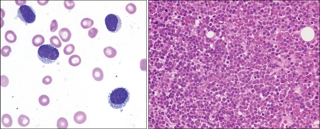
A 73-year-old man presented with abdominal distension and general weakness for 3 weeks. One month prior to visit, brown-colored patchy skin lesion on left flank had appeared. Axillary lymph nodes were palpable on physical examination. Peripheral blood examination showed bicytopenia, leukoerythroblastic features and abnormal cells. Bone marrow aspiration revealed numerous medium-sized tumor cells with cytoplasmic microvacuoles localized along the cell membrane and pseudopodia (Left). Bone marrow biopsy showed diffuse infiltration of tumor cells (Right). Tumor cells expressed CD4, CD56, CD43, CD68, CD7, CD33, TdT, and were negative for CD34, CD117, CD3, CD5, CD20, myeloperoxidase and EBV. The diagnosis of blastic plasmacytoid dendritic cell neoplasm was made and the patient received chemotherapy but expired soon after, due to disseminated disease. The characteristic morphology with necklace-like cytoplasmic microvacules localized along the cell border can be a diagnostic clue to blastic plasmacytoid dendritic cell neoplasm, even in the era of high technology medicine.


 PDF
PDF ePub
ePub Citation
 Print
Print


 XML Download