Abstract
We report a case of synchronous occurrence of KIT-positive acute myeloid leukemia (AML) and gastrointestinal stromal tumor (GIST). A 63-year-old woman was hospitalized for dizziness, and abdominal computed tomography revealed an exophytic gastric mass and hepatic metastasis. The patient was diagnosed with GIST and was administered imatinib (400 mg/day) for the metastatic unresectable tumor. After 2 weeks of imatinib treatment, the patient developed pancytopenia, which persisted even after the drug was discontinued, thereby necessitating bone marrow biopsy. Biopsy examination indicated AML, and karyotyping revealed a complex karyotype. We did not observe point mutations at residues D816 and N822 of KIT. Therefore, the patient received standard induction chemotherapy, but on the 18th day after completion of chemotherapy, she died of septic shock and multi-organ failure. Since KIT plays an important role in both GIST and AML, we consider that both these malignancies may have been associated with each other.
Gastrointestinal stromal tumors (GISTs) are the most common mesenchymal tumors of the gastrointestinal tract. Most of the GISTs occur in the stomach (60%), jejunum and ileum (30%), and the duodenum (5%). GISTs comprise a biological spectrum of small benign nodules to fully malignant sarcomas [1]. The development of GIST is associated with the mutational activation of receptor tyrosine kinases, namely, the platelet-derived growth factor receptor-a (PDGFRA) or KIT [1-3].
KIT is a type III receptor protein tyrosine kinase that is involved in the development and maintenance of erythropoietic stem cells, mast cells, melanocytes, germ cells, and interstitial cells of Cajal [4]. Over-expression and/or constitutive expression of KIT alone or in conjunction with a stem cell factor (SCF) induces several solid tumors, including small-cell lung carcinoma, neuroblastoma, pancreatic tumors, and several hematological malignancies [5]. Moreover, mutations involving the KIT exons 8 and 17 and to a less extent, the KIT exons 9 and 11 have been identified in these malignancies.
Previous studies have shown an association between GIST and other malignancies such as hepatocellular carcinoma, adenocarcinoma, mastocytosis, and lymphoma/leukemia [6-11], but synchronous occurrence of GIST and leukemia has not been reported. Here, we report the first case of synchronous development of KIT-positive acute myeloid leukemia (AML) and GIST. The potential nonrandom association and causal relationship between GIST and AML need to be investigated.
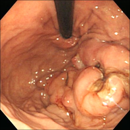
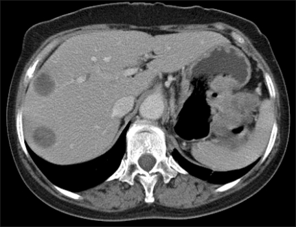
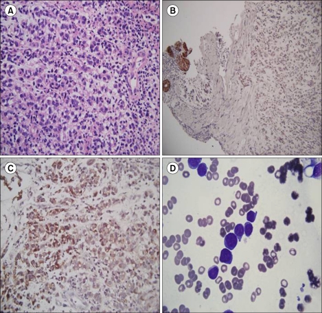
A 63-year-old woman was hospitalized after experiencing dizziness and fatigue for 2 months. Her physical examination was unremarkable, with the exception of pale conjunctivae. The initial laboratory tests revealed a white cell count of 3.08×10⁹/L, a hemoglobin level of 6.3 g/dL, and a platelet count of 80×109/L. Endoscopy revealed an ulcerofungating mass lesion of about 6.0×6.0 cm in the posterior wall of the high body-fundus (Fig. 1). Abdominal computed tomography revealed an exophytic gastric mass and hepatic metastasis (Fig. 2). The histopathologic analysis showed poorly differentiated malignant-cell infiltrates with epithelioid features in the myxoid stroma. Immunochemical staining of these cells was positive for CD117 and CD 34, thereby indicating GIST (Fig. 3A-C). However, KIT tyrosine kinase mutation in the gastric samples was not examined. The patient initially received imatinib mesylate (400 mg/day) to treat the metastatic unresectable tumor.
On the 14th day of imatinib treatment, complete blood cell count showed a white cell count of 1.87×10⁹/L, hemoglobin level of 8.7 g/dL, and a platelet count of 29×109/L. Pancytopenia persisted even after discontinuation of imatinib, and bone marrow biopsy revealed a markedly hypercellular marrow with infiltrates of myeloblasts (26%), thereby indicating AML with maturation (Fig. 3D). The karyotyping showed 39-47,XX,add(4)(q12),der(5;22)(p10;q10),+der (5;22),add(12)(p11.2),-17,-19,-20,+1-2marcp [19]/46, XX [1]. Immunophenotyping was positive for CD117, CD33, CD34, CD38, CD56, and CD7. We did not observe point mutations at residues D816 and N822 of KIT. Therefore, the patient underwent chemotherapy with idarubicin (12 mg/m2/day) for 3 days and cytarabine (200 mg/m2/day) for 7 days, but on the 18th day after the completion of chemotherapy, she died of septic shock and multi-organ failure.
The development of GIST is believed to be associated with the mutational activation of receptor tyrosine kinases, namely, KIT or PDGFRA [1]. Up to 75% of the activating KIT mutations that usually affect the juxtamembrane domain (exon 11) induce ligand-independent phosphorylation. A small number of GISTs, especially those of the gastric cells with epithelioid morphology, show PDGFRA mutations, which are mostly observed in the tyrosine kinase domain (exon 18) [1-3].
KIT is also an important pathogenetic factor in AML. KIT expression is significantly higher in AML blasts than in normal bone marrow cells, and one-third of the AML blasts co-express KIT and SCF, thereby suggesting autocrine and paracrine stimulation of KIT by SCF [5]. Moreover, KIT mutation involving exon 8 was observed in 20% of the patients with inv(16); KIT mutation involving exon 17 was expressed in a patient with t(8;21).
Since KIT plays a role in both GIST and AML, these malignancies may be associated. In 1997, a case of leukemia was reported, in which the disease developed after jejunal GIST [12]. The jejunal GIST was treated surgically, and the patient underwent chemotherapy with fluorouracil; however, a month later, the patient was diagnosed with acute promyelocytic leukemia. Leukemia was considered as the second malignancy, which developed after chemotherapy for GIST. However, in our case, GIST and AML were diagnosed synchronously. To the best of our knowledge, this is the first case report in which KIT-positive AML and GIST were synchronously observed. The results of an epidemiologic analysis indicated a nonrandom association between AML and GIST [9, 10]. Agaimy et al. [9] reported that lymphoma/leukemia developed in 7% of GIST patients. Miettinen et al. [10] examined the long-term follow-up data of 1892 GIST patients; among these, 6 patients had AML and 3 had chronic myeloid leukemia. In these patients, the leukemia developed 1.7-21 years after the GIST (median interval, 6 years). Moreover, the risk of AML was found to be significantly higher for women, as observed in our case.
The common pathogenesis of KIT activation or mutation in GIST and AML has still not been elucidated. However, the fact that KIT tyrosine kinase activation or mutation plays an important role in both GIST and AML corroborates that the 2 tumors are associated and do not develop incidentally as independent tumors. The possible causes for their association are as follows. First, the activated KIT pathway is involved in leukemogenesis in GIST patients. The hematopoietic progenitor cells are dependent on the KIT-signaling pathway. The KIT mutation in AML is structurally and functionally similar to that in GIST. For example, the KIT mutations involving exon 8 and the internal tandem duplications involving KIT exon 11 occur in both AML and GIST. Second, the circulating KIT ligand, SCF, which is detected in GIST patients, has a humoral effect on bone marrow precursors; this probably leads to myeloproliferation and ultimately to the development of leukemia.
Here, we report a case of synchronous occurrence of KIT-positive AML and GIST. These 2 tumors are considered to be nonrandomly associated rather than being double primary tumors. For appropriate treatment of these 2 malignancies, further investigations such as molecular therapeutic targeting of KIT signaling pathway and its impact on the response to tyrosine kinase inhibitors, are required.
References
1. Hirota S, Isozaki K, Moriyama Y, et al. Gain-of-function mutations of c-kit in human gastrointestinal stromal tumors. Science. 1998; 279:577–580. PMID: 9438854.
2. Heinrich MC, Corless CL, Duensing A, et al. PDGFRA activating mutations in gastrointestinal stromal tumors. Science. 2003; 299:708–710. PMID: 12522257.
3. Kitamura Y, Hirota S. Kit as human oncogenic tyrosine kinase. Cell Mol Life Sci. 2004; 61:2924–2931. PMID: 15583854.
4. Beghini A, Ripamonti CB, Cairoli R, et al. KIT activating mutations: incidence in adult and pediatric acute myeloid leukemia, and identification of an internal tandem duplication. Haematologica. 2004; 89:920–925. PMID: 15339674.
5. Hassan HT. c-Kit expression in human normal and malignant stem cells prognostic and therapeutic implications. Leuk Res. 2009; 33:5–10. PMID: 18639336.

6. Liu SW, Chen GH, Hsieh PP. Collision tumor of the stomach: a case report of mixed gastrointestinal stromal tumor and adenocarcinoma. J Clin Gastroenterol. 2002; 35:332–334. PMID: 12352297.
7. Felekouras E, Athanasios P, Vgenopoulou S, et al. Coexistence of hepatocellular carcinoma (HCC) and c-Kit negative gastrointestinal stromal tumor (GIST): a case report. South Med J. 2008; 101:948–951. PMID: 18708986.

8. Herbers AH, Keuning JJ. Staging for CLL-type non-Hodgkin's lymphoma reveals a gastrointestinal stromal tumour. Neth J Med. 2005; 63:74–75. PMID: 15766012.
9. Agaimy A, Wünsch PH, Sobin LH, Lasota J, Miettinen M. Occurrence of other malignancies in patients with gastrointestinal stromal tumors. Semin Diagn Pathol. 2006; 23:120–129. PMID: 17193825.

10. Miettinen M, Kraszewska E, Sobin LH, Lasota J. A nonrandom association between gastrointestinal stromal tumors and myeloid leukemia. Cancer. 2008; 112:645–649. PMID: 18041070.

11. Lee JW, Yang WS, Chung SY, et al. Aggressive systemic mastocytosis after germ cell tumor of the ovary: C-KIT mutation documentation in both disease states. J Pediatr Hematol Oncol. 2007; 29:412–415. PMID: 17551405.
12. Yoshioka K, Yamaguchi M, Kasamatsu Y, et al. Acute promyelocytic leukemia following leiomyosarcoma of the jejunum. Arch Intern Med. 1997; 157:1392–1393. PMID: 9201018.

13. Roskoski R Jr. Signaling by Kit protein-tyrosine kinase--the stem cell factor receptor. Biochem Biophys Res Commun. 2005; 337:1–13. PMID: 16129412.

Fig. 1
Esophagogastroenteroscopy shows an ulcerofungating mass lesion in the posterior wall of the gastric fundus. The mass is covered with normal mucosa-like submucosal tumor, but it has a large ulceration at the centre.

Fig. 2
Abdominal computed tomography shows an 8×7.3 cm exophytic gastric mass in the posterior wall of the gastric body containing a large area of cavitation and variable hepatic metastasis.

Fig. 3
(A) Poorly differentiated malignant-cell infiltrates with epithelioid features in the myxoid stroma. (B) Immunohistochemical staining of cytokeratins (CK MNF116) is negative. (C) Immunohistochemical staining of KIT is positive. (D) The uniform population of primitive myeloblasts is indicative of acute myeloid leukemia.


 PDF
PDF ePub
ePub Citation
 Print
Print


 XML Download