

 PDF
PDF ePub
ePub Citation
 Print
Print


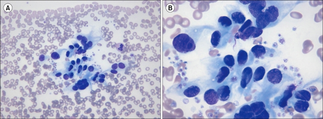
A blood sample from a 36-year-old woman was tested for anemia. The peripheral blood film shows microcytic, hypochromic erythrocytes; mild anisocytosis; marked poikilocytosis, comprising elliptocytes, teardrop cells, and target cells; and a cluster of endothelial cells (Wright stain; A, ×400 B, ×1,000). The endothelial cells are medium- to large-sized cells and contain oval-grooved nucleus; distinct or indistinct nucleolus; and a moderate amount of cytoplasm, which appears pale blue in color and frayed at the tapered ends of the cells. The probability of detecting endothelial cells is high, if the blood films are made using the first drop of blood from a needle. This characteristic is particularly prominent when the needles are reused or barbed. Increased numbers of endothelial cells are present in a variety of conditions that are characterized by vascular injury or vessel formation (e.g., coronary angioplasty, unstable angina, acute myocardial infarction, stable coronary disease, peripheral atherosclerosis, antineutrophil cytoplasmic antibody-associated vasculitis, systemic lupus erythematosus, Behçet's disease, rickettsial infection, cytomegalovirus infection, sickle cell anemia, allogeneic stem cell transplantation, cancer, and thrombotic thrombocytopenic purpura), but even in such conditions, they are very sparse. Gynecologic ultrasonography performed 4 months later revealed a huge uterine myoma. The myomatous uterus has high numbers of arterioles and venules.
 XML Download