Abstract
Background
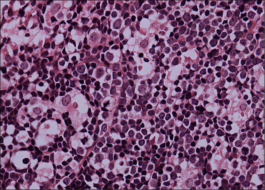
The bone marrow biopsy sections of acute leukemia patients occasionally reveal a proliferation of large mononuclear cells that accompany the leukemic blasts, and this proliferation shows a starry sky pattern. We characterized these large mononuclear cells by performing immunohistochemistry with 12 different antibodies. The clinical characteristics were examined and then we determined their difference from hemophagocytic lymphohistiocytosis (HLH) and malignant histiocytic disorders.
Methods
Of the 200 acute leukemic bone marrow biopsy samples, 11 ALL and 10 AML cases showed large mononuclear cell proliferations. The panel of antibodies used for immunohistochemistry included those against the mononuclear phagocyte system, and immunohistochemistry was performed on the patients' initial specimens and the complete remission specimens. 10 normal specimens, 4 initial CML specimens and their complete hematologic response specimens were included as controls.
Results
The large mononuclear cells showed immunohistochemical results consistent with histiocytes. They were negative for the markers of dendritic cells the histiocytes and cytokines that are involved in the pathogenesis of HLH and vascular proliferation. Histiocyte proliferation was not observed in the complete remission specimens and in the initial and complete hematological response specimens of the CML patients and the normal bone marrow specimens. None of the cases fulfilled the criteria of HLH, and all 5 ALL cases, for which the immunophenotype results were available, showed a B cell phenotype.
Conclusion
We characterized the large mononuclear cell proliferations as reactive histiocyte proliferations and we differentiated these from those of secondary HLH and malignant histiocytic disorders. A proportion of the large mononuclear cells showed negative results for all 12 antibodies and they showed characteristics that were suggestive of small fat cells. The pathophysiology and the prognostic effect of the reactive histiocyte proliferation accompanying acute leukemia require further study.
Go to : 
References
1. Frisch B, Bartl R. Acute leukemias. Frisch B, Bartl R, editors. Biopsy interpretation of bone and bone marrow. 2nd ed.Oxford, England: Oxford University Press;2006. p. 209–25.
2. Henter JI, Horne A, Arico M, et al. HLH-2004: diagnostic and therapeutic guidelines for hemophagocytic lymphohistiocytosis. Pediatr Blood Cancer. 2007; 48:124–31.

3. Jaffe ES. Histiocytic and dendritic cell neoplasms. In: Jaffe ES, Harris NL, Stein H, Vardiman JM, eds. World health organization classification of tumours. Pathology and genetics of tumors of haematopoietic and lymphoid tissues. Lyon, France: IARC Press. 2006. 273–89.
4. Clark BS, Dawson PJ. Histiocytic medullary reticulosis presenting with a leukemic blood picture. Am J Med. 1969; 47:314–7.

5. Kim SJ, Cho HI, Ahn HS. Two cases of virus associated hemophagocytic syndrome after chemotherapy of acute myelogenous leukemia. Korean J Hematol. 1990; 25:281–5.
6. Yoon SY, Cho YJ, Lee KN. Histiocytosis occurring in a patient with B-lineage acute lymphoblastic leukemia. Korean J Clin Pathol. 1994; 14:391–6.
7. Lee HK, Kim YG, Han KJ, Shim SI. Two cases of reactive histiocytosis with peroxidase positivity in acute myelogenous leukemia. Korean J Clin Pathol. 1996; 16:447–52.
8. Yoon HR, Ko YH, Kim SH. Peripheral T cell lymphoma associated with hemophagocytic histiocytosis mimicking malignant histiocytosis. Korean J Clin Pathol. 1997; 17:934–43.
9. Falini A, Pileri S, DeSolas I, et al. Peripheral T-cell lymphoma associated with hemophagocytic syndrome. Blood. 1990; 75:434.
10. Jaffe ES, Costa J, Fauci AS, Cossman J, Tsokos M. Malignant lymphoma and erythrophagocytosis simulating malignant histiocytosis. Am J Med. 1983; 75:741–9.

11. Shumichi K, Hiroto I, Hiroto T, Shotai K. Phagocytosis of terminally differentiated acute promyelocytic leukemia cells by marrow histiocytes during treatment with all-trans retinoic acid. Leuk Lymphoma. 2003; 44:2147–50.
12. Vacca A, Ribatti D, Ruco L, et al. Angiogenesis extent and macrophage density increase simultaneously with pathological progression in B-cell non-Hodgkin's lymphomas. Br J Cancer. 1999; 79:965–70.

13. Mehler PS, Howe SE. Serous fat atrophy with leucopenia in severe anorexia nervosa. Am J Hematol. 1995; 49:171–2.
Go to : 
 | Fig. 1.Bone marrow biopsy specimen of acute leukemia showing prominent starry sky pattern (H&E stain, ×400). |
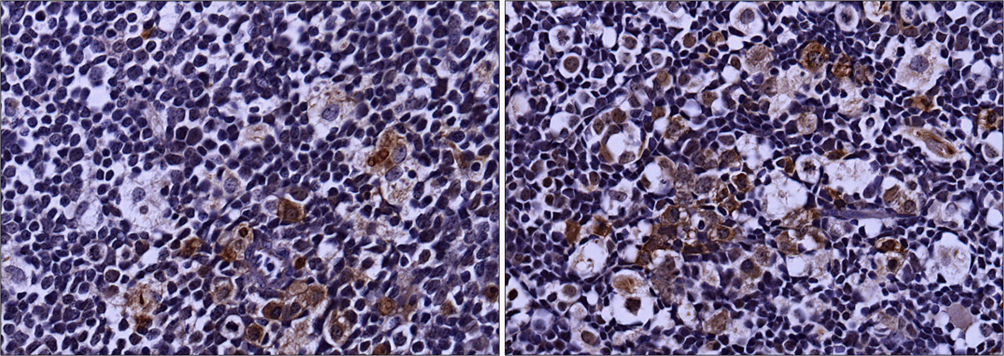
 | Fig. 2.Histiocytes showing positive staining with anti-CD68 monoclonal antibody (Immunohistochemical stain, ×400). |
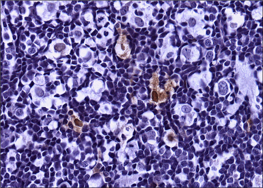
 | Fig. 3.Histiocytes showing positive staining with S-100 monoclonal antibody (Immunohistochemical stain, ×400). |
Table 1.
Antibodies used in the immunohistochemical staining
Table 2.
Clinical characteristics of ALL patients with histiocytosis
Table 3.
Clinical characteristics of AML patients with histiocytosis


 PDF
PDF ePub
ePub Citation
 Print
Print


 XML Download