Abstract
Acquired von Willebrand syndrome (AvWS) is a relatively rare acquired bleeding disorder similar to inherited von Willebrand disease in terms of laboratory findings, and occurs without a personal or family history of bleeding. A 23-year-old man with no previous disease history and no family history of hemorrhagic diathesis was referred to our hospital because of recurrent epistaxis and intramuscular hematoma. He was diagnosed as having AvWS because of an almost complete absence of ristocetin cofactor activity (vWF:RCo) despite normal vWF antigen level. Furthermore, anti-vWF antibody was detected in his serum using home-brewed ELISA. Finally, the amyloid deposit was found in muscle biopsy. He was diagnosed with AvWS which is associated with amyloidosis. AvWS should be considered in patients with current bleeding diatheses and no past history of bleeding.
References
1. Franchini M, Lippi G. Acquired von Willebrand syndrome: an update. Am J Hematol. 2007; 82:368–75.

2. Van Genderen PJ, Michiels JJ. Acquired von Willebrand Disease. Baillieres Clin Haematol. 1998; 11:319–30.
3. Krebs M, Meyer M, Quehenberger P, et al. Massive postoperative intramuscular bleeding in acquired von Willebrand's disease. Ann Hematol. 2002; 81:394–6.
4. Siaka C, Rugeri L, Caron C, Goudemand J. A new ELISA assay for diagnosis of acquired von Willebrand Syndrome. Haemophilia. 2003; 9:303–8.

5. Niiya M, Niiya K, Takazawa Y, et al. Acquired type 3-like von Willebrand syndrome preceded full-blown systemic lupus erythematosus. Blood Coagul Fibrinolysis. 2002; 13:361–5.

6. Federici AB, Rand JH, Bucciarelli P, et al. Acquired von Willebrand Syndrome: data from an international registry. Thromb Haemost. 2000; 84:345–9.

7. Collins P, Budde U, Rand JH, Federici AB, Kessler CM. Epidemiology and general guidelines of the management of acquired haemophilia and von Willebrand syndrome. Haemophilia. 2008; 14:49–55.

8. Hoshino Y, Hatake K, Muroi K, et al. Bleeding Tendency Caused by the Deposit of Amyloid Substance in the Perivascular Region. Internal Med. 1993; 32:879–81.

9. Fricke WA, Brinkhous KM, Garris JB, Roberts HR. Comparison of inhibitory and binding characteristics of an antibody causing acquired von Willebrand syndrome: an assay for von Willebrand factor binding by antibody. Blood. 1985; 66:562–9.

10. Van Genderen PJ, Vink T, Michiels JJ, van't Veer MB, Sixma JJ, van Vliet HH. Acquired von Willebrand disease caused by an autoantibody selectively inhibiting the binding of von Willebrand factor to collagen. Blood. 1994; 84:3378–84.

11. Tefferi A, Hanson CA, Kurtin PJ, Katzmann JA, Dalton RJ, Nichols WL. Acquired von Willebrand's disease due to aberrant expression of platelet glycoprotein Ib by marginal zone lymphoma cells. Br J Haematol. 1997; 96:850–3.

12. Scrobohaci ML, Daniel MT, Levy Y, Marolleau JP, Brouet JC. Expression of GpIb on plasma cells in a patient with monoclonal IgG and acquired von Willebrand disease. Br J Haematol. 1993; 84:471–5.

13. Denis CV, Christophe OD, Oortwijn BD, Lenting PJ. Clearance of von Willebrand factor. Thromb Haemost. 2008; 99:271–8.

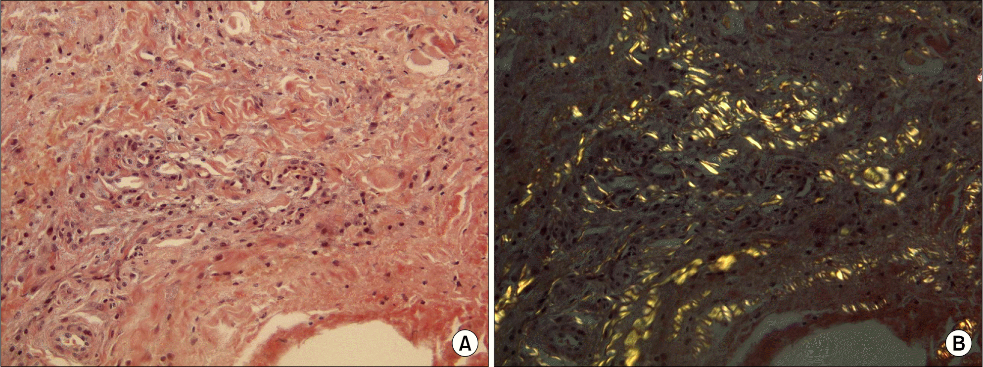
Fig. 1.
Microscopic findings of Extensor muscle. (A) Amyloid deposition showing Congo red staining (Congo red stain, ×200). (B) Apple-green birefringence in polarized light (Congo red stain, ×200).

Table 1.
Laboratory data of patient


 PDF
PDF ePub
ePub Citation
 Print
Print


 XML Download