

 PDF
PDF ePub
ePub Citation
 Print
Print


Abstract
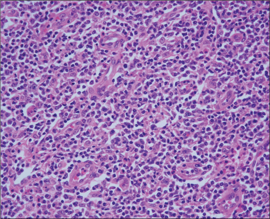
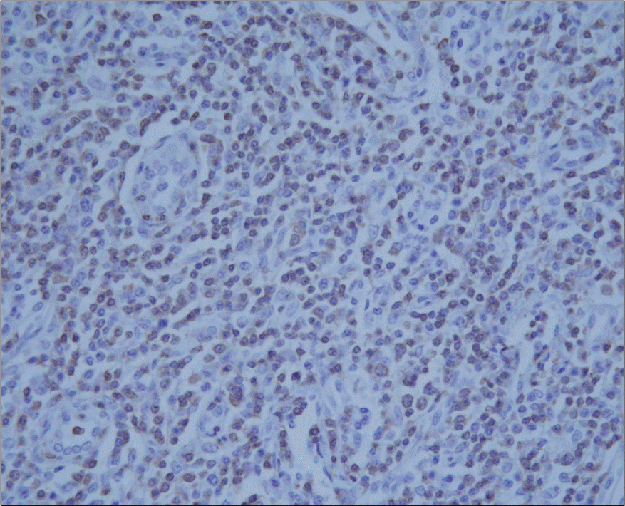
Systemic lupus erythematosus (SLE) patients tends to have a higher risk of developing lymphoid malignancies. The majority of such tumors are of a B cell origin. However, it is known that the T cell lymphoma subtypes in SLE patients are quite rare. Here, we describe a case of peripheral T cell lymphoma, unspecified (PTCL-U) that occurred in a 50-year-old female SLE patient. The lymphoma was located at the bilateral cervical and mediastinal lymph nodes. The staging workup revealed no evidence of any other organ involvement. Epstein-Barr virus messenger RNA was detected in the serum, but not in the lymph nodes. She received front-line chemotherapy with the CHOP regimen and she achieved complete remission. She then subsequently received high-dose chemotherapy with autologous peripheral stem cell transplantation. The patient currently remains in a clinical and serological state of remission for the SLE and PTCL until the time of this report 18 months after chemotherapy, and this was followed by autologous peripheral blood stem cell transplantation.
References
1. A clinical evaluation of the International Lymphoma Study Group classification of non-Hodgkin's lymphoma. The Non-Hodgkin's Lymphoma Classification Project. Blood. 1997; 89:3909–18.
2. Anderson JR, Armitage JO, Weisenburger DD. Epidemiology of the non-Hodgkin's lymphomas: distributions of the major subtypes differ by geographic locations. Non-Hodgkin's Lymphoma Classification Project. Ann Oncol. 1998; 97:717–20.
3. Jones JF, Shurin S, Abramowsky C, et al. T-cell lymphomas containing Epstein-Barr viral DNA in patients with chronic Epstein-Barr virus infections. N Engl J Med. 1988; 318:733–41.

4. Chang KL, Weiss LM. The association of the Epstein-Barr virus with malignant lymphoma. Biomed Pharmacother. 1996; 50:459–67.
5. King JK, Costenbader KH. Characteristics of patients with systemic lupus erythematosus (SLE) and non-Hodgkin's lymphoma (NHL). Clin Rheumatol. 2007; 26:1491–4.
6. Grulich AE, Vajdic CM. The epidemiology of non-Hodgkin lymphoma. Pathology. 2005; 37:409–19.
7. Ekström Smedby K, Vajdic CM, Faslster M, et al. Autoimmune disorders and risk of non-Hodgkin lymphoma subtypes: a pooled analysis within the InterLymph Consortium. Blood. 2008; 111:4029–38.
8. Suvajdzic N, Stojanovic-Milenkovic R, Tomasevic Z, Cemerikic-Martinovic V, Mihaljevic B, Atkinson HD. ALK-negative T-cell anaplastic large cell lymphoma associated with systemic lupus erythematosus. Med Oncol. 2003; 20:409–12.
9. Graninger WB, Smolen JS. Should the clinician have interest in the deregulation of apoptosis in autoimmunity? Br J Rheumatol. 1997; 36:1244–6.
10. Xu Y, Wiernik PH. Systemic lupus erythematosus and B-cell hematologic neoplasm. Lupus. 2001; 10:841–50.
11. Huggins ML, Todd I, Powell RJ. Reactivation of Epstein-Barr virus in patients with systemic lupus erythematosus. Rheumatol Int. 2005; 25:183–7.
12. Huh J, Cho K, Heo DS, Kim JE, Kim CW. Detection of Epstein-Barr virus in Korean peripheral T-cell lymphoma. Am J Hematol. 1999; 60:205–14.
13. Dupuis J, Emile JF, Mounier N, et al. Prognostic significance of Epstein-Barr virus in nodal peripheral T-cell lymphoma, unspecified: a Groupe d'Etude des Lymphomes de l'Adulte (GELA) study. Blood. 2006; 108:4163–9.
14. Ansell SM, Kurtin PJ, Stenson M, et al. Evaluation of the proliferative index as a prognostic factor in diffuse large cell lymphoma: correlation with the International Index. Leuk Lymphoma. 1999; 34:529–37.
15. Snowden JA, Patton WN, O'Donnell JL, Hannah EE, Hart DN. Prolonged remission of longstanding systemic lupus erythematosus after autologous bone marrow transplant for non-Hodgkin's lymphoma. Bone Marrow Transplant. 1997; 19:1247–50.
 XML Download