Abstract
Lymphomas of mucosa-associated lymphoid tissue (MALT) comprise 7% of all newly diagnosed non-Hodgkin's lymphomas. Helicobacter pylori (H. pylori) negative gastric MALT lymphomas account for 28 to 45% of gastric MALT lymphomas. H. pylori infection has a close relationship with most gastric low-grade B cell lymphomas of the MALT type. Monoclonal gammopathy can be seen in 36% of the patients and negatively associated with responses to eradication of H. pylori in gastric MALT lymphoma. Here, we describe a case of H. pylori negative MALT lymphoma that arose from the stomach with massive plasmacytic differentiation mimicking an extramedullary plasmacytoma with monoclonal gammopathy, and that was cured by total gastrectomy, chemotherapy and radiotherapy.
References
1. Nakamura S, Yao T, Aoyagi K, Iida M, Fujishima M, Tsuneyoshi M. Helicobacter pylori and primary gastric lymphoma. A histopathologic and immunohistochemical analysis of 237 patients. Cancer. 1997; 79:3–11.
2. Wöhrer S, Streubel B, Bartsch R, Chott A, Raderer M. Monoclonal immunoglobulin production is a frequent event in patients with mucosa-associated lymphoid tissue lymphoma. Clin Cancer Res. 2004; 10:7179–81.

3. Isaacson P, Wright DH. Malignant lymphoma of mucosa-associated lymphoid tissue. A distinctive type of B-cell lymphoma. Cancer. 1983; 52:1410–6.

4. The Non-Hodgkin's Lymphoma Classification Project. A clinical evaluation of the international lymphoma study group classification of non-Hodgkin's lymphoma. Blood. 1997; 89:3909–18.
5. Oh SY, Kim WS, Kim JH, et al. Extra-gastric MALT lymphoma: analysis of 50 cases. Korean J Med. 2000; 59:261–7.
7. Zucca E, Roggero E. Biology and treatment of lymphoma: the state of the art in 1996. A workshop at the 6th international conference on malignanant lymphoma mucosa-associated lymphoid tissue. Ann Oncol. 1996; 7:782–92.
8. Tieblemount C, Bastion Y, Berger F, et al. Mucosa-associated lymphoid tissue gastrointestinal and non gastrointestinal lymphoma behavior: analysis of 108 patients. J Clin Oncol. 1997; 15:1624–30.
9. Allez M, Mariette X, Linares G, Bertheau P, Jian R, Brouet JC. Low-grade MALT lymphoma mimicking Waldenström's macroglobulinemia. Leukemia. 1999; 13:484–5.

10. Kim GL, Son SH, Song HS, Kwon KY, Jeon DS, Kim JR. Case of malignant lymphoma with monoclonal gammopathy of IgM, lambda type. Korean J Hematol. 1992; 27:155–60.
11. Joo YD, Lee WS, Kim YJ, et al. A case of low grade MALT lymphoma in the mediastinum with clinical appearance of Wandenström's macroglobulinemia. Korean J Hematol. 2004; 39:172–6.
12. Wöhrer S, Troch M, Streubel B, et al. Pathology and clinical course of MALT lymphoma with plasmacytic differentiation. Ann Oncol. 2007; 18:2020–4.

Fig. 1.
Results of serum protein electrophoresis showed an abnormal band of protein between β and γ fractions (arrows, panel A & B). The band was identified by immunoelectrophoresis as an IgM lambda light chain component (panel C).
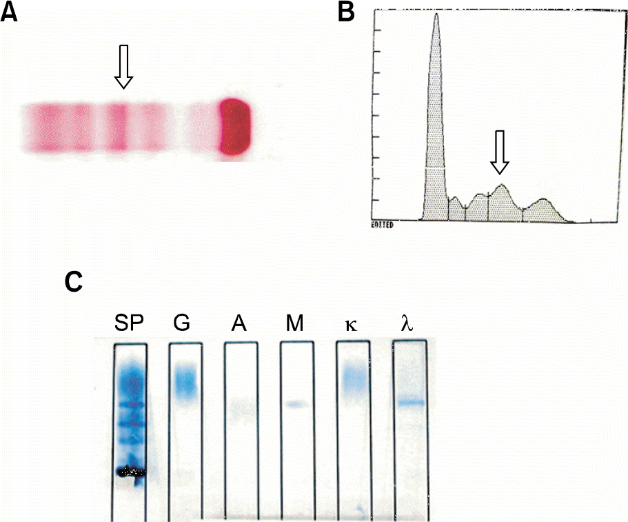
Fig. 2.
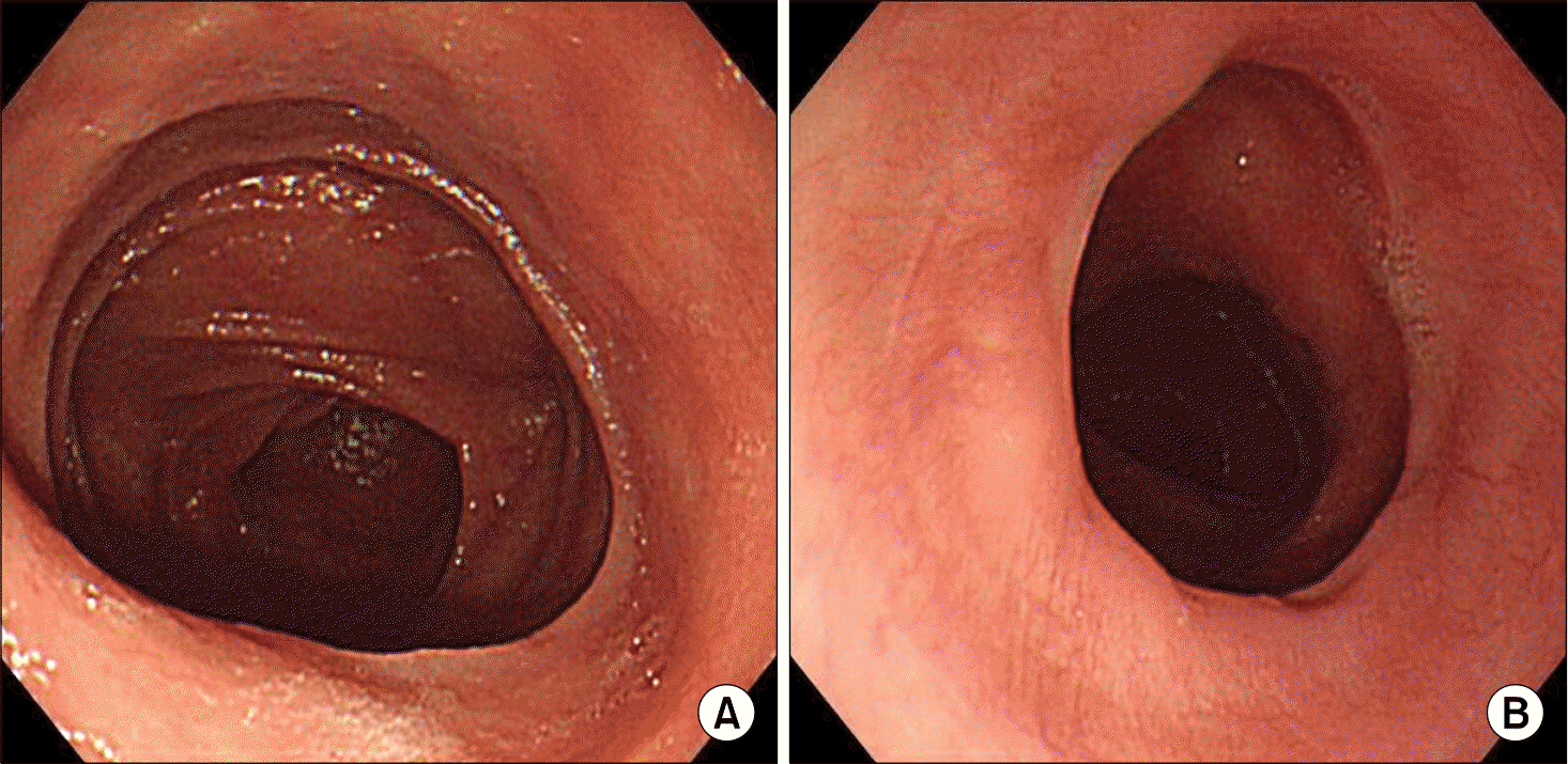
Esophago-Gastro-Duode-noscopic findings revealed multiple shallow ulcerations spread on entire stomach. (A, gastric fundus; B, gastric mid-body).
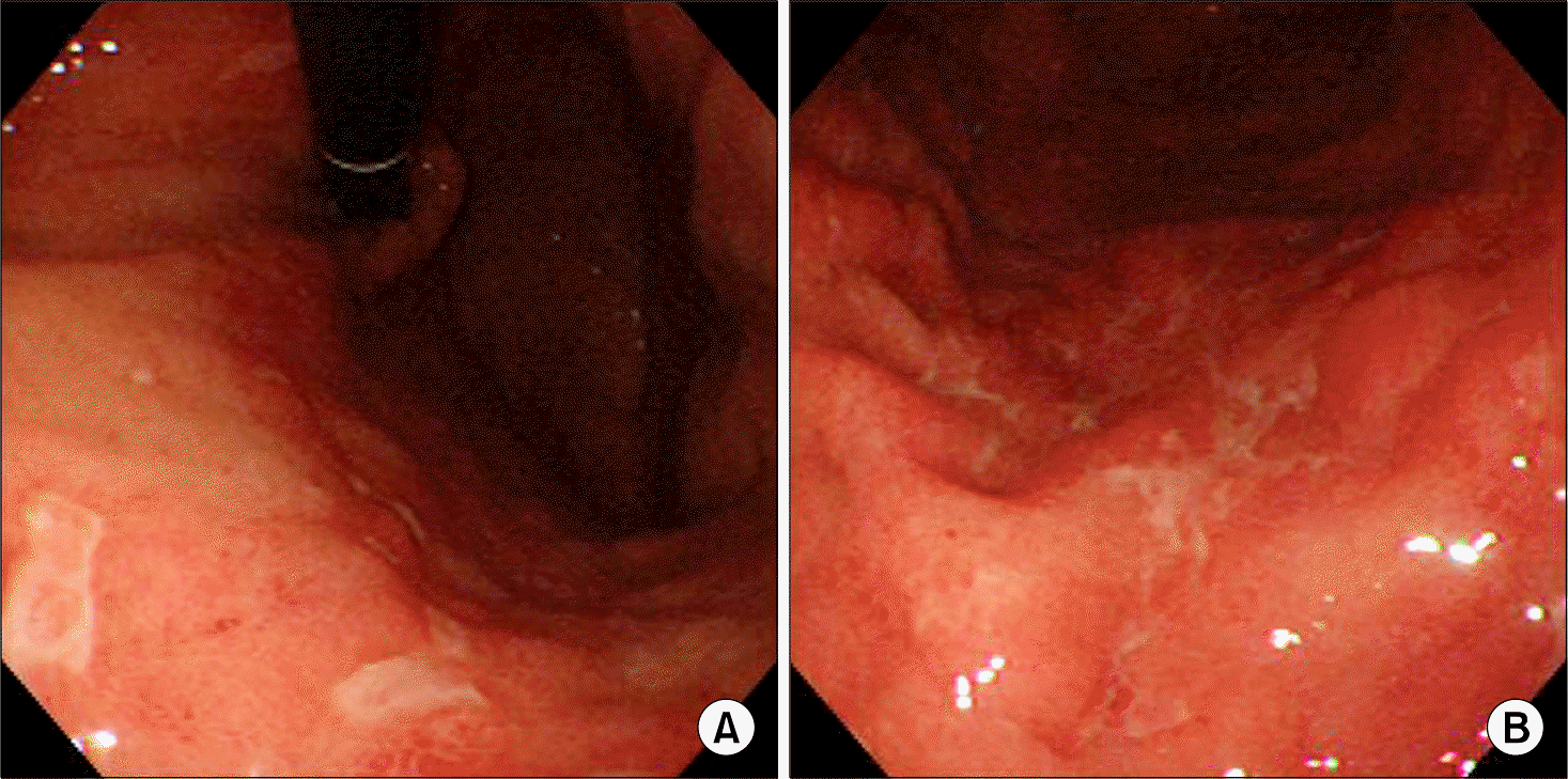
Fig. 3.
Endoscopic Ultrasonographic finding showed that the gastric lesions were usually limited only to mucosa and submucosa. However, some focal area (arrows) showed infiltrations reached up to lamina propria.
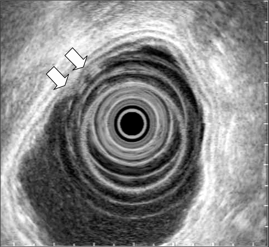
Fig. 4.
The pathology of total gastrectomy specimen. (A) Pathologic gross findings of the stomach showed multiple ulcerations (arrows) similar to endoscopic findings. (B) H&E stain (×200) of the stomach shows differentiated or undifferentiated plasma cells packed in mucosa and submucosa layer just like plasmacytoma.
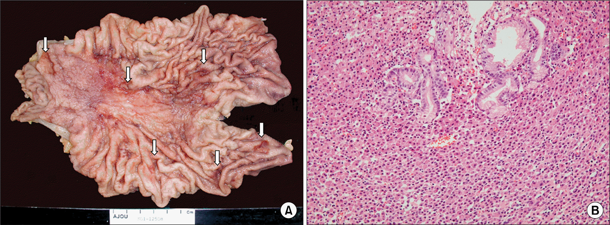
Fig. 5.
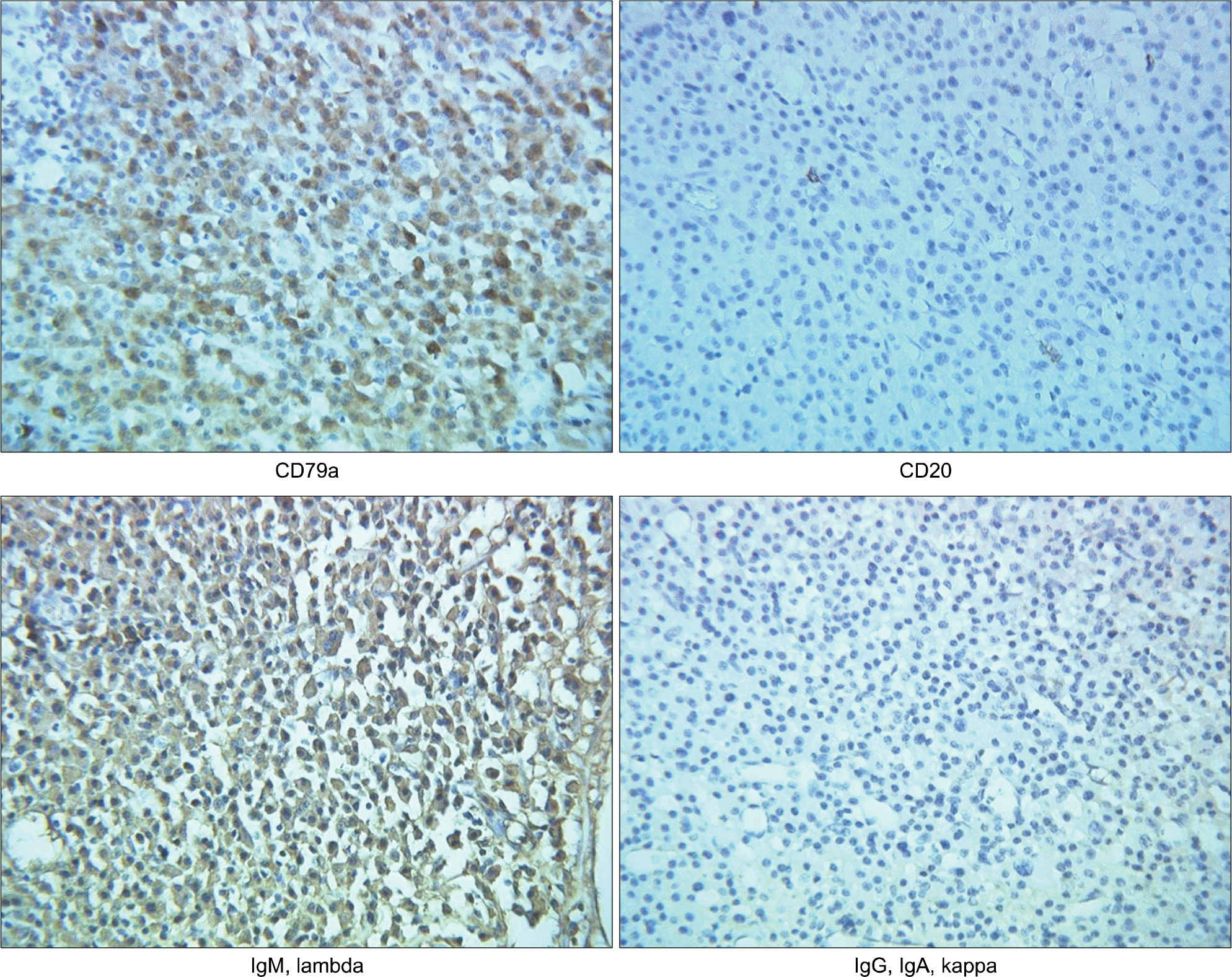
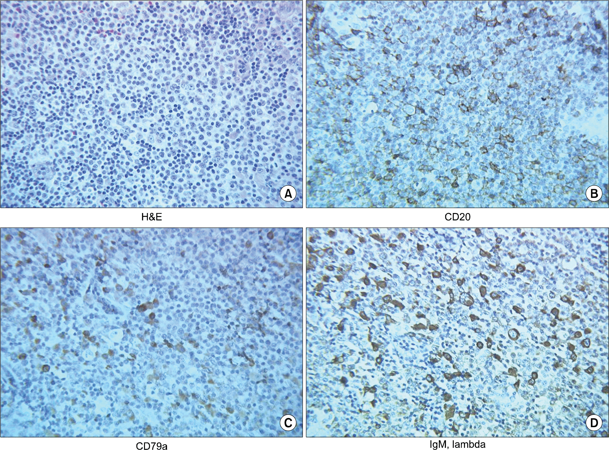
Immunohistochemical stains (×200) in stomach showed that the putative cells expressed CD79a and Ig M, lambda, but did not express CD20. According to strong expression of CD79a, common marker of plasma cells, these findings suggest rather extramedullary plasmacytoma of the stomach than gastric MALT lymphoma.


 PDF
PDF ePub
ePub Citation
 Print
Print


 XML Download