Abstract
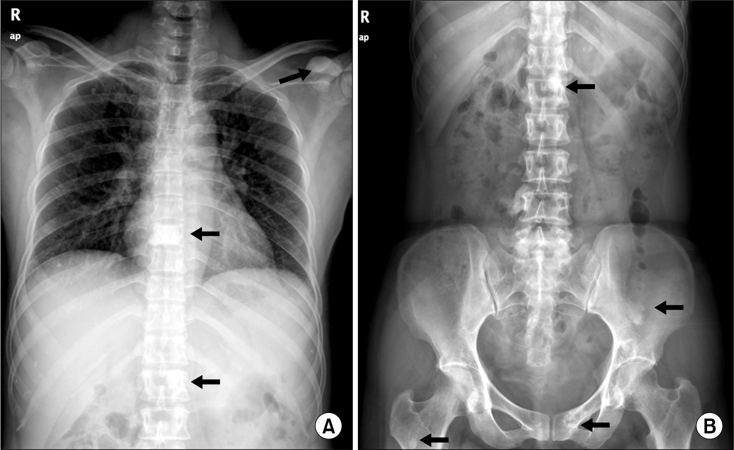
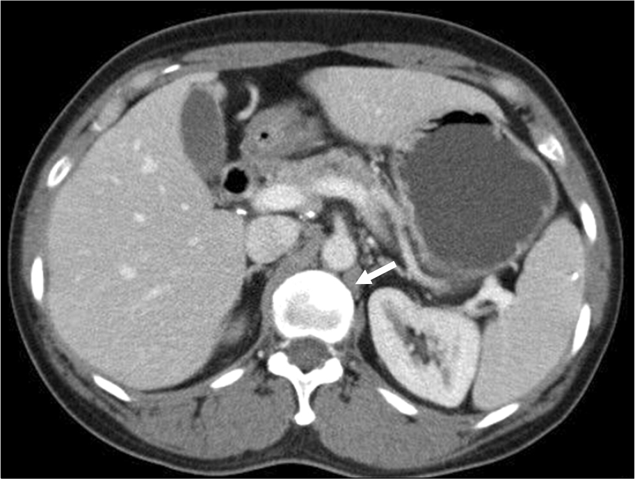
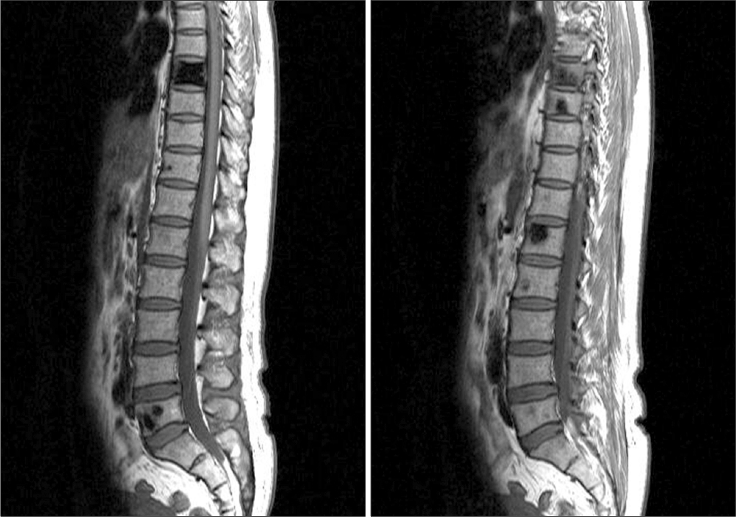
Osteosclerotic myeloma is a rare entity, characterized by single or multiple osteosclerotic bone lesions and usually accompanied by a polyneuropathy syndrome (POEMS). Multiple myeloma with osteosclerotic lesions without polyneuropathy is exceedingly rare. We report a case of multiple myeloma associated with multifocal osteosclerotic lesions without any evidence of POEMS. A 48-year-old woman presented with incidentally found osteosclerosis of 8th thoracic vertebra on a plain chest film. Bone survey, CT scan, MR scan, and radioisotope scintigraphy revealed multiple localized osteoclerosis; serum protein immunofixation showed IgG, lambda monoclonal gammopathy. A biopsy of T8 vertebral body disclosed plasma cell myeloma. Given that there was no organ or tissue damage other than multifocal osteosclerosis, the patient was placed on close observation with regular examination. This case indicates that although rare, multiple myeloma should be included in the differential diagnosis of sclerotic bone lesions.
Go to : 
References
1. Barlogie B, Shaughnessy J, Espstein J, et al. Plasma cell myeloma. Lichtman MA, Beutler E, Kipps TJ, editors. Williams Hematology. 7th ed.New York: USA, McGraw-Hill Medical;2006. p. 1501–33.
2. Schey S. Osteosclerotic myeloma and ‘POEMS'syndrome. Blood Rev. 1996; 10:75–80.
3. Dispenzieri A. POEMS syndrome. Hematology Am Soc Hematol Educ Program. 2006. 360–7.
4. Sharnoff JG, Belsky H, Melton J. Plasma cell leukemia or multiple myeloma with osteosclerosis. Am J Med. 1954; 17:582–4.

5. Hall FM, Gore SM. Osteoslcerotic myeloma variants. Skeletal Radiol. 1998; 17:101–5.
6. Lacy MQ, Gertz MA, Hanson CA, Inwards DJ, Kyle RA. Multiple myeloma associated with diffuse osteosclerotic bone lesions: a clinical entity distinct from osteosclerotic myeloma (POEMS syndrome). Am J Hematol. 1997; 56:288–93.

7. Mulleman D, Gaxatte C, Guillerm G, et al. Multiple myeloma presenting with widespread osteosclerotic lesions. Joint Bone Spine. 2004; 71:79–83.

8. The International Myeloma Working Group. Criteria for the classification of monoclonal gammopathies, multiple myeloma and related disorders: a report of the International Myeloma Working Group. Br J Haematol. 2003; 121:749–57.
9. Park SY, Lee JS, Lee KS, et al. A case of multiple myeloma with sclerosis and cryoglobulinemia. Korean J Hematol. 1985; 20:103–8.
10. Lee CK, Kim HK, Lee DN. A case of multiple myeloma sssociated with osteosclerosis. Korean J Clin Pathol. 1991; 11:103–8.
Go to : 
 | Fig. 1.Plain images show multifocal bony sclerosis. (A) A thoracic spine image shows sclerotic changes in vertebral bodies (T8, L1) and coracoid process of left scapula (arrows). (B) A lumbar spine image shows osteosclerotic lesions in pelvic bone and femur (arrows). |


 PDF
PDF ePub
ePub Citation
 Print
Print


 XML Download