Abstract
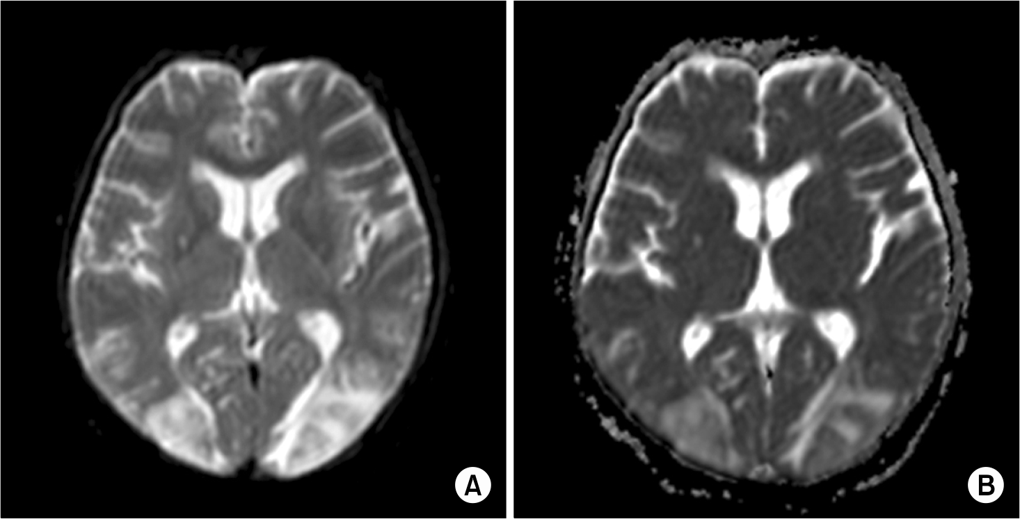
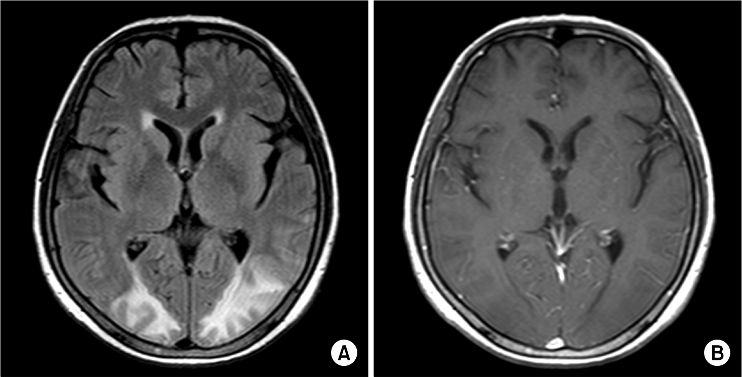
Reversible posterior leukoencephalopathy syndrome (RPLS) is a distinctive clinicoradiological entity that's characterized by headache, confusion, seizure and frequent visual disturbances. It is associated with certain neuro-radiological findings, and predominantly white matter abnormalities of the parieto-occipital lobes. RPLS has been identified mostly in patients with malignant hypertension, pre-eclampsia and renal insufficiency and in those patients who are using immunosuppressive agents or cytotoxic drugs. We report here on a case of RPLS in a patient who was undergoing chemotherapy. A 49-year-old woman presented with abrupt mental changes and visual disturbances five days after the administration of a chemotherapeutic agent. MRI showed hyper-intense signals on the magnetic resonance (MR) diffusion images in the bilateral temporal, parietal and occipital lobes. The clinical manifestations completely resolved after one week of treatment that consisted of blood pressure control, a negative intake-output balance and the best supportive care. These radiological changes and the reversible clinical manifestations were consistent with RPLS.
References
1. Hinchey J, Chaves C, Appignani B, et al. A reversible posterior leukoencephalopathy syndrome. N Engl J Med. 1996; 334:494–500.

2. Greenwood MJ, Dodds AJ, Garricik R, Rodriguez M. Posterior leukoencephalopathy in association with the tumour lysis syndrome in acute lymphoblastic leukaemia–a case with clinicopathological correlation. Leuk Lymphoma. 2003; 44:719–21.

3. Bartynski WS, Boardman JF. Distinct imaging patterns and lesion distribution in posterior reversible encephalopathy syndrome. Am J Neuroradiol. 2007; 28:1320–7.

4. Tam CS, Galanos J, Seymour JF, Pitman AG, Stark RJ, Prince HM. Reversible posterior leukoencephalopathy syndrome complicating cytotoxic chemotherapy for hematologic malignancies. Am J of Hematol. 2004; 77:72–6.

5. Edvinsson L, Owman C, Sjöberg NO. Autonomic nerves, mast cells, and amine receptors in human brain vessels. A histochemical and pharmacological study. Brain Res. 1976; 115:377–93.

6. Faraci M, Lanino E, Dini G, et al. Severe neurologic complications after hematopoietic stem cell transplantation in children. Neurology. 2002; 59:1895–904.

7. Serkova NJ, Christians U, Benet LZ. Biochemical mechanisms of cyclosporine neurotoxicity. Mol Interv. 2004; 4:97–107.

8. Lai R, Abrey LE, Rosenblum MK, DeAngelis LM. Treatment-induced leukoencephalopathy in primary CNS lymphoma: a clinical and autopsy study. Neurology. 2004; 62:451–6.

9. Shah-Khan FM, Pinedo D, Shah P. Reversible posterior leukoencephalopathy syndrome and antineoplastic agents: a review. Oncol Rev. 2007; 1:152–61.

10. Mizutani T. Leukoencephalopathy caused by antineoplastic drugs. Brain Nerve. 2008; 60:137–41.


 PDF
PDF ePub
ePub Citation
 Print
Print


 XML Download