

 PDF
PDF ePub
ePub Citation
 Print
Print


Abstract
Myelofibrosis is usually observed in association with hematologic malignancies or metastatic solid tumors, but it has rarely been reported in patients who suffer with autoimmune disorders. Autoimmune myelofibrosis is a distinct clinicopathologic entity and it can occur alone or in association with autoimmune disorders, and the final result is chronic peripheral cytopenia. Primary autoimmune myelofibrosis, in which the autoimmune myelofibrosis is not preceded by a well-defined autoimmune disease, has recently been defined as a distinct clinicopathologic syndrome. We report here on a case of an 18-year-old woman who was diagnosed with primary autoimmune myelofibrosis, and she manifested peripheral pancytopenia, positivity for autoantibodies and Grade III myelofibrosis without having any preceding autoimmune or hematologic disorders.
Go to : 
References
1. Tefferi A. Pathogenesis of myelofibrosis with myeloid metaplasia. J Clin Oncol. 2005; 23:8520–30.

2. McCarthy DM. Fibrosis of the bone marrow: content and causes. Br J Haematol. 1985; 59:1–7.
3. Kiss E, Gál I, Simkovics E, et al. Myelofibrosis in systemic lupus erythematosus. Leuk Lymphoma. 2000; 39:661–5.
4. Ramakrishna R, Kyle PW, Day PJ, Manoharan A. Evans' syndrome, myelofibrosis and systemic lupus erythematosus: role of procollagens in myelofibrosis. Pathology. 1995; 27:255–9.
5. Marie I, Levesque H, Cailleux N, et al. An uncommon association: Sjögren's syndrome and autoimmune myelofibrosis. Rheumatology. 1999; 38:370–1.
6. Muslimani A, Ahluwalia MS, Palaparty P, Daw HA. Idiopathic myelofibrosis associated with dermatomyositis. Am J hematol. 2006; 81:559–60.
7. Bass RD, Pullarkat V, Feinstein DI, Kaul A, Winberg CD, Brynes RK. Pathology of autoimmune myelofibrosis. A report of three cases and a review of the literature. Am J Clin Pathol. 2001; 116:211–6.
8. Kuter DJ, Bain B, Mufti G, Bagg A, Hasserjian RP. Bone marrow fibrosis: pathophysiology and clinical significance of increased bone marrow stromal fibres. Br J Haematol. 2007; 139:351–62.
9. Pullarkat V, Bass RD, Gong JZ, Feinstein DI, Brynes RK. Primary autoimmune myelofibrosis: definition of a distinct clincopathologic syndrome. Am J Hematol. 2003; 72:8–12.
10. Kasahara S, Tsurumi H, Yoshikawa T, et al. Plasma cell leukemia with myelofibrosis. J Clin Exp Hemato-pathol. 2008; 48:71–3.
11. Arellano-Rodrigo E, Esteve J, GinéE , Panés J, Cervantes F. Idiopathic myelofibrosis associated with ulcerative colitis. Leuk Lymphoma. 2002; 43:1481–3.
12. Stéphan JL, Galambrun C, Dutour A, Freycon F. Myelofibrosis: an unusual presentation of vitamin D-deficient rickets. Eur J Pediatr. 1999; 158:828–9.
13. Mesa RA, Schwager S, Radia D, et al. The myelofibrosis symptom assessment form (MFSAF): an evidencebased brief inventory to measure quality of life and symptomatic response to treatment in myelofibrosis. Leuk Res. 2009; 26:1199–203.
14. Paquette RL, Meshkinpour A, Rosen PJ. Autoimmune myelofibrosis. A steroid responsive cause of bone marrow fibrosis associated with systemic lupus erythematosus. Medicine (Baltimore). 1994; 73:145–52.
15. Norton A, Roberts I. Management of Evans syndrome. Br J Haematol. 2006; 132:125–37.
Go to :
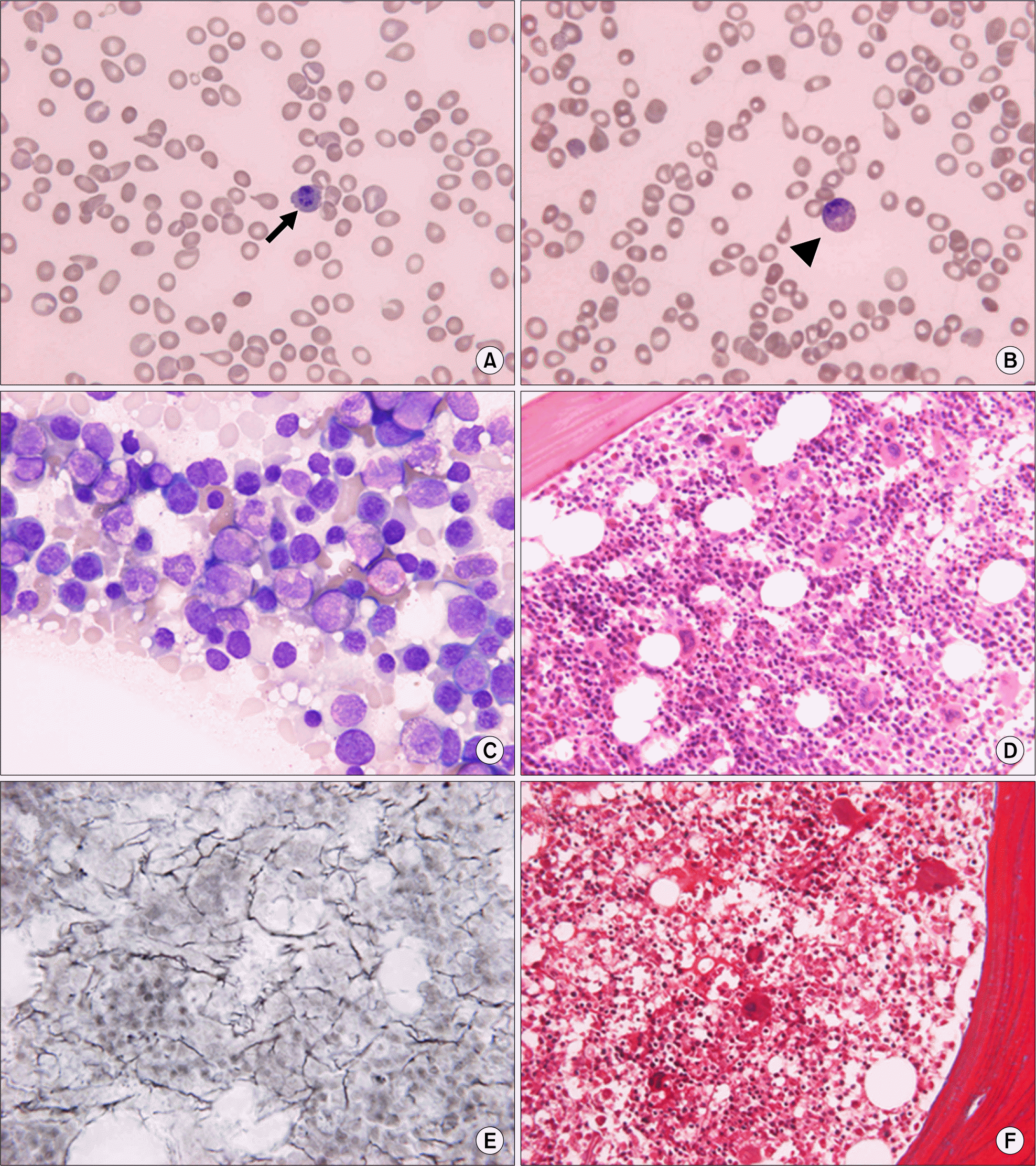 | Fig. 1.Blood and bone marrow findings of the patient at diagnosis. (A, B) Peripheral blood smears showed marked anisopoikilocytosis with tear drop cells and spherocytes, and leukoerythroblastosis with a nucleated RBC (arrow) and a myelocyte (arrowhead) (Wright-Giemsa stain, ×1,000). (C) The touch print smear showed erythroid hyperplasia without myelodysplastic features (Wright-Giemsa stain, ×1,000). (D) The bone marrow biopsy section showed normocellularity with erythroid and megakaryocytic hyperplasia (Hematoxylin & eosin stain, ×400). (E) The reticulin stain showed diffuse reticulin fiber network with scattered coarse fibers (Reticulin silver stain, ×400). (F) Negative trichrome stain (Masson's trichrome stain, ×400). |
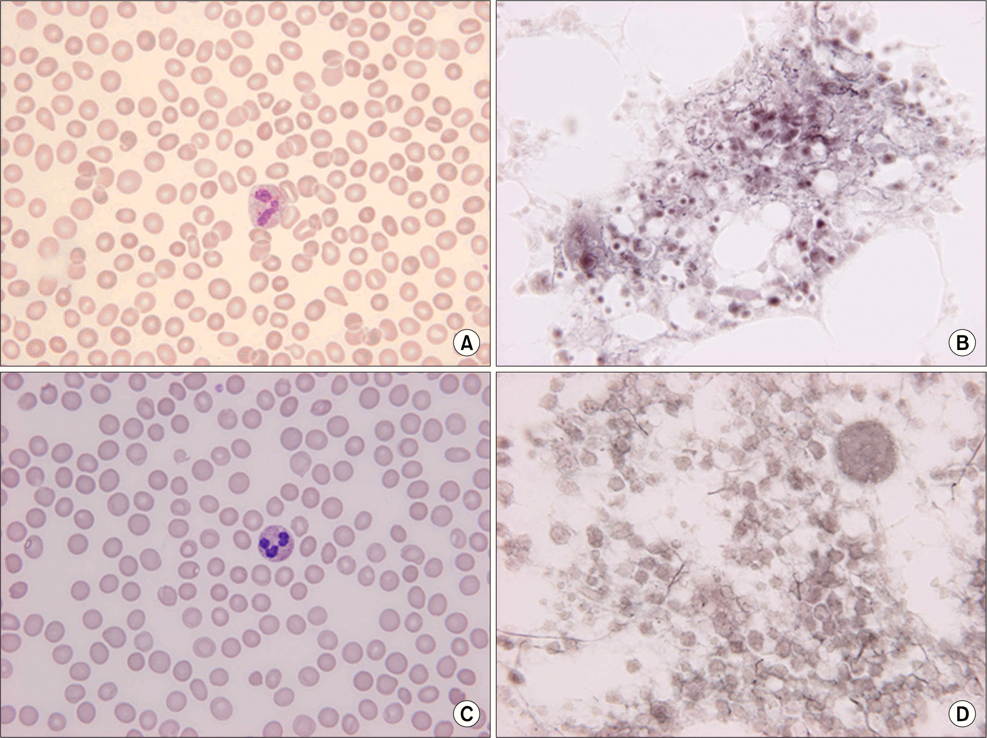 | Fig. 2.Findings of blood smears and the reticulin stain of bone marrow biopsy in 40 days (A, B) and in 4 months (C, D) after treatment. (A) Mild poikilocytosis with tear drop cells. (B) Fine fiber network throughout most of the section and no coarse fibers. (C) More decreased tear drop cells than those observed in Fig. 2A. (D) Occasional fine individual fibers and foci of a fine fiber network. (A and C, Wright-Giemsa stain, ×1,000; B and D, Reticulin silver stain, ×400). |
Table 1.
The changes of CBC, blood smear findings and bone marrow fibrosis after steroid treatment
| Pretreatment | After 40 days | After 4 M | After 13 M | |
|---|---|---|---|---|
| Hgb (g/dL) | 5.7 | 12.7 | 13.1 | 11.6 |
| Hct (%) | 18.2 | 39.1 | 40.0 | 38.8 |
| WBC (/μL) | 2,860 | 4,280 | 8,480 | 10,180 |
| Platelet (103/μL) | 4 | 81 | 113 | 172 |
| Tear drop cells/HPF∗ | 19 | 4 | 2 | 0 |
| Spherocytes/HPF | 2 | 0.8 | 0.2 | 0 |
| Leukoerythroblastosis | + | − | − | − |
| BM fibrosis† | Grade 3 | Grade 2 | Grade 1 | NA |
Table 2.
The changes of the results of autoantibodies after steroid treatment
 XML Download