Abstract
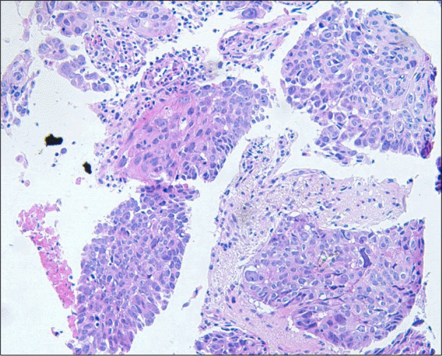
Chronic lymphocytic leukemia (CLL) can be characterized by the accumulation of small mature lymphocytes in the peripheral blood, bone marrow and other lymphoid tissues. It is well known that the risk of secondary malignancy is high in patients with CLL. A secondary malignancy in a patient with CLL may influence the prognosis as well as the treatment of CLL. As CLL is a rare disease in Korea, there have been only a few reported Korean cases of CLL with secondary malignancy. We experienced the case of a 73-year-old man who suffered from CLL with basal cell carcinoma of the skin and non-small cell lung cancer. At first, he presented with excessive lymphocytosis (>100,000/mm3), anemia, thrombocytopenia and splenomegaly, and he was diagnosed with CLL according to the bone marrow biopsy. Simultaneously he had basal cell carcinoma on his face. Seven months later, he began to feel chest discomfort and his chest X-ray showed a mass like lesion on the left upper lung. It was proven to be non-small cell lung cancer by bronchoscopic biopsy.
References
1. Wintrobe MM. Chronic lymphocytic leukemia. Lee GR, Bithell T C, Foster J, Athens J, Lukens J, editors. Wintrobe's clinical hematology. 9th ed.Philadelphia, United States of America: Lea and Febiger;2006. p. 2034–53.
2. Ahn YO, Koo HH, Park BJ, Yoo KY, Lee MS. Incidence estimation of leukemia among Koreans. J Korean Med Sci. 1991; 6:299–307.

3. Kipps TJ. Chronic Lymphocytic Leukemia and Related Diseases. Lichtman Marshall A., Ernest Beutler, Kipps Thomas J., Uri Seligsohn, Kenneth Kaushansky, editors. Williams Hematology. 7th ed.New York, United States of America: McGraw-Hill;2006. p. 1362.
4. Parekh K, Rusch V, Kris M. The clinical course of lung carcinoma in patients with chronic lymphocytic leukemia. Cancer. 1999; 86:1720–23.

5. Dasanu CA, Alexandrescu DT. Risk for second non-lymphoid neoplasms in chronic lymphocytic leukemia. MedGenMed. 2007; 9:35.
6. Nam SH, Kwon JM, Mun YC, et al. A case of coexistent chronic lymphocytic leukemia and multiple myeloma. Korean J Hematol. 2005; 40:41–4.

7. Lee JL, Kang HJ, Oh HA, et al. A case of pure red cell aplasia associated with b-cell chronic lymphocytic leukemia. Korean J Hematol. 2002; 37:60–4.
8. Lee SA, Kim YJ, Lim KC, et al. A case of adenocarcinoma of the lung in patient with chronic lymphocytic leukemia. J Korean Cancer Assoc. 1998; 30:613–9.
9. Schollkopf C, Rosendahl D, Rostgaard K, Pipper C, Hjalgrim H. Risk of second cancer in chronic lymphocytic leukemia. Int J Cancer. 2007; 121:151–6.
10. Potti A, Ganti AK, Koch M, Mehdi SA, Levitt R. Identification of HER-2/neu overexpression and the clinical course of lung carcinoma in non-smokers with chronic lymphocytic leukemia. Lung Cancer. 2001; 34:227–32.

11. Wiernik PH. Second neoplasms in patients with chronic lymphocytic leukemia. Curr Treatment Options in Oncology. 2004; 5:215–23.

12. Ravandi F, O'Brien S. Immune defects in patients with chronic lymphocytic leukemia. Cancer Immunol Immunother. 2006; 55:197–209.

13. Cheson BD, Vena DA, Barrett J, Freidlin B. Second malignancies as a consequence of nucleoside analog therapy for chronic lymphoid leukemias. J Clin Oncol. 1999; 17:2454–60.

14. Ahn YJ, Kim YA, Hwang KT, Heo SC, Jung IM, Chung JK. Synchronous triple primary cancer with concomitant IPMN. J Korean Surg Soc. 2008; 75:70–5.
Fig. 1.
(A) The patient has diffuse sclerotic brownish patch with a firm nodule on the left face. (B) The microscopic findings of a nodular lesion show that tumor cells with pleomorphic, hyperchromatic and oval nuclei are seen budding from the undersurface of the epidermis (H&E stain, ×200).
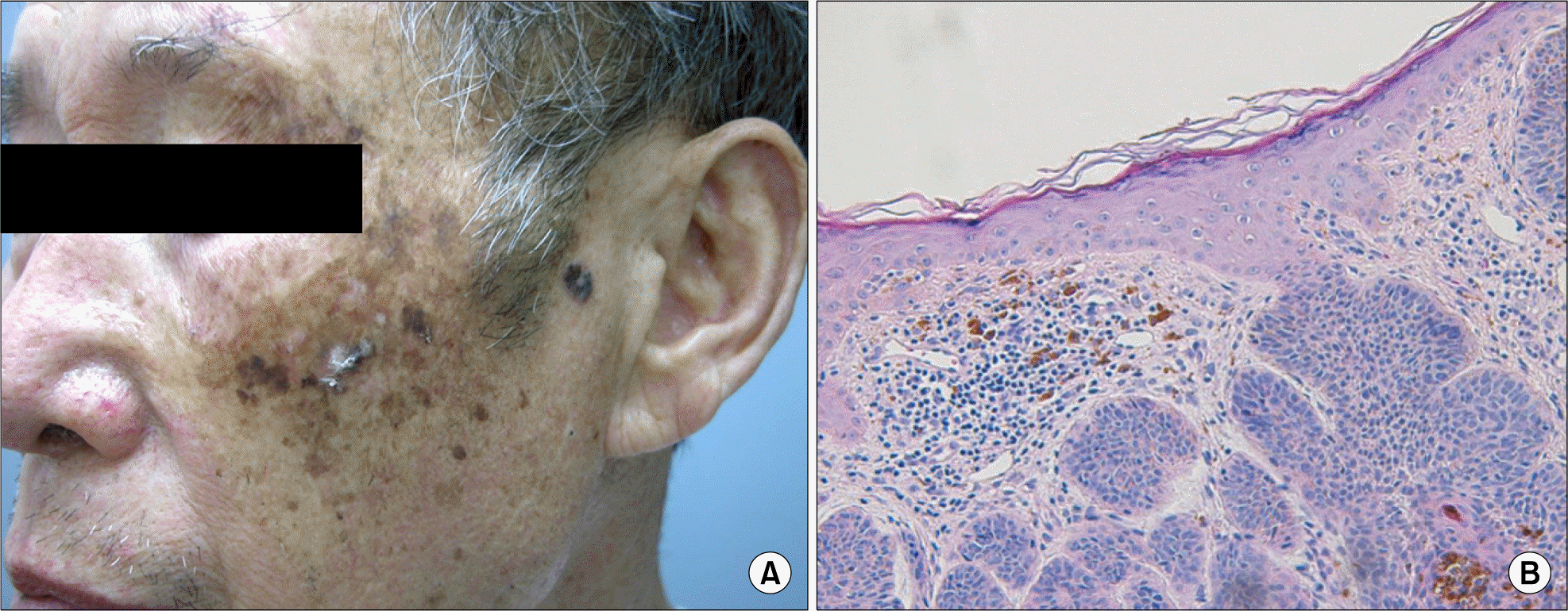
Fig. 2.
At the time of diagnosis of CLL, the chest x-ray shows subtle fibrotic scars in left upper lung field (A). And after 7 months of diagnosis of CLL, the chest x-ray shows a large mass in the left upper lung field with broad base in the mediastinum (B).
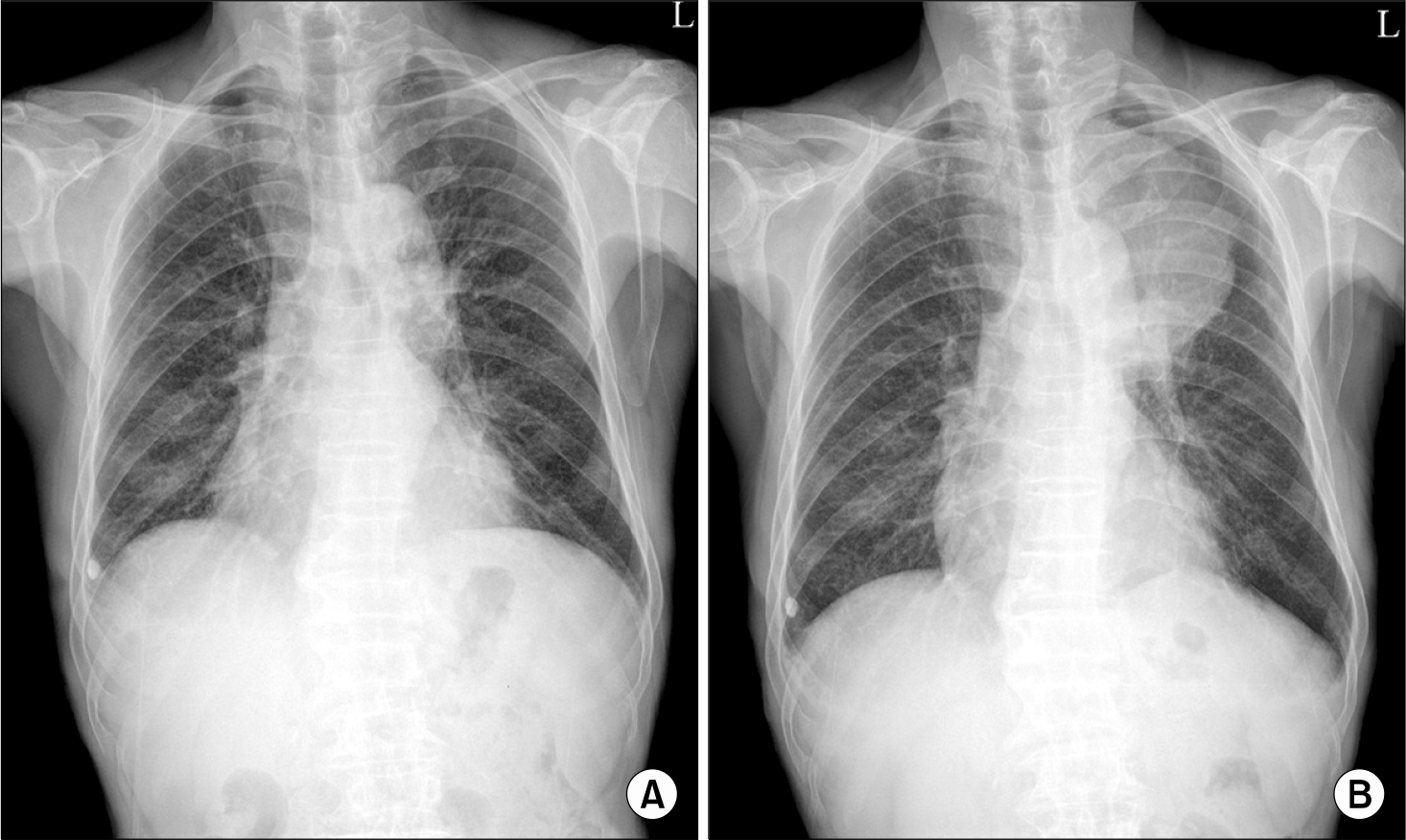
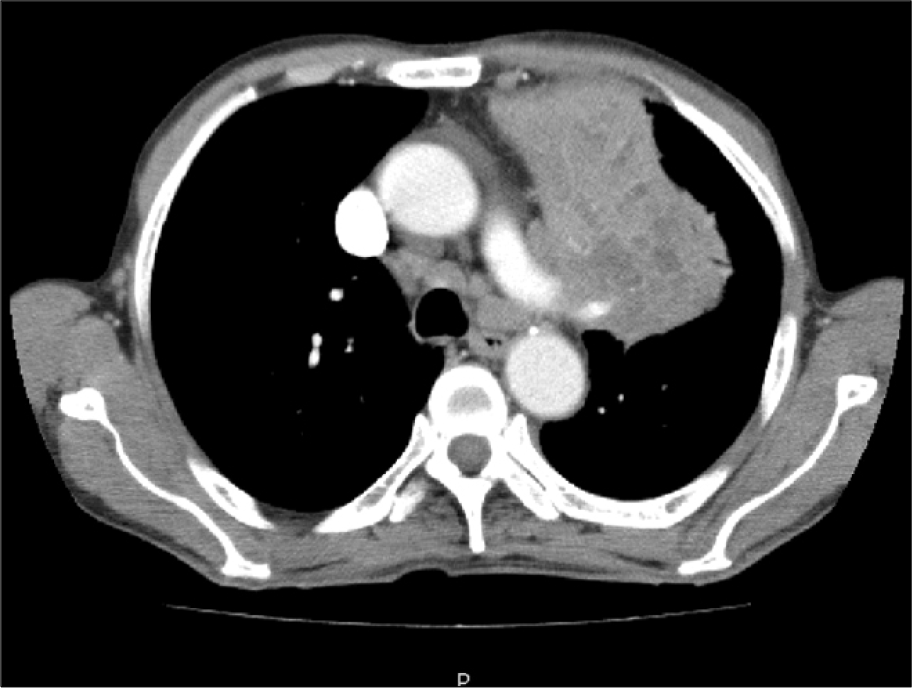
Fig. 3.
The chest CT shows that a large mass with multiple nonenhancing areas in the left upper lobe is in contact with left pulmonary artery.


 PDF
PDF ePub
ePub Citation
 Print
Print


 XML Download