Abstract
Fanconi anemia is an autosomal recessive disease that's characterized by congenital anomalies, defective hematopoiesis and a high risk of developing acute myeloid leukemia and certain solid tumors. The clinical phenotype is extremely variable; therefore, the diagnosis is frequently delayed until the pancytopenia appears. Chromosomal instability, especially on exposure to an alkylating agent, may be seen in affected patients and it is the basis for a diagnostic test. This cellular phenotype can be demonstrated in cultured T cells, B cells, fibroblasts and fetal cells cultured from both amniotic fluid and chorionic villi. But somatic mosaicism may make the diagnosis of Fanconi anemia difficult because of inconclusive chromosome breakage studies. If the test is negative in lymphocytes and yet the clinical setting is highly suspicious, then the skin fibroblasts must be assessed. Because skin fibroblasts are somatic cells, a definitive test can be performed on primary skin fibroblasts. In this report we describe a case of Fanconi anemia that was diagnosed with the use of cultured skin fibroblasts, and this was despite the normal breakage studies in the peripheral blood.
REFERENCES
1). Fanconi G. Familial constitutional panmyelocytop-athy, Fanconis anemia (F.A.).I. Clinical aspects. Semin Hematol. 1967. 4:233–40.
2). Auerbach AD., Allen RG. Leukemia and preleukemia in Fanconi anemia patients. A review of literature and report of International Fanconi Anemia Registry. Cancer Genet Cytogenet. 1991. 51:1–12.
3). Alter BP. Fanconi's anemia and it's variability. Br J Hematol. 1993. 85:9–14.
4). Auerbach AD., Alder B., Chaganti RS. Prenatal and postnatal diagnosis and carrier detection of Fanconi anemia by cytogenetic method. Pediatrics. 1981. 67:128–35.
5). Auerbach AD. Fanconi anemia diagnosis and the diepoxybutane (DEB) test. Exp Hematol. 1993. 21:731–3.
6). Auerbach AD., Rogatko A., Schroeder-Kurth TM. International Fanconi Anemia Registry: relation of clinical symptoms to diepoxybutane sensitivity. Blood. 1989. 73:391–6.

7). Mankad A., Taniguchi T., Cox B, et al. Natural gene therapy in monozygotic twins with Fanconi anemia. Blood. 2006. 107:3084–90.

8). Gregory JJ Jr., Wagner JE., Verlander PC, et al. Somatic mosaicism in Fanconi anemia: evidence of geneotypic reversion in lymphohematopoietic stem cells. Proc Natl Acad Sci USA. 2001. 98:2532–7.
10). Auerbach AD. Diagnosis of Fanconi anemia by Diepoxybutane analysis. Current Protocols in Human Genetics. 2003. 37:1–15.

11). Taniguchi T., D'Andrea AD. Molecular pathogenesis of Fanconi anemia: recent progress. Blood. 2006. 107:4223–33.

12). Joenje H., Patel KJ. The emerging genetic and molecular basis of Fanconi anemia. Nat Rev Genet. 2001. 2:446–57.
13). Tischkowitz MD., Hodgson SV. Fanconi anemia. J Med Genet. 2003. 40:1–10.
14). Cho B., Jeong DC., Kim HK., Kook H., Whang TJ. A case of allogeneic bone marrow transplantation for Fanconi anemia using low-dose cyclophosphamide/TN1 as conditioning regimen. Korean J Hematopoietic Stem Cell Transplant. 1997. 2:155–9.
15). D'Andrea AD., Grompe M. Molecular biology of Fanconi anemia: implications for diagnosis and therapy. Blood. 1997. 90:1725–36.
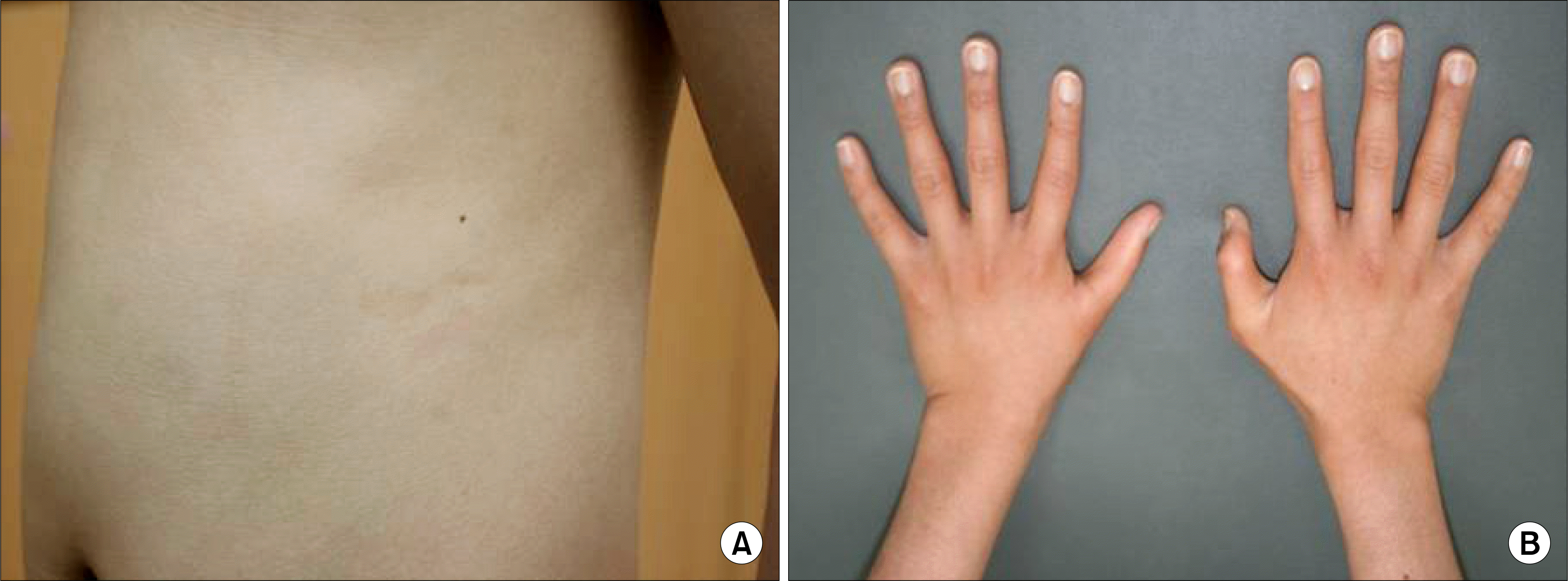
Fig. 1
(A) 4 Café au lait spots were shown in his back. (B) Thumb and ulnar anomalies are noted at both hands.

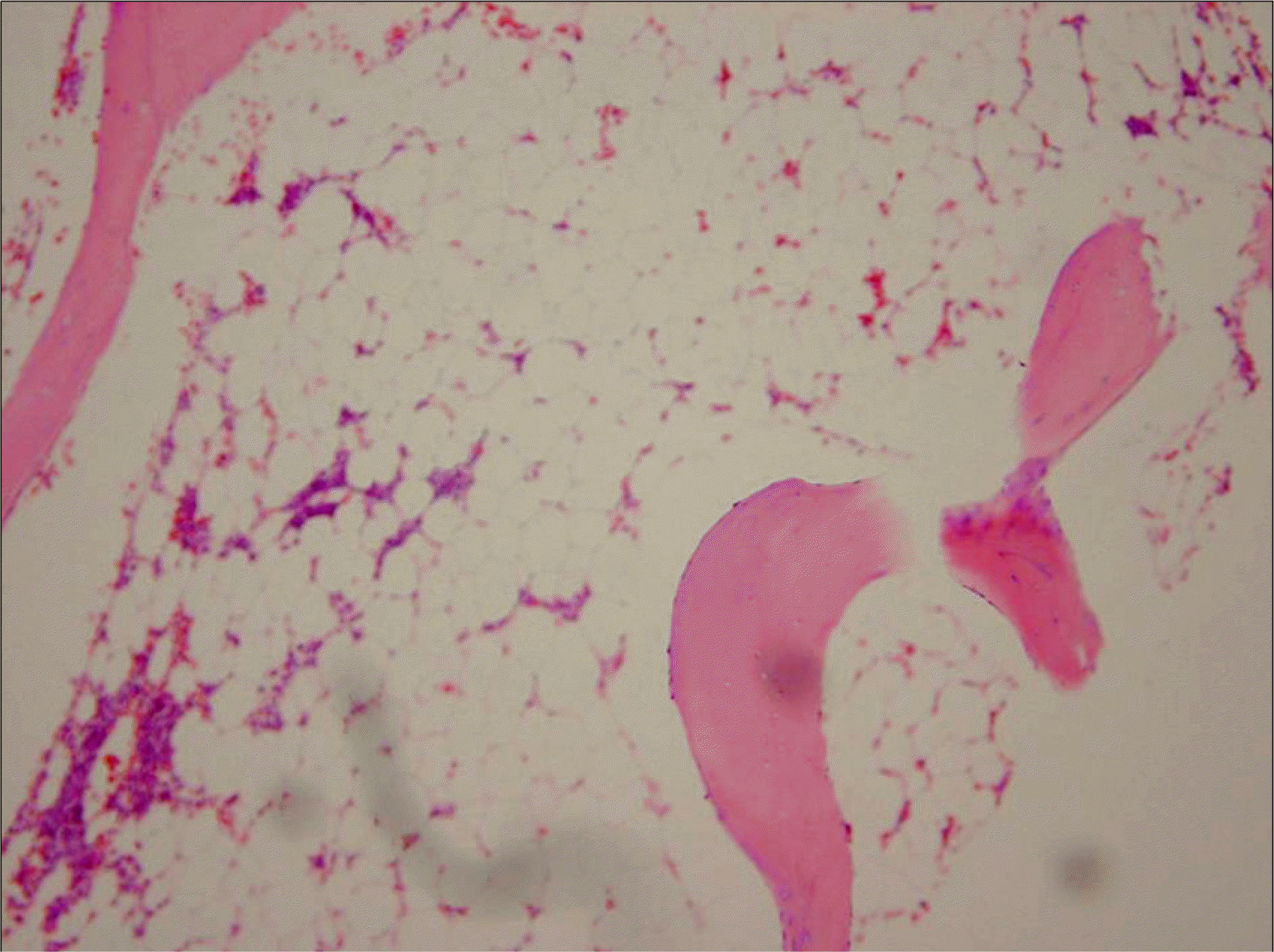
Fig. 2
Bone marrow biopsy shows markedly reduced hematopoiesis and increased fat. The cellularity was about 10%, which was acellular for his age (H&E stain, ×100).

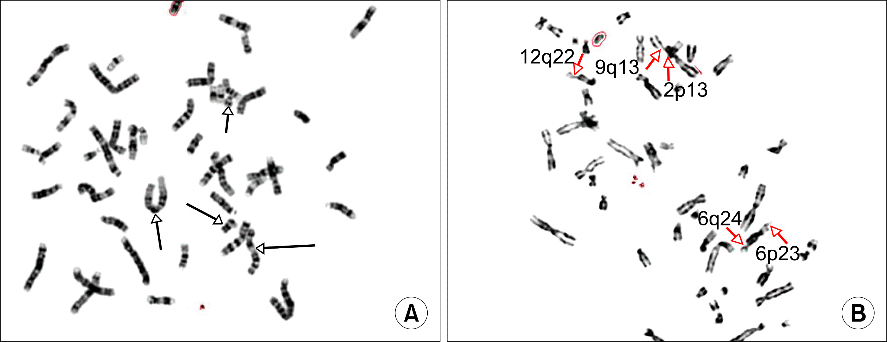
Fig. 3
Cytogenetic findings of induced chromosome breakage test with DEB (diepoxybutane). (A) No chromatid breakage is noted in peripheral blood lymphocyte culture. (B) Open arrowheads indicate chromatid breakages in skin fibroblast culture. We studied 50 cells from MMC and DEB stress test in this patient. Major and minor breaks (total breaks/cell: 0.58) were observed, especially in DEB stress test. These results were significantly different compared with a parallel control and exceed the normal limits.


 PDF
PDF ePub
ePub Citation
 Print
Print


 XML Download