Abstract
Granulocytic sarcoma is a localized tumor that's composed of immature granulocytic cells and this is more common in patient with 8;21 translocation. We present here a case in a 64-year-old man who was diagnosed with acute myelogenous leukemia (erythroleukemia) that had a complex hyperdiploid karyotype. While he underwent chemotherapy, he developed nausea, vomiting, headache and dysarthria. After several diagnostic work-ups, granulocytic sarcoma in the cerebellum and leptomeningeal metastasis of his leukemia were found on the magnetic resonance imaging and the cerebrospinal fluid cytology.
REFERENCES
1). Krishnamurthy M., Nusbacher N., Elguezabal A., Seligman BR. Granulocytic sarcoma of the brain. Cancer. 1977. 39:1542–6.

2). Domingo-Claros A., Larriba I., Rozman M, et al. Acute erythroid neoplastic proliferations. A biological study based on 62 patients. Haematologica. 2002. 87:148–53.
3). Rappaport H. Tumors of the hematopoietic system, in Atlas of Tumor Pathology, Section III, Fascicle 8. Armed Forces Institute of Pathology, Washington DC. 1967. 241–7.
5). Menasce LP., Banerjee SS., Beckett E., Harris M. Extra-medullary myeloid tumour (granulocytic sarcoma) is often misdiagnosed: a study of 26 cases. Histopathology. 1999. 34:391–8.

6). Neiman RS., Barcos M., Berard C, et al. Granulocytic sarcoma: a clinicopathologic study of 61 biopsied cases. Cancer. 1981. 48:1426–37.

7). Nishimura S., Kyuma Y., Kamijo A., Maruta A. Isolated recurrence of granulocytic sarcoma manifesting as extra- and intracranial masses - case report. Neurol Med Chir (Tokyo). 2004. 44:311–6.
8). Meltzer JA., Jubinsky PT. Acute myeloid leukemia presenting as spinal cord compression. Pediatr Emerg Care. 2005. 21:670–2.

9). Tallman MS., Hakimian D., Shaw JM., Lissner GS., Russell EJ., Variakajis D. Granulocytic sarcoma is associated with the 8;21 translocation in acute myeloid leukemia. J Clin Oncol. 1993. 11:690–7.

10). Park KU., Lee DS., Lee HS., Kim CJ., Cho HI. Granulocytic sarcoma in MLL-positive infant acute myelogenous leukemia: fluorescence in situ hybridization study of childhood acute myelogenous leukemia for detecting MLL rearrangement. Am J Pathol. 2001. 159:2011–6.
11). Alvarez S., Cigudosa JC. Gains, losses and complex karyotypes in myeloid disorders: a light at the end of the tunnel. Hematol Oncol. 2005. 23:18–25.

12). Schoch C., Haferlach T., Bursch S, et al. Loss of genetic material is more common than gain in acute myeloid leukemia with complex aberrant karyotype: a detailed analysis of 125 cases using conventional chromosome analysis and fluorescence in situ hybridization including 24-color FISH. Genes Chromosomes Cancer. 2002. 35:20–9.

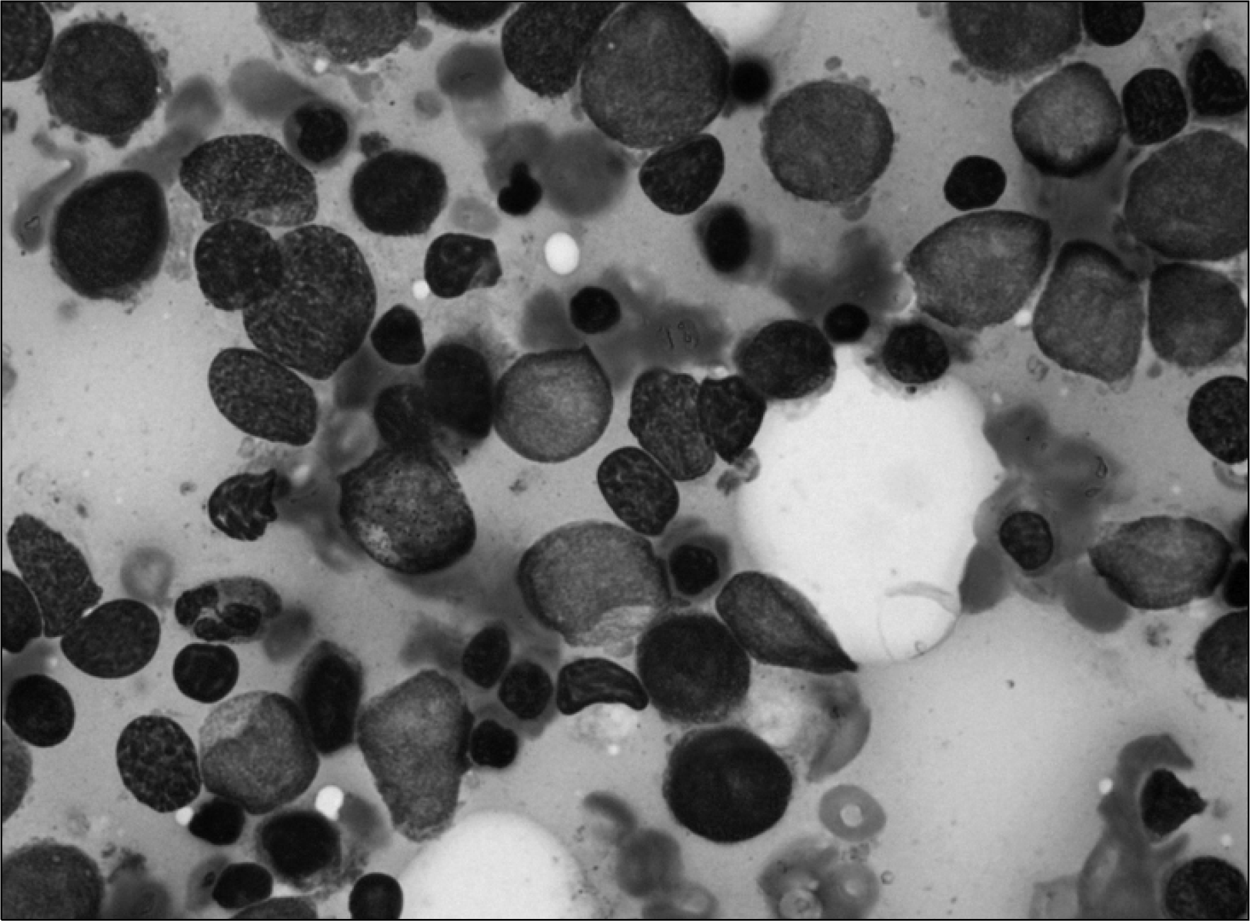
Fig. 1
The touch preparation of bone marrow biopsy demonstrating moderately hypercellular marrow with marked proliferation of severe dysplastic erythroid precursors (80% of all nucleated cells) and blasts. (Wright-Giemsa stain, 1,000×, adapted from Department of Laboratory Medicine, Pusan National University Hospital).


 PDF
PDF ePub
ePub Citation
 Print
Print


 XML Download