Abstract
Patients with acquired immunodeficiency virus (AIDS) are at increased risk for B-cell neoplasms and plasma cell dyscrasias. Plasmablastic lympomas (PBLs) were originally described exclusively in HIV-positive patients who presented with jaw or oral mucosa involvement. Recent studies report that this neoplasm also occurs in patients without HIV infection and may involve sites other than the head and neck. Until now, only sinus, oral mucosa, bone, testicles, gastrointestinal and pulmonary manifestations have been reported. In this report we describe a 41-year-old male with AIDS who mistook plasmablastic lymphoma of the pelvic cavity for perianal abscess. The patient presented with multiple involvement of bone and highly aggressive progression of pelvic mass without the involvement of bone marrow.
REFERENCES
1). Franceschi S., Dal Maso L., La Vecchia C. Advances in the epidemiology of HIV-associated non-Hodgkin's lymphoma and other lymphoid neoplasms. Int J Cancer. 1999. 83:481–5.

3). Mounier N., Spina M., Gisselbrecht C. Modern management of non-Hodgkin lymphoma in HIV-in-fected patients. Br J Hematol. 2007. 136:685–98.

4). Folk GS., Abbondanzo SL., Childers EL., Foss RD. Plasmablastic lymphoma: a clinicopathologic correlation. Ann Diagn Pathol. 2006. 10:8–12.

5). Mounier N., Spina M., Gabarre J, et al. AIDS-related non-Hodgkin lymphoma: final analysis of 485 patients treated with risk-adapted intensive chemotherapy. Blood. 2006. 107:3832–40.

6). Besson C., Goubar A., Gabarre J, et al. Changes in AIDS-related lymphoma since the era of highly active antiretroviral therapy. Blood. 2001. 98:2339–44.

7). Delecluse HJ., Anagnostopoulos I., Dallenbach F, et al. Plasmablastic lymphomas of the oral cavity: a new entity associated with the human immunodeficiency virus infection. Blood. 1997. 89:1413–20.

8). Lin O., Gerhard R., Zerbini MC., Teruya-Feldstein J. Cytologic features of plasmablastic lymphoma. Cancer. 2005. 105:139–44.

9). Schichman SA., McClure R., Schaefer RF., Mehta P. HIV and plasmablastic lymphoma manifestating in sinus, testicles, and bones: a further expansion of the disease spectrum. Am J Hematol. 2004. 77:291–5.
10). Pruneri G., Graziadei G., Ermellino L., Baldini L., Neri A., Buffa R. Plasmablastic lymphoma of the stomach. A case report. Haematologica. 1998. 83:87–9.
11). Lin Y., Rodrigues GD., Turner JF., Vasef MA. Plasmablastic lymphoma of the lung: report of a unique case and review of the literature. Arch Pathol Lab Med. 2001. 125:282–5.
12). Choe PG., Song JS., Cho JH, et al. Malignancies in patients with human immunodeficiency virus infection in South Korea. Infect Chemother. 2006. 38:367–73.
Fig. 1
There was about 8.4×6.3cm sized slightly enhanced low density mass (arrow) in the anus and lower rectum and 7.4×5.9cm sized enhanced mass (arrow) in the pelvic cavity of left side (B).

Fig. 2
(A) Histologic findings of perianal mass showed the diffuse proliferation of large atypical cells with starry sky pattern is seen in the submucosal layer (H&E stain, ×40). (B) Tumor cells had moderate amount of basophilic cytoplasm and eccentric nuclei with a prominent nucleolus (H&E stain, ×400). In immunohistochemical stains and in situ hybridization findings of perianal mass, each tumor cells were negative for CD20 (C) and positive for CD45 (D), CD138 (E), and EBER (F). (C, D, E: avidin-biotin complex method ×400; F: in situ hybridization, ×200).

Fig. 3
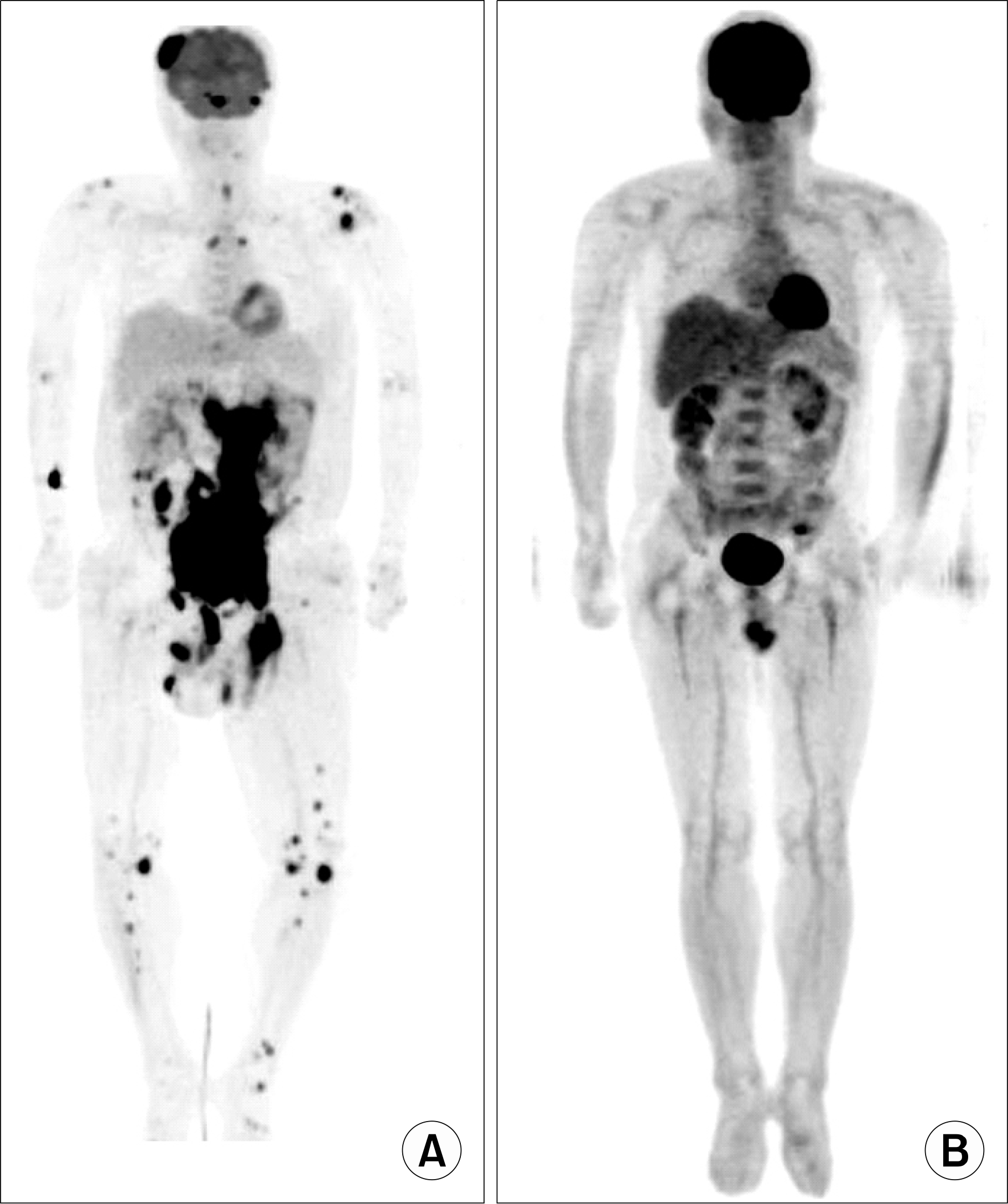
PET-CT prior to chemotherapy (A) showed that hy-permetabolic lesions were involving in right parietal bone with destruction, sphenoid sinus, entire abdomen, left inguinal lesion and multiple bone lesions (both clavicle, both sternoclavicular junction, left humeral, both knee, both femur, left humeral head). And PET-CT after chemotherapy (B) revealed completely remitted status.


 PDF
PDF ePub
ePub Citation
 Print
Print


 XML Download