

 PDF
PDF ePub
ePub Citation
 Print
Print


Abstract
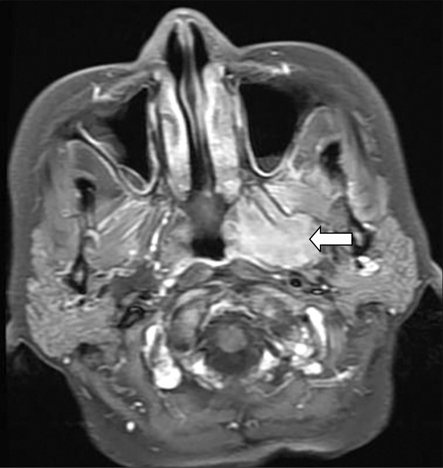
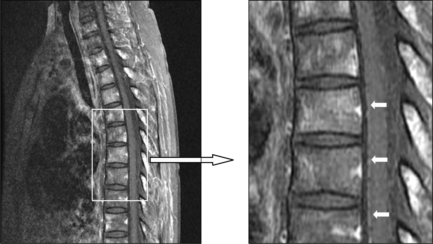
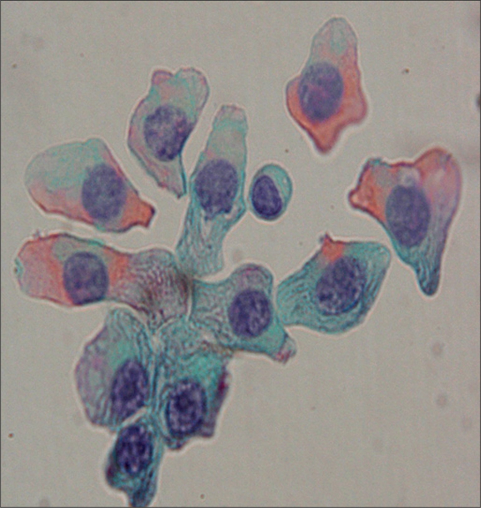
Central nervous system (CNS) myelomatosis, which is the presence of monoclonal plasma cells in the cerebrospinal fluid (CSF), is extremely rare. We report a case of CNS myelomatosis developed in a 45-year-old woman with multiple myeloma in complete response, which was achieved by allogeneic peripheralblood stem cell transplantation using a reduced-intensity conditioning regimen consisting of melphalan, fludarabine, and antithymocyte globulin. Two months after the transplant, she developed a moderatemotor and sensory weakness in both lower extremities. Atypical plasma cells were found in the CSF, and immunofixation revealed monoclonal light chain in the CSF. She was given three courses of weekly intra-thecal chemotherapy consisting of methotrexate, cytarabine, and dexamethasone, which cleared the CSF. This case indicates that the allogeneic transplantation could not control CNS myelomatosis, despite successfully treating the bone marrow myeloma.
REFERENCES
1). Brenner B., Carter A., Tatarsky I., Gruszkiewicz J., Peyser E. Incidence, prognostic significance and therapeutic modalities of central nervous system involvement in multiple myeloma. Acta Haematol. 1982. 68:77–83.

2). Fassas ABT., Muwalla F., Berryman T, et al. Myeloma of the central nervous system: association with high-risk chromosomal abnormalities, plasmablastic morphology and extramedullary manifestations. Br J Haematol. 2002. 117:103–8.
3). Kim YM., Jeon MY., Chi JG. Cytologic features and distribution of primary sites of malignant cells in cerebrospinal fluid. Korean J Cytopathol. 2000. 11:65–73.
4). Nieuwenhuizen L., Biesma DH. Central nervous system myelomatosis: review of the literature. Eur J Haematol. 2008. 80:1–9.
5). Sasser RL., Yam LT., Li C. Myeloma with involvement of the serous cavities. Cytologic and immu-nochemical diagnosis and literature review. Acta Cytol. 1990. 34:479–85.
6). Péter A. The plasma cells of the cerebrospinal fluid. J Neurol Sci. 1967. 4:227–39.
7). Moulopoulos LA., Granfield CA., Dimopoulos MA., Kim EE., Alexanian R., Libshitz HI. Extraosseous multiple myeloma: imaging features. AJR Am J Roentgenol. 1994. 161:1083–7.
8). Umapathi T., Chaudhry V. Toxic neuropathy. Curr Opin Neurol. 2005. 18:574–80.
9). de la Fuente J., Prieto I., Albo C., Sopeña B., Somoli-nos N., Martinez C. Plasma cell myeloma presented as myelomatous meningitis. Eur J Haematol. 1994. 53:244–5.
10). Cavanna L., Invernizzi R., Berte R., Vallisa D., Buscarini L. Meningeal involvement in multiple myeloma: report of a case with cytologic and immunocytochemical diagnosis. Acta Cytol. 1996. 40:571–75.
11). Spiers AS., Halpern R., Ross SC., Neiman RS., Harawi S., Zipoli TE. Meningeal myelomatosis. Arch Intern Med. 1980. 140:256–9.
12). Patriarca F., Zaja F., Silvestri F, et al. Meningeal and cerebral involvement in multiple myeloma patients. Ann Hematol. 2001. 80:758–62.
13). Patriarca F., Prosdocimo S., Tomadini V., Vasciaveo A., Bruno B., Fanin R. Efficacy of bortezomib therapy for extramedullary relapse of myeloma after autologous and non-myeloablative allogeneic transplantation. Haematologica. 2005. 90:278–9.
 XML Download