

 PDF
PDF ePub
ePub Citation
 Print
Print


Abstract
Background:
The iron chelating agents (ICA) have various biological effects besides iron chelation. We investigated the immunomodulatory effects of Deferasirox (DFS) compared to Deferoxamine (DFO).
Methods:
Spleen cells (SP) were obtained from 5 week-old C57/BL6 (H-2b). The cytotoxicity of ICAs was examined using the CCK8 method. For the cell proliferation assay, SP were cultured with irradiated in addition to 10, 50, 100μM of DFS or DFO and 200ng/mL of cyclosporin A (CSA). Cytokines and nitrite levels were evaluated from supernatants by ELISA.
Results:
The viability of ICA was reported to be over 100%. Both DFS and DFO inhibited cell proliferation in a manner comparable to CSA. Cell proliferation without iron was reduced at the concentration of 100μM of DFO. With iron treatment, the reduction of the stimulation index was dependent on DFO concentrations. DFS decreased the proliferation without reference to the concentrations. After stimulation of phytohemagglutinin, the nitrite concentrations increased with iron. With lipopolysaccharides, the nitrite levels were higher in DFO with iron than control, but similar in DFS regardless of iron treatment. The levels of interleukin-2 were not different. Interleukin-10 was more abundantly produced in 50μM of DFO compared to DFS. Transforming growth factor-β was higher in DFS than DFO at the low concentration, but opposite at the high concentration.
REFERENCES
1). Heeney MM., Andrews NC. Iron homeostasis and inherited iron overload disorders: an overview. Hematol Oncol Clin N Am. 2004. 18:1379–403.

2). Oppenheimer SJ. Iron and its relation to immunity and infectious disease. J Nutr. 2001. 131(Suppl):S616–S35.
3). Gabutti V., Borgna-Pignatti C. Clinical manifestations and therapy of transfusional haemosiderosis. Bail-lieres Clin Haematol. 1994. 7:919–40.
4). Marx JJ. Iron and infection: competition between host and microbes for a precious element. Best Pract Res Clin Haematol. 2002. 15:411–26.
5). Cohen AR. New advances in iron chelation therapy. Hematology Am Soc Hematol Educ Program. 2006. 42–7.
6). Polson RJ., Jenkins R., Lombard M, et al. Mechanism of inhibition of mononuclear activation by the iron-chelating agent deferrioxamine. Immunology. 1990. 71:176–81.
7). Kontoghiorghes GJ., Eracleous E., Economides C., Kolnagou A. Advances in iron overload therapies. Prospects for effective use of deferiprone (L1), deferoxamine, the new experimental chelators ICL670, GT56-252, L1NA11 and their combinations. Curr Med Chem. 2005. 12:2663–81.
8). Kontoghiorghes GJ., Pattichi K., Hadjigavriel M., Kolnagou A. Transfusional iron overload and chelation therapy with deferoxamine and deferiprone (L1). Transfus Sci. 2000. 23:211–23.
9). Hileti D., Panayiotidis P., Hoffbrand V. Iron chelators induce apoptosis in proliferative cells. Br J Haematol. 1995. 89:181–7.
10). Kim BM., Choi JY., Kim YJ., Woo HD., Chung HW. Desferrioxamine (DFX) has genotoxic effects on cultured human lymphocytes and induces the p53-mediated damage response. Toxicology. 2007. 229:226–35.
11). Soyano A., Chinea M., Romano EL. The effect of des-ferrioxamine on the proliferative response of rat lymphocytes stimulated with various mitogens in vitro. Immunopharmacology. 1984. 8:163–9.
12). Bowern N., Ramshaw IA., Badenoch-Jones P., Doherty PC. Effect of an iron-chelating agent on lymphocyte proliferation. Aust J Exp Biol Med Sci. 1984. 62:743–54.
13). Whitley WD., Hancock WW., Kupiec-Weglinishi JW., DeSousa M., Tilney NL. Iron chelation suppress mononuclear cell activation, modifies lymphocyte migration patterns, and prolongs rat cardiac allograft survival in rats. Transplantation. 1993. 56:1182–8.
14). Hileti D., Panayiotidis P., Hoffbrand AV. Iron chelators induce apoptosis in proliferating cells. Br J Haematol. 1995. 89:181–7.
15). Cory JG., Lasater L., Sato A. Effect of iron-chelating agents on inhibitors of ribunucleotide reductase. Biochem Pharmacol. 1981. 30:979–84.
16). Carutenuto P., Pontesilli O., Cambier JC., Hayward AR. Desferoximine blocks IL-2 receptor expression on human T lymphocytes. J Immunol. 1986. 136:2342–7.
17). Crichton RR., Wilmet S., Legssyer R., Ward RJ. Molecular and cellular mechanisms of iron homeostasis and toxicity in mammalian cells. J Inorganic Bio-chem. 2002. 91:9–18.
18). Emerit J., Beaumont C., Trivin F. Iron metabolism, free radicals, and oxidative injury. Biomed Pharmac-other. 2001. 55:333–9.
19). Fritsche G., Larcher C., Schennach H., Weiss G. Regulatory interactions between iron and nitric oxide metabolism for immune defense against Plasmo-diuim falciparum infection. J Infect Dis. 2001. 183:1388–94.
20). Legssyer R., Josse C., Piette J., Ward RJ., Crichton RR. Changes in function of iron-loaded alvelolar marcro-phages after in vivo administration of desferriox-amine and/or chloroquine. J Inorganic Biochem. 2003. 94:36–42.
21). Golenser J., Domb A., Mordechai-Daniel T., Leshem B., Luty A., Kremsner P. Iron chelators: correlation between effects on Plasmodium spp. and immune functions. J Parasitol. 2006. 92:170–7.
22). Yoon G., Kim HJ., Yoon YS., Cho H., Lim IK., Lee JH. Iron chelation-induced senescence-like growth arrest in hepatocyte cell lines: association of transforming growth factor beta1 (TGF-beta1)-mediated p27Kip1 expression. Biochem J. 2002. 366:613–21.
Fig. 1
Cell viability according to concentrations of iron chelating agents (ICA) by CCK 8 method. (A) There is no difference in cell viability among various concentrations of ICA without ferric chloride (n=5). (B) The viability is significantly diminished at 20μM ferric chloride, but there are no difference in viability among ICA in spite of presence of iron (n=5) (∗: P<0.05). Abbreviations: DFS, deferasirox; DFO, deferoxamine; FeCl3, ferric chloride.
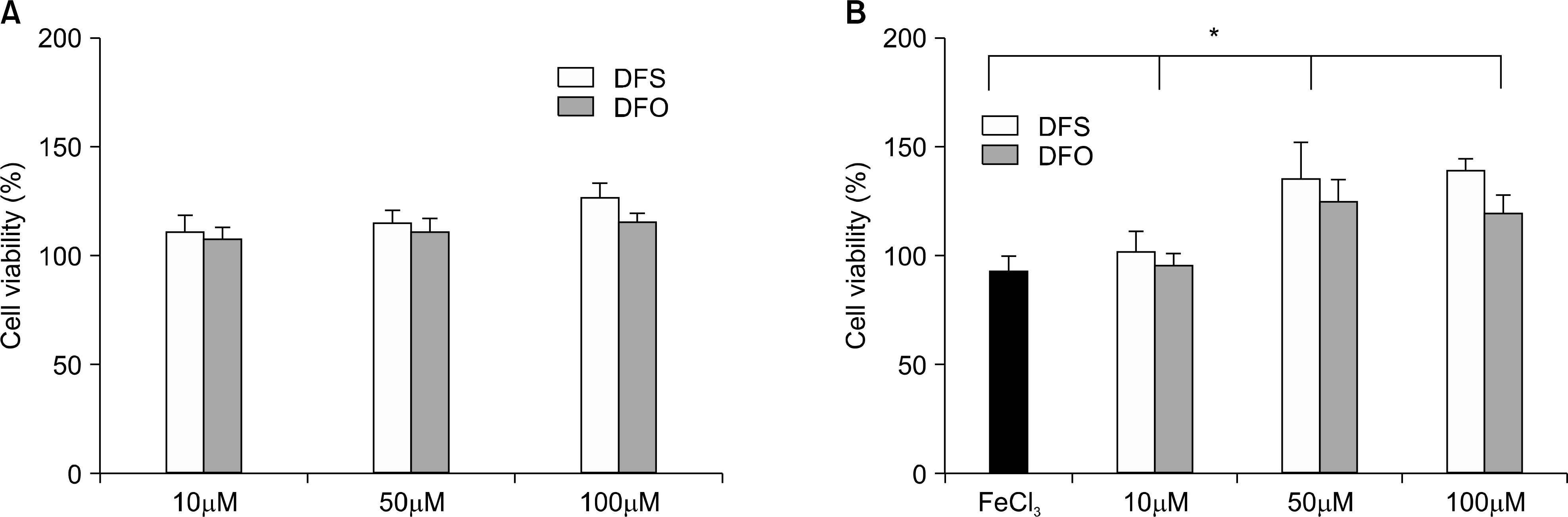
Fig. 2
Stimulation index (SI) of C57/BL6 spleen cells (SP) and irradiated BALB/c SP under 5μg/mL of phytohemagglutinin with 1:1 ratio according to the concentration of iron chelating agents (ICA) (n=7) without (A) and with (B) ferric chloride. There are significant differences between cyclosporin A (CSA) and ICA with or without ferric chloride (†: P<0.05). (A) The high level (100μM) of deferoxamine (DFO) exerts profound inhibition of cell proliferation than deferasirox (DFS) (∗: P<0.05), although it is not different at 10 and 50μM of DFO. Deferasirox (DFS) showed no difference in SI among any concentrations. (B) The decrement of SI is dependent on concentration in DFO (†: P<0.01), but is not in DFS. There is a significant difference in SI between DFO and DFS at 10μM (∗: P<0.01).

Fig. 3
The levels of nitrite in supernatants from cell proliferation assay with or without ferric chloride according to the concentration of iron chelating agents by non-specific mitogen (A; phytohemagglutinin (PHA), B; lipopolysaccharide (LPS)). All nitrite levels after stimulation are not different according to PHA or LPS. (A) There is lower nitrite in 10μM of Deferoxamine (DFO) and Deferasirox (DFS) among ICA without iron, and higher at 10μM of DFS than other ICA with iron (†: P<0.01). (B) The levels of nitrite are not different among ICA without iron, but higher only in DFO with iron (‡: P<0.01). ∗: P<0.05 in each ICA concentration according to presence with iron. Abbreviations: DFS, deferasirox; DFO, deferoxamine.
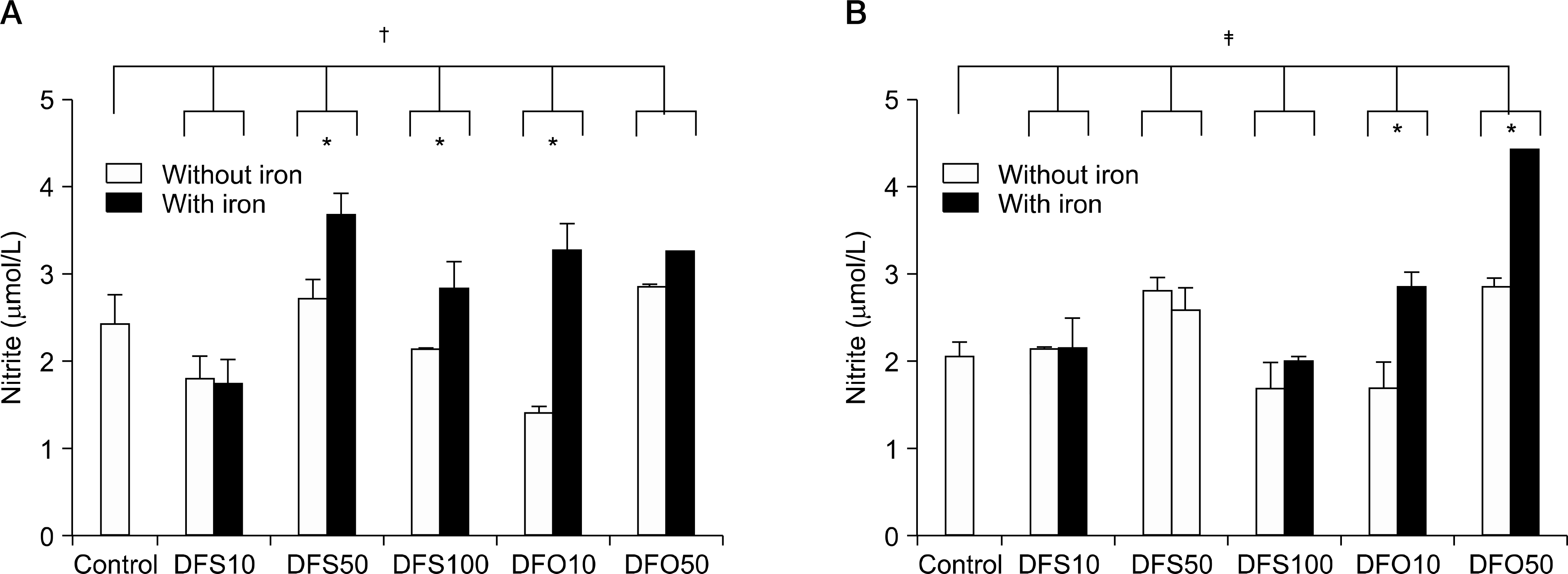
Table 1.
The level of cytokines in supernatants from cell proliferation assay according to the concentration of iron chelating agents without ferric chloride (n=4∼6)
| (pg/mL) | Control | CSA | 10μM | 50μM | 100μM | |||
|---|---|---|---|---|---|---|---|---|
| DFO | DFS | DFO | DFS | DFO | DFS | |||
| IL-2 | 49.7±7.2 | 17.9±10.4† | 41.4±16.3 | 46.9±7.3 | 36.9±14.3 | 40.8±12.0 | 55.7±9.9 | 25.1±16.5 |
| IL-10 | 18.2±7.0 | 15.5±2.8 | 25.1±5.0 | 12.8±7.8 | 14.4±0.1∗ | 7.4±4.7 | 12.8±5.2 | 10.3±5.3 |
| TGF-β | 1,575.8±473.4 | 1,424.8±466.7 | 1,248.8±82.2 | 1,567.0±472.3∗ | 1,222.2±82.1 1 | 1,133.4±151.9 | 1,362.9±402.3∗ | 1,279.9±39.7 |
Table 2.
The level of cytokines in supernatants from cell proliferation assay according to the concentration of iron chelatingagents with ferric chloride (n=4∼6)
| (pg/mL) | Control | CSA | 10μM | 50μM | 100μM | |||
|---|---|---|---|---|---|---|---|---|
| DFO | DFS | DFO | DFS | DFO | DFS | |||
| IL-2 | 42.4±10.6 | 29.8±16.8 | 30.5±17.3 | 41.7±12.4 | 62.6±23.8 | 33.41±15.3 | 49.3±32.3 | 47.9±23.3 |
| IL-10 | 21.1±8.0 | 19.2±5.9 | 7.6±3.4 | 13.2±12.1 | 41.0±11.5 | 8.06±3.4 | 7.3±1.7 | 30.5∗±11.3 |
| TGF-β | 1,066.6±53.6 | 1,376.8±317.2 | 1,355.0±276.0 | 1,672.7±372.1† | 1,374.7±252.7 1 | 1,498.9±352.1 | 1 1,202.0±2.7 | 1,225.9±66.8 |
 XML Download