Abstract
Background:
Although engraftment following murine allogeneic bone marrow transplantation (BMT) is most commonly confirmed by H2 typing using flow cytometry, recipient mice can be seriously injured during peripheral blood (PB) sampling. Therefore, we developed an alternative DNA-based assay that does not require the large volume of PB necessary for flow cytometry.
Methods:
A minute volume of PB from the tail vein was used to evaluate the engraftment by PCR amplification of a microsatellite in the class II Eb gene. Dilution experiments were performed to evaluate the sensitivity of this assay for detecting donor cells in mixed cell populations compared with flow cytometry analysis.
Results:
Early engraftment and mixed chimerism were confirmed, based on the length variation of the microsatellite in the class II Eb gene. The degree of donor chimerism in the donor-recipient cell mixture could be estimated semiquantitatively in a dilution experiment. The sensitivity of this assay by the naked eye approached 10% of the degree of donor chimerism.
REFERENCES
1). Exner BG., Domenick MA., Bergheim M., Mueller YM., Ildstad ST. Clinical applications of mixed chimerism. Ann N Y Acad Sci. 1999. 872:377–85. discussion 385-6.

2). Cho SG., Shuto Y., Soda Y, et al. Anti-NK cell treatment induces stable mixed chimerism in MHC-mismatched, T cell-depleted, nonmyeloablative bone marrow transplantation. Exp Hematol. 2004. 32:1246–54.

3). Mapara MY., Kim YM., Wang SP., Bronson R., Sachs DH., Sykes M. Donor lymphocyte infusions mediate superior graft-versus-leukemia effects in mixed compared to fully allogeneic chimeras: a critical role for host antigen-presenting cells. Blood. 2002. 100:1903–9.

4). Luznik L., Slansky JE., Jalla S, et al. Successful therapy of metastatic cancer using tumor vaccines in mixed allogeneic bone marrow chimeras. Blood. 2003. 101:1645–52.

5). Exner BG., Acholonu IN., Bergheim M., Mueller YM., Ildstand ST. Mixed allogeneic chimerism to induce tolerance to solid organ and cellular grafts. Acta Haematol. 1999. 101:78–81.

6). Mapara MY., Kim YM., Marx J., Sykes M. Donor lymphocyte infusion-mediated graft-versus-leukemia effects in mixed chimeras established with a non-myeloablative conditioning regimen: extinction of graft-versus-leukemia effects after conversion to full donor chimerism. Transplantation. 2003. 76:297–305.
7). Sykes M., Preffer F., McAfee S, et al. Mixed lympho-haemopoietic chimerism and graft-versus-lymphoma effects after non-myeloablative therapy and HLA-mismatched bone-marrow transplantation. Lancet. 1999. 353:1755–9.
8). Li H., Kaufman CL., Boggs SS., Johnson PC., Patrene KD., Ildstad ST. Mixed allogeneic chimerism induced by a sublethal approach prevents autoimmune diabetes and reverses insulitis in nonobese diabetic (NOD) mice. J Immunol. 1996. 156:380–8.
9). Nikolic B., Takeuchi Y., Leykin I., Fudaba Y., Smith RN., Sykes M. Mixed hematopoietic chimerism allows cure of autoimmune diabetes through allogeneic tolerance and reversal of autoimmunity. Diabetes. 2004. 53:376–83.

10). Cho SG., Min SY., Park MJ, et al. Immunoregulatory effects of allogeneic mixed chimerism induced by nonmyeloablative bone marrow transplantation on chronic inflammatory arthritis and autoimmunity in interleukin-1 receptor antagonist-deficient mice. Arthritis Rheum. 2006. 54:1878–87.

11). Saha BK. Typing of murine major histocompatibility complex with a microsatellite in the class II Eb gene. J Immunol Methods. 1996. 194:77–83. Acad Sci U S A 1993;90: 5312-6.

12). Saha BK., Shields JJ., Miller RD., Hansen TH., Shreffler DC. A highly polymorphic microsatellite in the class II Eb gene allows tracing of major histocompatibility complex evolution in mouse. Proc Natl.
13). Gardiner N., Lawler M., O'Riordan J., DeArce M., Humphries P., McCann SR. Persistent donor chimaerism is consistent with disease-free survival following BMT for chronic myeloid leukaemia. Bone Marrow Transplant. 1997. 20:235–41.

14). Gardiner N., Lawler M., O'Riordan J., De'Arce M., McCann SR. Donor chimaerism is a strong indicator of disease free survival following bone marrow transplantation for chronic myeloid leukaemia. Leukemia. 1997. 11(Suppl 3):512–5.
15). Lawler M., Humphries P., McCann SR. Evaluation of mixed chimerism by in vitro amplification of dinucleotide repeat sequences using the polymerase chain reaction. Blood. 1991. 77:2504–14.

16). Park SJ., Min WS., Yang IH, et al. Effects of mixed chimerism and immune modulation on GVHD, disease recurrence and survival after HLA-identical marrow transplantation for hematologic malignancies. Korean J Intern Med. 2000. 15:224–31.
17). Buno I., Nava P., Simon A, et al. A comparison of fluorescent in situ hybridization and multiplex short tandem repeat polymerase chain reaction for quantifying chimerism after stem cell transplantation. Haematologica. 2005. 90:1373–9.
18). O'Neill PA., Lawler M., Pullens R, et al. PCR amplification of short tandem repeat sequences allows serial studies of chimaerism/engraftment following BMT in rodents. Bone Marrow Transplant. 1996. 17:265–71.
Fig. 1
Assessing the degree of mixed chimerism in an arbitrary cell mixture using PCR amplification of a microsatellite in the class II Eb gene of the murine MHC. Donor (D, C57BL/6, H-2kb mice) and recipient (R, BALB/c, H-2kd) splenocytes were mixed in vitro in various proportions (D to R=0 to 100, 5 to 95, 10 to 90, 25 to 75, 50 to 50, 75 to 25, 90 to 10, 95 to 5, and 100 to 0) in dilution experiments. PCR amplification of the class II Eb gene and flow cytometry analysis were performed using the cell mixtures. The DNA fragments amplified from the donor and recipient were 107 and 139bp, respectively. The lanes show the results for artificial mixtures of donor and recipient splenocytes.

Fig. 2
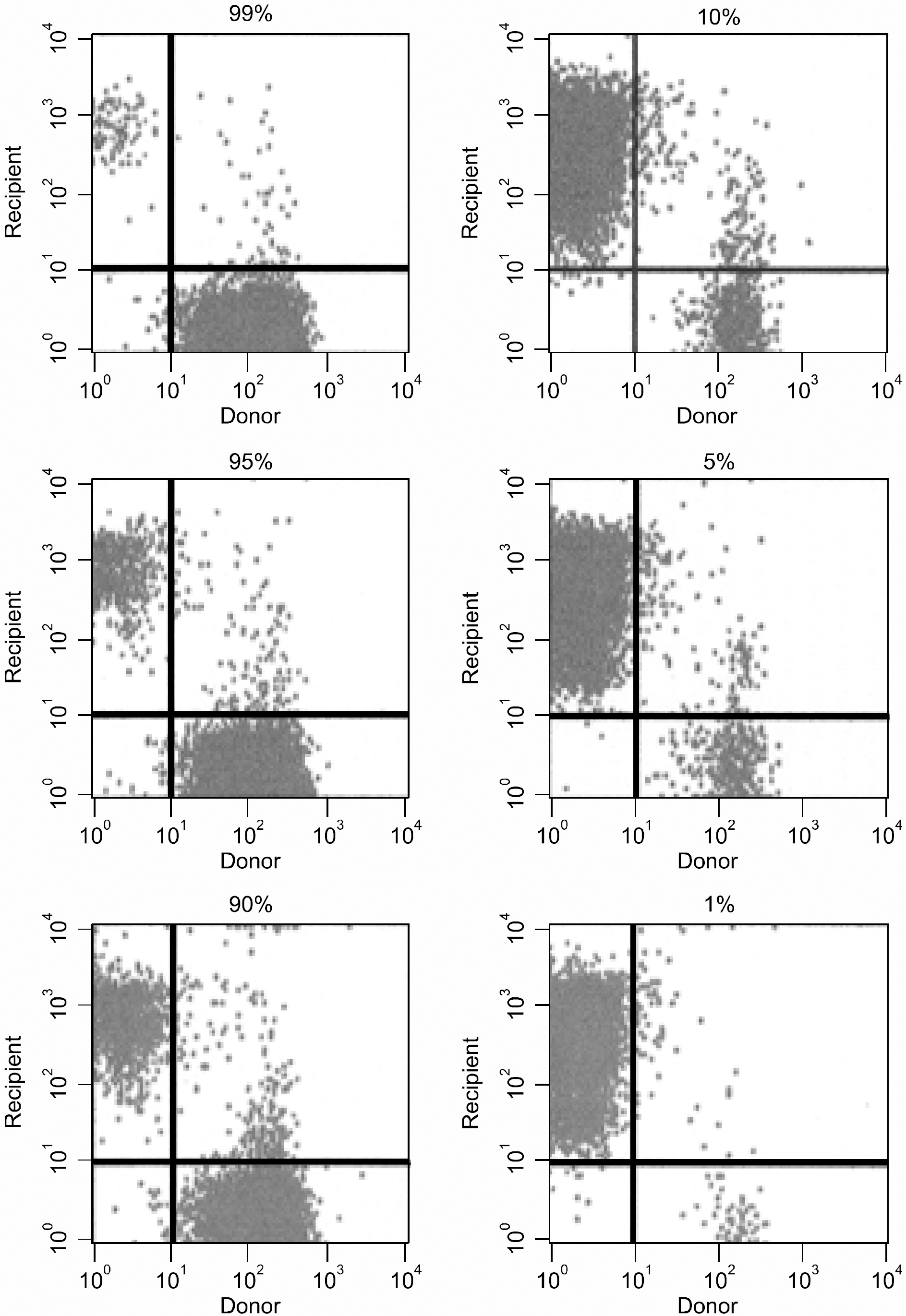
Assessing the degree of mixed chimerism in arbitrary cell mixtures using flow cytometry. Donor (C57BL/6, H-2kb mice) and recipient (BALB/c, H-2kd) splenocytes were mixed in vitro in various proportions (D to R=99 to 1, 95 to 5, 90 to 10, 10 to 90, 5 to 95, and 1 to 99) for dilution experiments. Donor (H-2Kb) and recipient (H-2Kd) cells were distinguished during lymphoid gating by staining with fluorescein isothiocyanate-labeled anti-H-2Kb and phycoerythrin-labeled anti-H-2Kd antibodies (PharMingen, San Diego, CA), respectively. Stained cells were analyzed using CellQuest software on a FACSCalibur flow cytometer (both from Becton Dickinson, Mountain View, CA). The percentage of donor-derived cells was calculated by dividing the number of donor cells by the total net number of donor plus recipient cells that showed positive staining.

Fig. 3
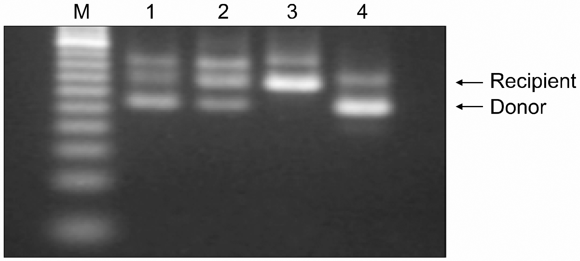
Donor chimerism in peripheral blood of an allogeneic mixed chimera following MHC-mismatched non-myeloablative BMT. Engraftment/mixed chimerism at 3 weeks post-BMT was evaluated using PCR amplification of a microsatellite in the class II Eb gene. Allogeneic mixed chimeric mice (lanes 1, 2, and 4) showed both the 107-bp (donor, C57BL/6, H-2b) and 139-bp (host, BALB/c, H-2d) fragments, whereas non-transplanted recipient mice as a negative control (lane 3) showed only the 107-bp (host, BALB/c, H-2d) fragment. The relative ratios of donor-derived cells in the PB from those mice were 76, 42, 0, and 89% of all lymphocytes, respectively. The DNA-based assay developed in this study roughly correlated with the results of the flow cytometry assay.

Fig. 4
Assessing NOD/Scid mice using PCR amplification of a microsatellite in the class II Eb gene of the murine MHC. The presence of NOD/Scid-derived cells can be detected using PCR amplification of a microsatellite in the class II Eb gene of the murine MHC, although NOD/Scid mice do not express the H-2 haplotype antigen. The amplified DNA fragments of the C57BL/6 (H-2b), BALB/c (H-2d), and NOD/Scid mice were 107, 139, and 80bp, respectively.


 PDF
PDF ePub
ePub Citation
 Print
Print


 XML Download