

 PDF
PDF ePub
ePub Citation
 Print
Print


Abstract
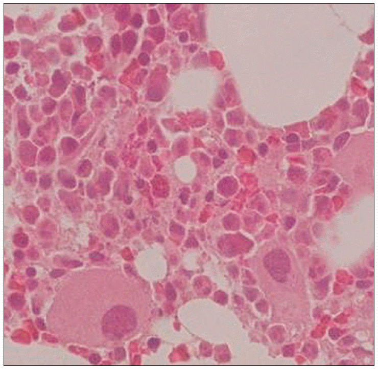
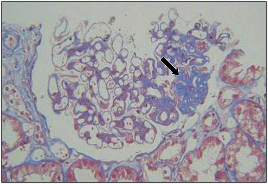
Polycythemia vera (PV) is a myeloproliferative disorder that results from clonal expansion of a transformed hematopoietic stem cell, and this is associated with a prominent overproduction of erythrocytes, and to a lesser extent, expansion of the granulocytic and megakaryocytic elements. Secondary polycythemia is occasionally associated with renal diseases such as renal tumors, cysts, hydronephorosis, renal transplantation, renal artery stenosis and Bartter's syndrome, and it is rarely associated with nephritic syndrome, nephrosclerosis, chronic glomerulonephritis (GN) and membranous nephropathy. Although cases of erythrocytosis with concomitant GN have occasionally been reported, there are few reports regarding PV. Focal segmental glomerulosclerosis (FSGS) is one of the most frequent forms of GN. However, its association with PV has rarely been described. We report here on one patient with concomitant PV and FSGS along with a review of the previously reported literature.
REFERENCES
1). Kwon MY., Eom HS., Lee SW., Kim MJ., Kim TS., Kwon KH. A case of chronic renal failure, caused by IgA nephropathy combined with polycythemia vera. Korean J Nephrol. 1999. 18:483–7.
2). Kim JH., Choi P., Jung YS, et al. A case of IgA nephropathy associated with polycythemia vera. Korean J Nephrol. 2002. 21:1006–10.
3). Chung J., Park PG., Song KI. IgA nephropathy in a patient with polycythemia vera. Clinical manifestation of chronic renal failure and heavy proteinuria. Am J Nephrol. 2002. 22:397–401.
4). Kosch M., August C., Hausberg M, et al. Focal sclerosis with tip lesions secondary to polycythaemia vera. Nephrol Dial Transplant. 2000. 15:1710–1.

5). Au WY., Chan KW., Lui SL., Lam CC., Kwong YL. Focal segmental glomerulosclerosis and mesangial sclerosis associated with myeloproliferative disorders. Am J Kidney Dis. 1999. 34:889–93.
6). Iyoda M., Ito J., Ajiro Y, et al. Focal segmental glomerulosclerosis in a patient with polycythemia vera. Nippon Jinzo Gakkai Shi. 2005. 47:828–33.
7). Michiels JJ., Bernema Z., Van Bockstaele D., De Raeye H., Schroyens W. Current diagnostic criteria for the chronic myeloproliferative disorders (MPD) essential thrombocythemia (ET), polycythemia vera (PV) and chronic idiopathic myelofibrosis (CIMF). Pathol Biol (Paris). 2006. 17:[Epub ahead of print].
8). Polycythemia vera: the natural history of 1,213 patients followed for 20 years. Gruppo Italiano Studio Policitemia. Ann Intern Med. 1995. 123:656–64.
9). Futrakul P., Siriviriyakul P., Patumraj S., Bunnag S., Kulaputana O., Futrakul N. A hemodynamically mediated mechanism of renal disease progression in severe glomerulonephritis or nephrosis. Clin Hemor-heol Microcirc. 2003. 29:183–7.
10). Ono T., Kanatsu K., Doi T, et al. Relationship of in-traglomerular coagulation and platelet aggregation to glomerular sclerosis. Nephron. 1991. 58:429–36.
11). Correa PN., Eskinazi D., Axelrad AA. Circulating erythroid progenitors in polycythemia vera are hypersensitive to insulin-like growth factor-1 in vitro: studies in an improved serum-free medium. Blood. 1994. 83:99–112.
12). Landolfi R., Marchioli R., Kutti J. European Collaboration on Low-Dose Aspirin in Polycythemia Vera Investigators. . Efficacy and safety of low-dose aspirin in polycythemia vera. N Engl J Med. 2004. 350:114–24.
13). Korbet SM. Angiotensin antagonists and steroids in the treatment of focal segmental glomerulosclerosis. Semin Nephrol. 2003. 23:219–28.
14). Dingli D., Larson DR., Plevak MF., Grande JP., Kyle RA. Focal and segmental glomerulosclerosis and plasma cell proliferative disorders. Am J Kidney Dis. 2005. 46:278–82.
 XML Download