Abstract
Essential thrombocythemia (ET) is a clonalmyeloproliferative disorder that can rarely transform into acute leukemia in 1∼5% of cases. A recent study has found that a significant proportion of leukemic cases from ET were associated with a cytogenetic abnormality (17p deletion). Herein, we report two cases of acute myeloid leukemic transformations harboring a 17p abnormality from a series of 119 ET patients. The first case, a 48-year-old female, developed acute myeloid leukemia with maturation (AML-M2) accompanying myelodysplasia was diagnosed 6.1 years after the initial diagnosis of ET. She was treated with hydroxyurea. Her karyotype showed a monosomy 17. The second case, a 61-year-old male, developed acute megakaryoblastic leukemia (AML-M7) with a very complex hyperdiploidy including addition of 17p13 that developed 6.5 years after the initial diagnosis. He was treated with hydroxyurea and anagrelide. The immunohistochemistry showed p53 overexpression in both cases. Our cases support the specificity of chromosome 17 abnormality and p53 overexpression in acute leukemic transformation from ET.
REFERENCES
1). Imbert M., Vardiman JW., Pierre R., Brunning RD., Thiele J., Flandrin G. Essential thrombocythaemia. Jaffe ES, Harris NL, Stein H, Vardiman JW, editors. Tumours of haematopoietic and lymphoid tissues. 1st ed.Lyon, France: IARC Press;2001. p. 39–41.
2). Sterkers Y., Preudhomme C., Lai JL, et al. Acute myeloid leukemia and myelodysplastic syndromes following essential thrombocythemia treated with hydroxyurea: high proportion of cases with 17p deletion. Blood. 1998. 91:616–22.

3). Finazzi G., Ruggeri M., Rodeghiero F., Barbui T. Second malignancies in patients with essential thrombocythaemia treated with busulphan and hydroxyurea: long-term follow-up of a randomized clinical trial. Br J Haematol. 2000. 110:577–83.

4). Lee JH., Kim TI., Kim GT, et al. A case of leukemic transformation in essential thrombocythemia treated with hydroxyurea. Korean J Hematol. 1997. 32:433–9.
5). Fruchtman SM., Petitt RM., Gilbert HS., Fiddler G., Lyne A. Anagrelide Study Group. Anagrelide: analysis of long-term efficacy, safety and leukemogenic potential in myeloproliferative disorders. Leuk Res. 2005. 29:481–91.

6). Lai JL., Preudhomme C., Zandecki M, et al. Myelo-dysplastic syndromes and acute myeloid leukemia with 17p deletion. An entity characterized by specific dysgranulopoiesis and a high incidence of p53 mutations. Leukemia. 1995. 9:370–81.
7). Merlat A., Lai JL., Sterkers Y, et al. Therapy-related myelodysplastic syndrome and acute myeloid leukemia with 17p deletion. A report on 25 cases. Leukemia. 1999. 13:250–7.

8). Bernasconi P., Boni M., Cavigliano PM, et al. Acute myeloid leukemia (AML) having evolved from essential thrombocythemia (ET): distinctive chromosome abnormalities in patients treated with pipobroman or hydroxyurea. Leukemia. 2002. 16:2078–83.

9). Radaelli F., Mazza R., Curioni E., Ciani A., Pomati M., Maiolo AT. Acute megakaryocytic leukemia in essential thrombocythemia: an unusual evolution? Eur J Haematol. 2002. 69:108–11.

10). Vianelli N., Baravelli S., Gugliotta L. Acute megaka-ryoblastic transformation of essential thrombocythemia. Haematologica. 1996. 81:288–9.
11). Geurts van Kessel A., dos Santos NR., Simons A, et al. Molecular cytogenetics of bone and soft tissue tumors. Cancer Genet Cytogenet. 1997. 95:67–73.

12). de Bruijn DR., dos Santos NR., Thijssen J, et al. The synovial sarcoma associated protein SYT interacts with the acute leukemia associated AF10. Oncogene. 2001. 20:3281–9.
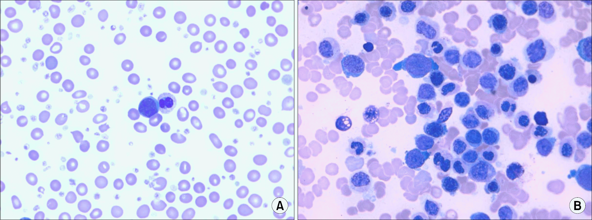
Fig. 1
(A) Peripheral blood smear of case 1 shows a pseudo-Pelger Huet anomaly in neutrophil, a blast, and numerous hypogranular platelets (Wright-Giemsa stain, ×1,000). (B) Bone marrow aspiration of case 1 shows dysgranulocytic changes such as pseudo-Pelger Huet anomaly, hypogranularity and bizarre nuclei and proliferation of blasts (Wright-Giemsa stain, ×1,000).

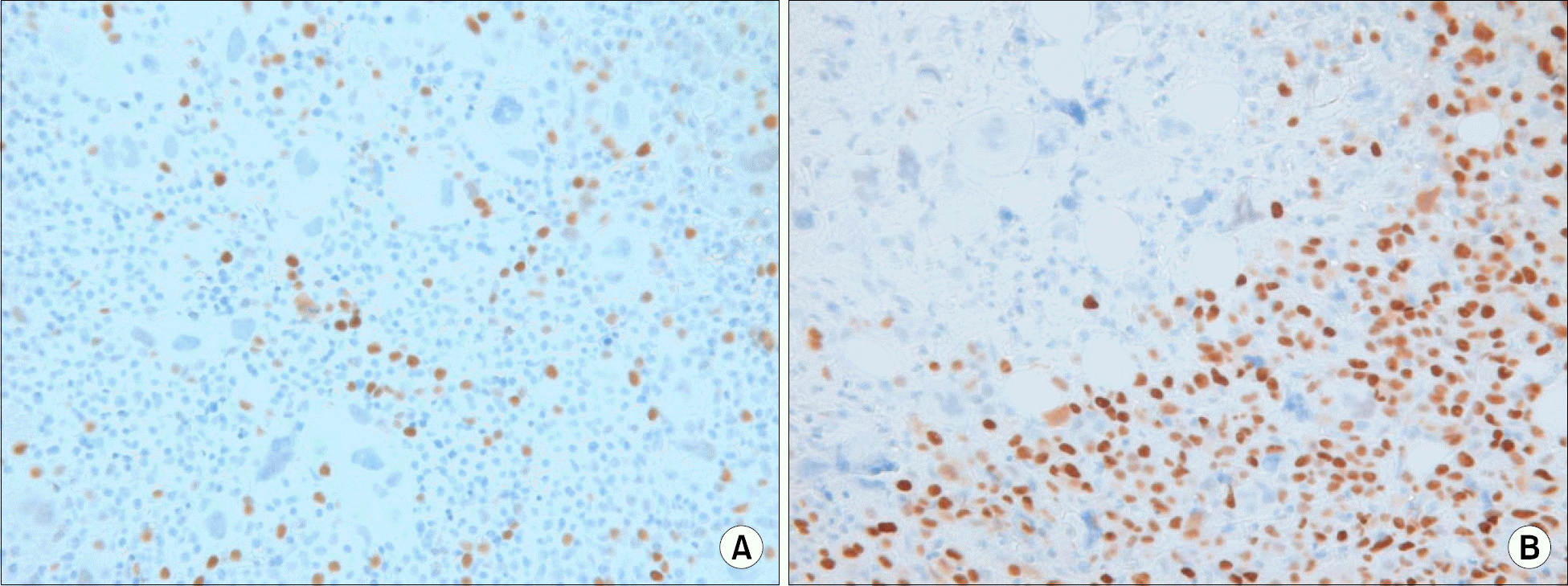
Fig. 2
(A) Leukemic cells reveal overexpression of p53 on the biopsy section in case 1 (Immunohistochemistry for p53, ×400). (B) Leukemic cells reveal overexpression of p53 on the biopsy section in case 2. Note the demarcation between leukemic area and hemopoietic area (Immunohistochemistry for p53, ×400).

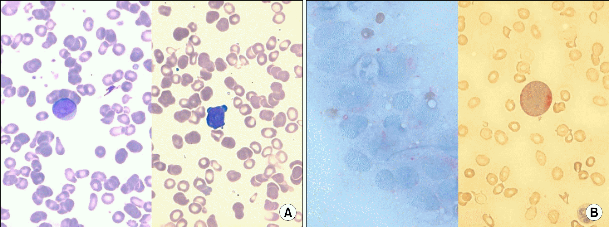
Fig. 3
(A) Peripheral blood smear of case 2 shows a blast (left). Bone marrow aspiration also shows a blast with cytoplasmic pseudopod formation (right) (Wright-Giemsa stain, ×1,000). (B) Bone marrow aspiration of case 2 shows positive reaction to acid phosphatase (left), and positive to alpha naphthyl acetate esterase (right) (Acid phosphatase stain and alpha naphthyl acetate esterase stain, respectively, ×1,000).

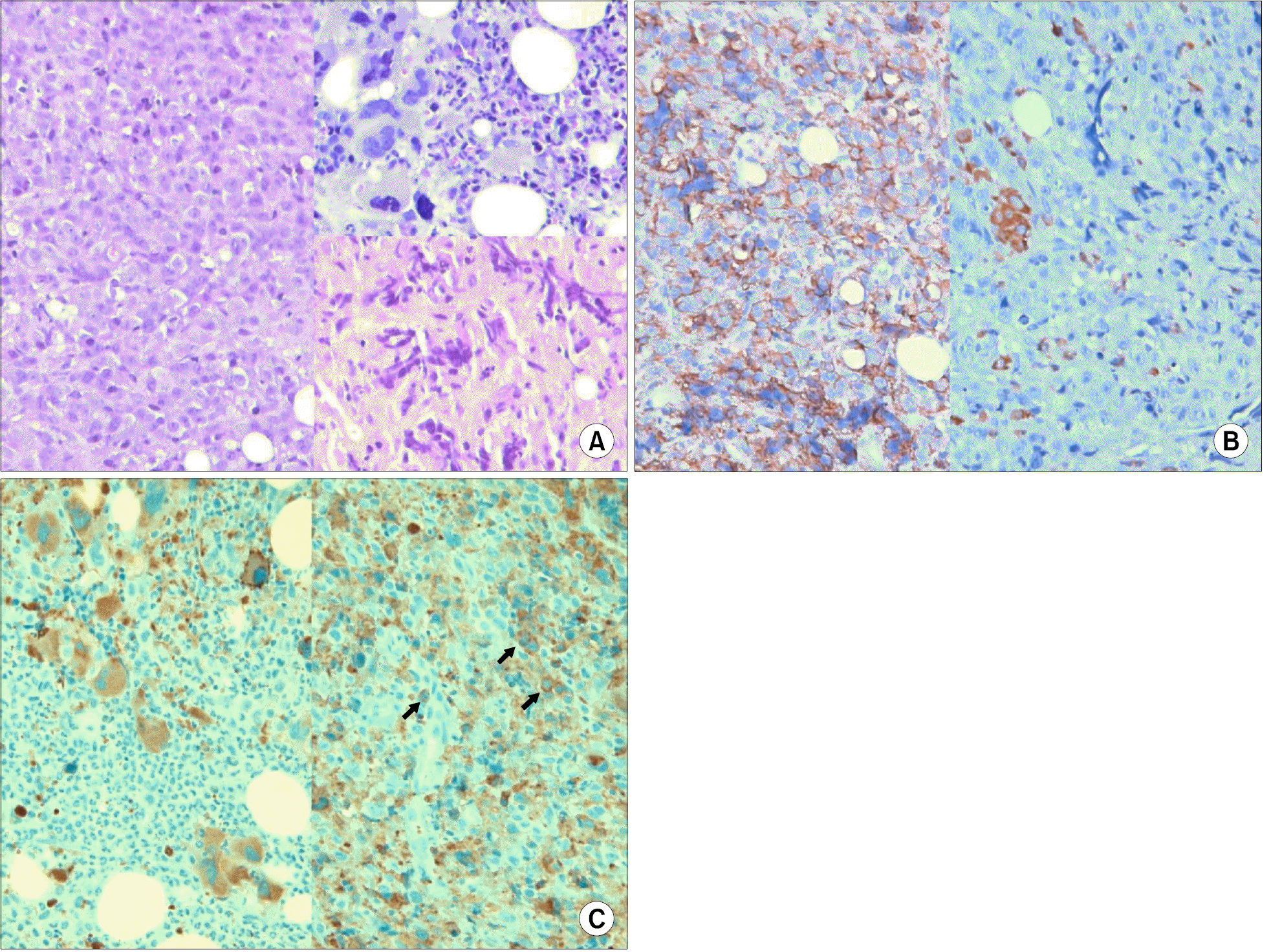
Fig. 4
(A) Bone marrow biopsy of case 2 shows diffuse infiltration of leukemic cells (left). Marked megakaryocytic hyperplasia is noted in hemopoietic area (right upper) and diffuse extensive fibrosis in fibrotic area (right lower) (H&E stain, ×400). (B) Leukemic cells reveal positive reaction to LCA (left), and negative reaction to MPO (right). (Immunohistochemistry for LCA and anti-MPO, respectively, ×400). (C) Megakaryocytes show positive reaction to CD61 but granulocytic cells reveal negative reaction in case 2 (left). Arrows indicate that leukemic cells are positive for CD61 in case 2 (right) (Immunohistochemistry for CD61, ×400).

Table 1.
Clinical and laboratory features of the two ET cases with evolution into AML
| Case 1 | Case 2 | |
|---|---|---|
| Gender/Age∗ | F/48 | M/61 |
| Interval between ET and AML (years) | 6.1 | 6.5 |
| Duration of HU treatment (years) | 6.1 | 6.1 |
| CBC at leukemic transformation | 39,100-7.4-969K | 9,800-8.2-431K |
| FAB subtype | M2 (AML with multilineage dysplasia by WHO classification) | M7 |
| Pseudo Pelger-Huet anomaly | Yes | No |
| Karyotype | 45,XX,t(6;14)(p21.3;q32.3), -17,add(20)(q11.2)[20] | 57,Y,der(X)t(X;18)(p11.2;q11.2),+6,+8,+8,+9,de (9)(q22)x2,+13,+14,+15,add(17)(p13), der(18)add(18)(p13.3)t(X;18),+19,+idic(21) (p11.2)x2,+mar[15]/46,XY[5] |
| p53 overexpression† | Yes | Yes |
| Induction chemotherapy | Ara-C and daunorubicin | ND |
| Clinical results | No response to chemotherapy, follow-up loss | Supportive care, death |
| Survival after diagnosis of leukemia (months) | 11.9 | 1.0 |


 PDF
PDF ePub
ePub Citation
 Print
Print


 XML Download