Abstract
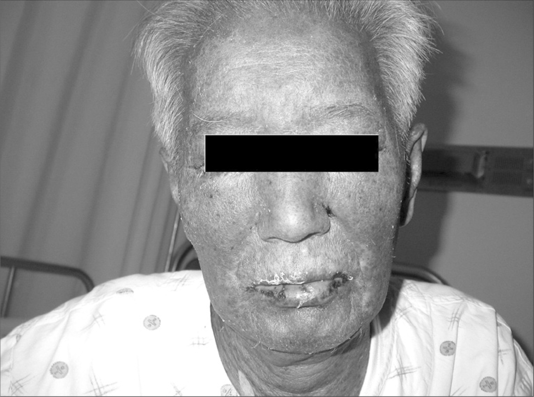
Many chemotherapeutic agents induce variable cutaneous adverse reactions. Among the side effects, Stevens-Johnson syndrome is rare, but a fatal complication. There are two prior reports of cytosine arabinoside (ARA-C) induced toxic epidermal necrolysis, which is considered in the continuum of Stevens-Johnson syndrome. The prior cases were female patients under 16 years old with acute lymphocytic leukemia. We treated a 77-year-old man with recurrent mantle cell lymphoma who developed Stevens-Johnson syndrome after high dose ARA-C therapy. This is the first case of ARA-C induced Stevens-Johnson syndrome in Korea.
REFERENCES
1). Abeloff MD., Armitage JO., Niederhuber JE., Kastan MB., McKenna WG. Clinical oncology. 3rd ed.Pennsylvania, USA: Elsevier;2004. p. 793–816.
2). Roujeau JC., Stern RS. Severe adverse cutaneous reactions to drugs. N Engl J Med. 1994. 331:1272–85.

3). Ozkan A., Apak H., Celkan T., Yuksel L., Yildiz I. Toxic epidermal necrolysis after the use of high-dose cytosine arabinoside. Pediatr Dermatol. 2001. 18:38–40.
4). Figueiredo MS., Yamamoto M., Kerbauy J. Toxic epidermal necrolysis after the use of intermediate dose of cytosine arabinoside. Rev Assoc Med Bras. 1998. 44:53–5.
5). Graves T., Hooks MA. Drug-induced toxicities associated with high-dose cytosine arabinoside infusions. Pharmacotherapy. 1989. 9:23–8.

6). Cetkovsk P., Pizinger K., Cetkovsk P. High-dose cytosine arabinoside-induced cutaneous reactions. J Eur Acad Dermatol Venereol. 2002. 16:481–5.
7). Khalili B., Bahna SL. Pathogenesis and recent therapeutic trends in Stevens-Johnson syndrome and toxic epidermal necrolysis. Ann Allergy Asthma Immunol. 2006. 97:272–80.

8). Hynes AY., Kafkala C., Daoud YJ., Foster CS. Controversy in the use of high-dose systemic steroids in the acute care of patients with Stevens-Johnson syndrome. Int Ophthalmol Clin. 2005. 45:25–48.

9). Severino G., Chillotti C., De Lisa R., Del Zompo M., Ardau R. Adverse reactions during imatinib and lansoprazole treatment in gastrointestinal stromal tumors. Ann Pharmacother. 2005. 39:162–4.

10). Hsiao LT., Chung HM., Lin JT, et al. Stevens-Johnson syndrome after treatment with STI571: a case report. Br J Haematol. 2002. 117:620–2.

11). Hiraki A., Aoe K., Murakami T., Maeda T., Eda R., Takeyama H. Stevens-Johnson syndrome induced by paclitaxel in a patient with squamous cell carcinoma of the lung: a case report. Anticancer Res. 2004. 24:1135–7.
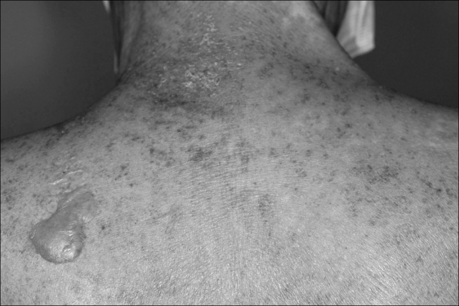
Fig. 1
Erythematous skin lesion with multiple petechia was showen in the patient's back. Single vesicle was also noted.

Fig. 2
(A) The epidermis of skin biopsy shows subepidermal blister formation with scattered apoptotic keratinocytes (H&E ×100). (B) The dermis of skin biopsy shows mild perivascular lymphohistocytic infiltration with mild erythrocyte extravasation (H&E, ×200).


 PDF
PDF ePub
ePub Citation
 Print
Print


 XML Download