Abstract
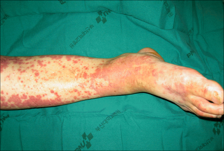
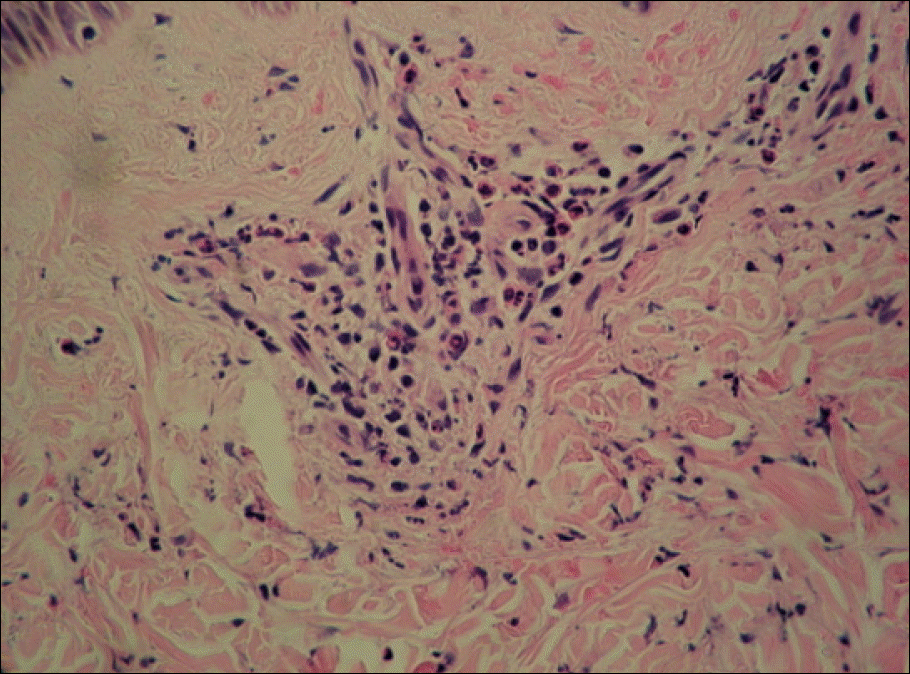
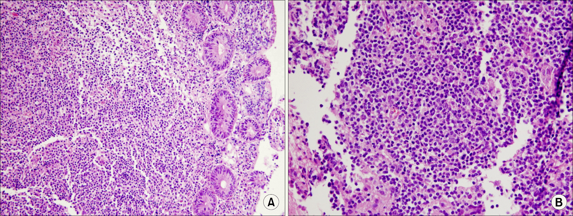
A 69-year-old female was referred to our institution due to abdominal pain and palpable purpura on both buttocks and legs. A skin biopsy of her purpura revealed granulocyte infiltration and leucocytoclasia around the arterioles and venuoles at the dermis, as well as an elevated serum immunoglobulin A level, hematuria and proteinuria. Therefore she was diagnosed with Henoch-Schönlein purpura. She had been diagnosed with diffuse large B cell lymphoma after a biopsy of her left inguinal lymph node 12 years ago and received 6 cycles of CHOP (cyclophosphamide, doxorubicin, vincristine and prednisone) chemotherapy, which was followed by a complete remission. Abdominal and chest CT revealed multiple lymph node enlargement and bowel wall thickening at the ileocecal area, and lesions were observed in a whole body PET CT scan. Recurrence of the diffuse large B cell lymphoma was confirmed by a biopsy of the ileocecal area via colonoscopy. The purpura was improved after oral prednisolone therapy and etoposide, oxaliplatin and ifosfamide salvage combination chemotherapy was used to treat the lymphoma.
REFERENCES
1). Roth DA., Wilz DR., Theil GB. Schönlein-Henoch syndrome in adults. Q J Med. 1985. 55:145–52.
2). Mills JA., Michel BA., Bloch DA, et al. The American College of Rheumatology 1990 criteria for the classification of Henoch-Schönlein purpura. Arthritis Rheum. 1990. 33:1114–21.

3). Pillebout E., Thervet E., Hill G., Alberti C., Vanhille P., Nochy D. Henoch-Schönlein purpura in adults: outcome and prognostic factors. J Am Nephrol. 2002. 13:1271–8.

4). Zurada JM., Ward KM., Grossman ME. Henoch-Schönlein purpura associated with malignancy in adults. J AM Acad Dermatol. 2006. 55(5 Suppl):S65–70.

5). Hayem G., Gomez MJ., Grossin M., Meyer O., Kahn MF. Systemic vasculitis and epithelioma. A report of three cases with a literature review. Rev Rhum Engl Ed. 1997. 64:816–24.
6). Garcias VA., Herr HW. Henoch-Schönlein purpura associated with cancer of prostate. Urology. 1982. 19:155–8.
7). Arrizabalaga P., Saurina A., Sole M., Blade J. Henoch-Schönlein IgA glomerulonephritis complicating myeloma kidneys: case report. Ann Hematol. 2003. 82:526–8.

8). Day C., Savage CO., Jones EL., Cockwell P. Henoch-Schönlein nephritis and non-Hodgkin's lymphoma. Nephrol Dial Transplant. 2001. 16:1080–1.

9). Ng JP., Murphy J., Chalmers EM., Hogg RB., Cumming RL., Peebles S. Henoch-Schönlein purpura and Hodgkin's disease. Postgrad Med J. 1988. 64:881–2.
10). Blanco R., Gonzalez-Gay MA., Ibanez D, et al. Henoch-Schönlein purpura as clinical presentation of a myelodysplastic syndrome. Clin Rheumatol. 1997. 16:626–8.

11). Pertuiset E., Liote F., Launay-Russ E., Kemiche F., Cerf-Payrastre I., Chesneau AM. Adult Henoch-Schö-nlein purpura associated with malignancy. Semin Arthritis Rheum. 2000. 29:360–7.

12). Greer JM., Longley S., Edwards NL., Elfenbein GJ., Panush RS. Vasculitis associated with malignancy. Experience with 13 patients and literature review. Medicine (Baltimore). 1988. 67:220–30.

13). Kurzrock R., Cohen PR., Markowitz A. Clinical manifestations of vasculitis in patients with solid tumors. A case report and review of the literature. Arch Intern Med. 1994. 154:334–40.

14). Magro CM., Crowson AN. A clinical and histologic study of 37 cases of immunoglobulin A-associated vasculitis. Am J Dermatopathol. 1999. 21:234–40.

15). Wooten MD., Jasin HE. Vasculitis and lymphoproliferative diseases. Semin Arthritis Rheum. 1996. 26:564–74.
Fig. 2
Abdominal computed tomography shows the enlargement of multiple mesenteric lymph nodes and mild thickening of bowel wall at ileocecal area.
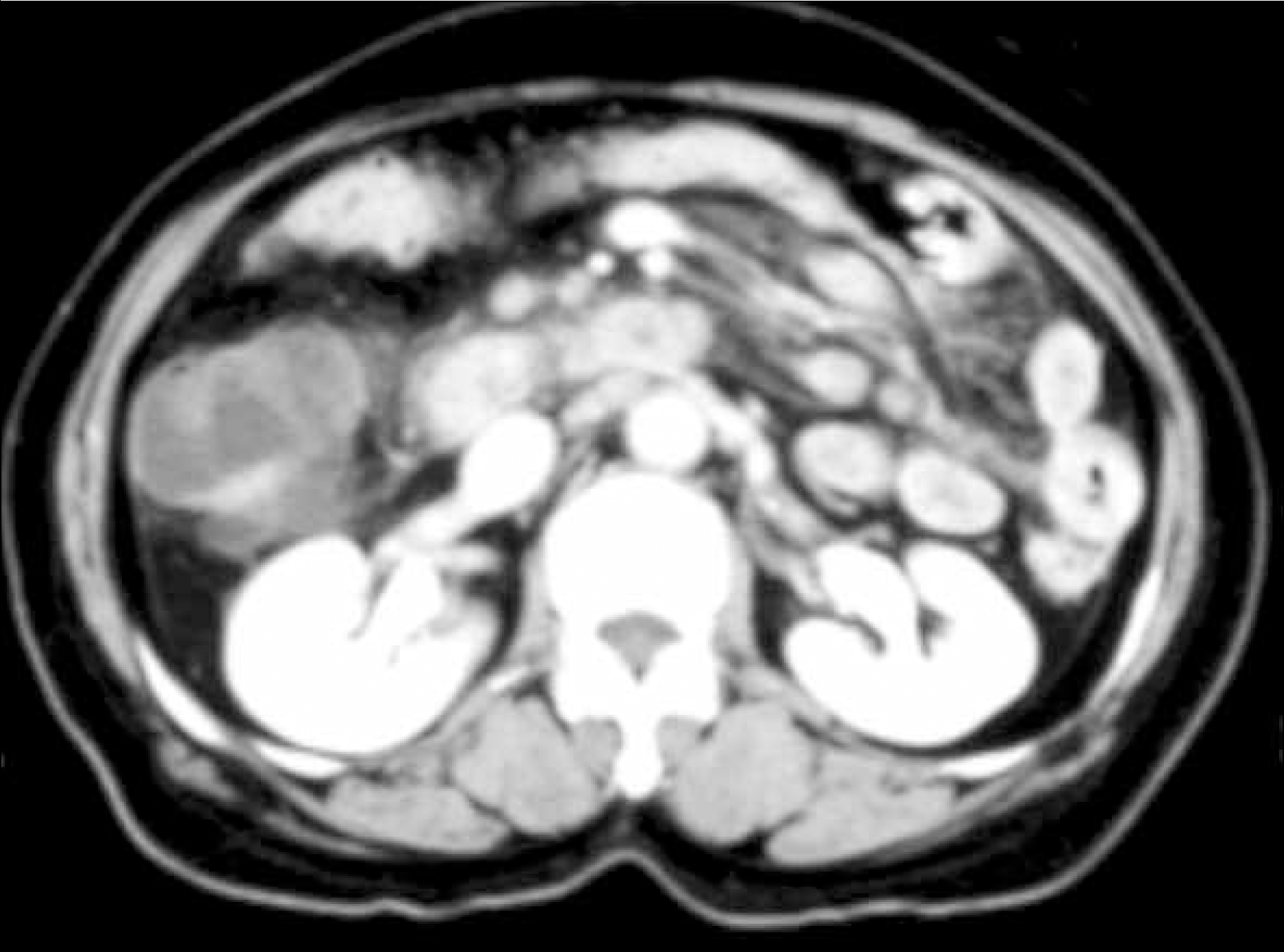
Fig. 3.
Chest computed tomography shows supraclavicular, bilateral paratracheal, subcarinal, right hilar lymph nodes enlargement, bilateral pleural effusion and lung involvement of lymphoma at right lower lobe.
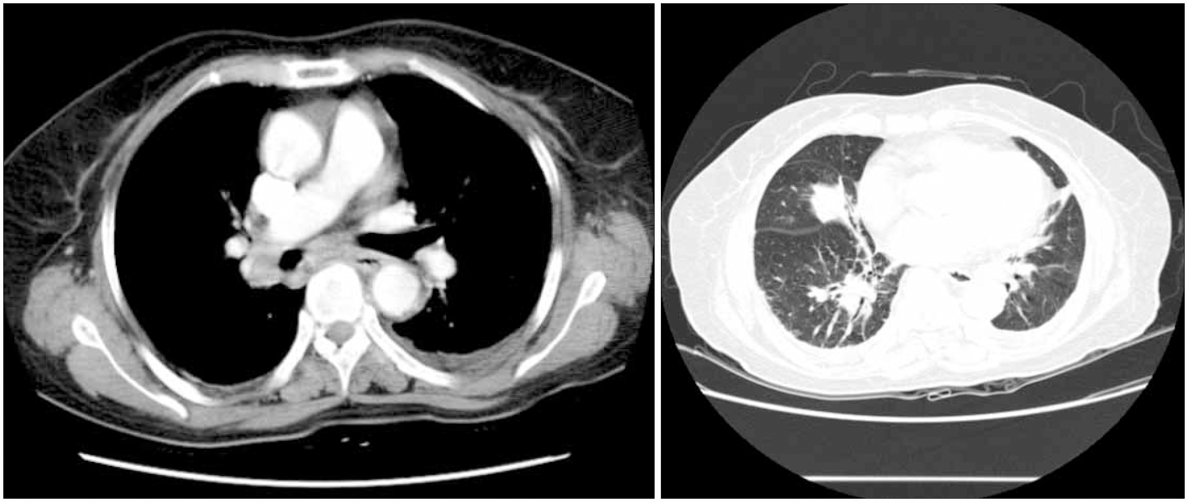
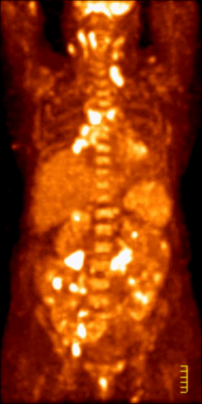
Fig. 4
Whole body positron emission tomography shows lymphoma involvement in right tonsil, both level IIA lymph nodes of the neck, left supraclavicular lymph node, lower lobe of right lung, spleen and ileocecal valve, portahepatis, portocaval, paraaortic, and aortocaval lymph nodes, lymph nodes around ileocolic artery and jejunum, retroperitoneal lymph nodes around right psoas muscle, right common iliac lymph node, right internal and external iliac lymph nodes, and both inguinal lymph nodes.


 PDF
PDF ePub
ePub Citation
 Print
Print


 XML Download