Abstract
Littoral cell angioma is a recently described vascular tumor of spleen with an unknown etiology. We present a case in a 74-year-old woman with severe anemia, thrombocytopenia and palpable splenomegaly. The computed tomography of abdomen showed well-defined multiple nodules in spleen. Splenectomy was performed and the histological and immunohistochemical features of splenic tumor were consistent with a littoral cell angioma. The patients had persistent anemia and thrombocytopenia after splenectomy. We suggest hepatic destruction or other autoimmune mechanism contributes persistent bicytopenia. This case illustrated the refractory bicytopenia combined with littoral cell angioma.
Fig. 1
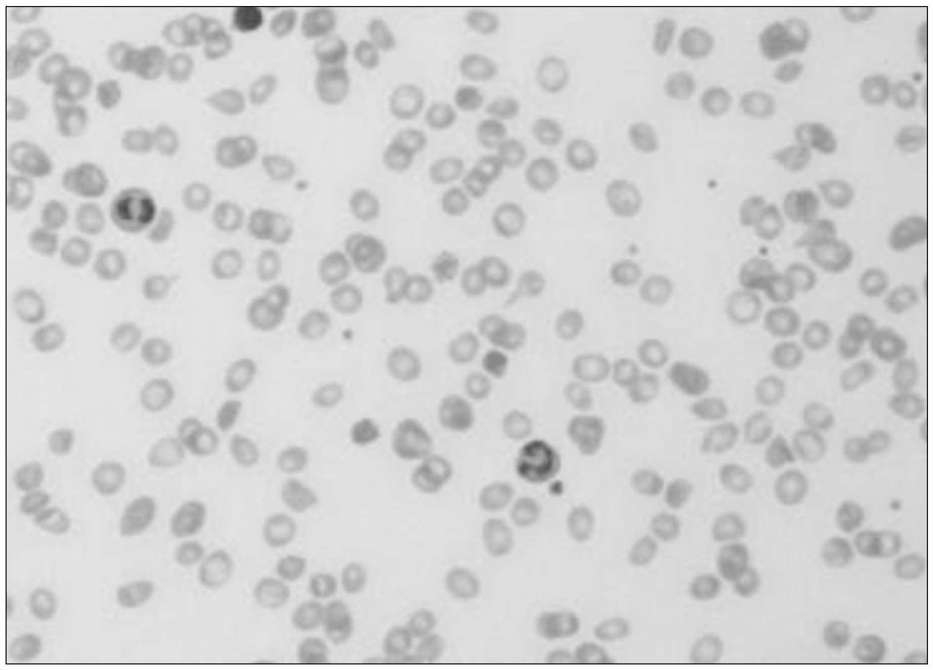
Peripheral blood smear showed macrocytic anemia and thrombocytopenia. Reticulocyte, tear-drop cell and stomatocyte were occasionally seen.

Fig. 2
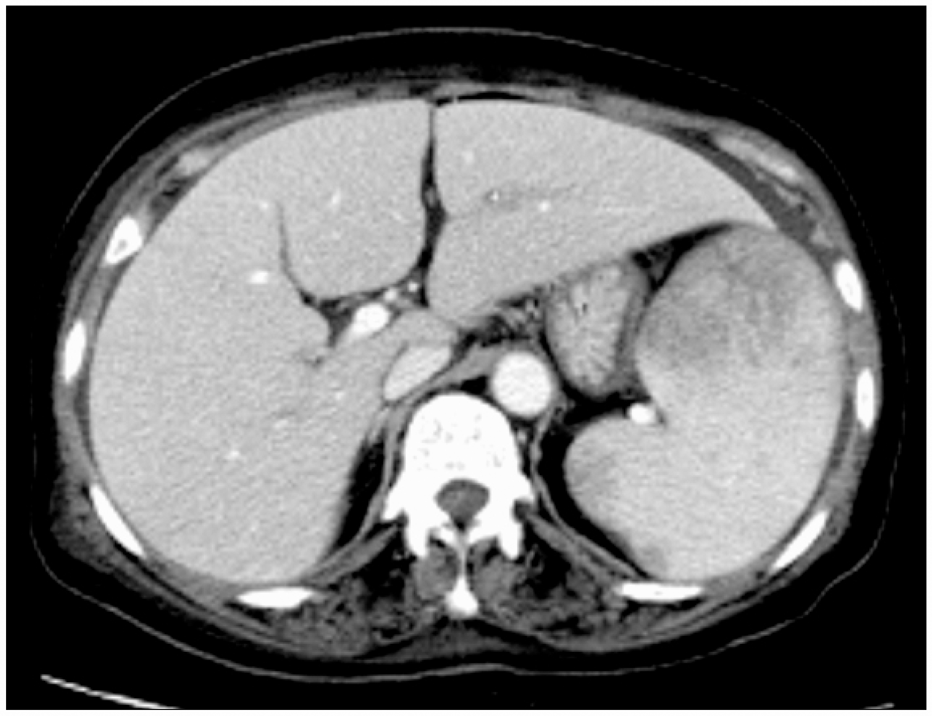
Computed tomography scan demonstrated hema-tosplenomegaly and several hypodense areas of spleen.

Fig. 3
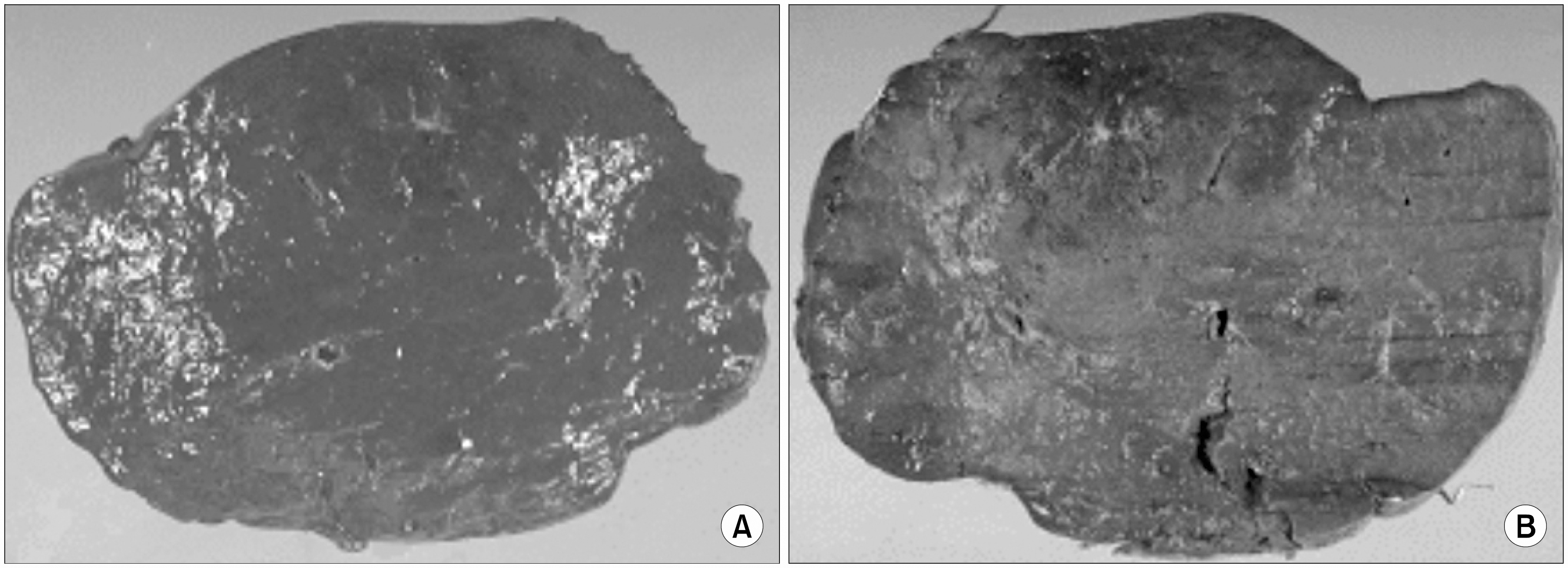
Grossly, multiple nodules intervening spleen parenchyma were found. The spleen was measuring 490g in weight and 17.0×12.0×6.0cm in dimensions. It was entirely replaced by tumor mass (A). The cut surface of the tumor was multinodular and dark brown in color. It was soft and spongy-like in consistency (B).


 PDF
PDF ePub
ePub Citation
 Print
Print


 XML Download