Abstract
Acute lymphoblastic leukemia with maturation (ALLm) has different disease characteristics that does typical ALL. ALLm is characterized by an increased number of mature appearing leukemic cells (>20% of ANCs in the BM) having differentiation beyond the prolymphocyte stage according to light microscopic (LM) examination. It also has a worse prognosis than typical ALL. We have recently experienced a case of ALLm and we report on this case along with a literature review. A 36 year old patient showed lymphoblasts and mature appearing leukemic cells that were counted up to 15.8% and 23.0%, respectively, of the WBCs on bone marrow examination. Despite their mature appearance, these cells showed positivity for Tdt, CD10, CD19 and HLA-DR on the immunophenotypic study. Differentiating the mature-appearing leukemic cells from the hematogones or mature lymphocytes is difficult, and only through immuno-phenotypic examination is it possible to discriminate ALLm from typical ALL. We suggest performing a leukemic marker study that includes CD38 to effectively differentiate mature appearing leukemic cells from hematogones, especially for the follow up of leukemia with mature appearing cells.
Go to : 
REFERENCES
1). Kim MS., Lee SO., Lim YG, et al. Argyrophilic nucleolar organizer regions in acute lymphoblastic leukemia. Korean J Clin Pathol. 1999. 19:479–85.
2). Kim Y., Kang CS., Lee EJ, et al. Acute lymphoblastic leukemia with maturation-a new entity with clinical significance. Leukemia. 1998. 12:875–81.

3). Bloomfield CD., Goldman AI., Alimena G, et al. Chromosomal abnormalities identify high-risk and low-risk patients with acute lymphoblastic leukemia. Blood. 1986. 67:415–20.

4). Hann IM., Scarffe JH., Palmer MK., Evans DI., Jones PH. Haemoglobin and prognosis in childhood acute lymphoblastic leukaemia. Arch Dis Chil. 1981. 56:684–6.

5). Thomas X., Le QH., Danaila C., Lheritier V., Ffrench M. Bone marrow biopsy in adult acute lymphoblastic leukemia: morphological characteristics and contribution to the study of prognostic factors. Leuk Res. 2002. 26:909–18.

6). Mckenna RW., Asplund SL., Kroft SH. Immuno-phenotypic analysis of hematogones (B-lymphocyte precursors) and neoplastic lymphoblasts by 4-color flow cytometry. Leuk Lymphoma. 2004. 45:277–85.

7). Mckenna RW., Washington LT., Aquino DB., Picker LJ., Kroft SH. Immunophenotypic analysis of hematogones (B-lymphocyte precursors) in 662 consecutive bone marrow specimens by 4-color flow cytometry. Blood. 2001. 98:2498–507.

8). Boucheix C., David B., Sebban C, et al. Immunop-henotype of adult acute lymphoblastic leukemia, clinical parameters, and outcome: an analysis of a prospective trial including 652 tested patients (LA LA87). French Group on Therapy for Adult Acute Lymphoblastic Leukemia. Blood. 1994. 84:1603–12.
9). Weir EG., Cowan K., LeBeau P., Borowitz MJ. A limited antibody panel can distinguish B-precursor acute lymphoblastic leukemia from normal B precursors with four color flow cytometry: implications for residual disease detection. Leukemia. 1999. 13:558–67.

10). Farahat N., Lens D., Zomas A., Morilla R., Matutes E., Catovsky D. Quantitative flow cytometry can distinguish between normal and leukaemi B-cell precursors. Br J Haematol. 1995. 91:640–6.
12). Hecker S., Sauerbrey A., Volm M. p53 expression and poor prognosis in childhood acute lymphoblastic leukemia. Anticancer Res. 1994. 14:2759–62.
13). Westbrook CA., Hooberman AL., Spino C, et al. Clinical significance of the BCR-ABL fusion gene in adult acute lymphoblastic leukemia: a Cancer and leukemia Group B Study (8762). Blood. 1992. 80:2983–90.

14). Giaccone G., Gazdar AF., Beck H., Zunino F., Cap-ranico G. Multidrug sensitivity phenotype of human lung cancer cells associated with topoisomerase II expression. Cancer Res. 1992. 52:1666–74.
15). Prosperi E., Sala E., Negri C, et al. Topoisomerase II alpha and beta in human tumor cells grown in vitro and in vivo. Anticancer Res. 1992. 12:2093–9.
Go to : 
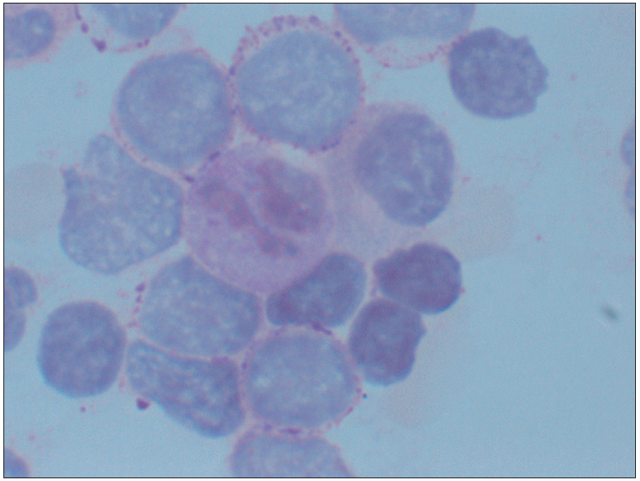
 | Fig. 1Bone marrow aspiration & biopsy section smear. Positive finding displaying dot or coarse granular pattern (Periodic acid-Schiff stain, ×1,000). |
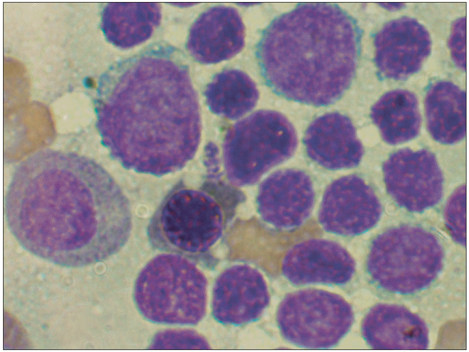
 | Fig. 2Bone marrow aspirate smears at time of first relapse demonstrate numerous mature appearing leukemic cells (Wright stain, ×1,000). |
Table 1.
Progression of patient course according to results of bone marrow study
| Days∗ | Bone marrow study | Cell count | Immunophenotypic study† | |
|---|---|---|---|---|
| Lymphoblasts | Mature appearing leukemic cells | |||
| 1 | ALL L1 | 78.2% | 0% | HLA-DR: 90% |
| CD10: 80% | ||||
| CD19: 90% | ||||
| 32 | Persistence | 7.2% | 0% | ND |
| 71 | Relapse | 15.8% | 23.0% | HLA-DR: 95.1% |
| Tdt: 21.0% | ||||
| CD10: 93.9% | ||||
| CD19: 89.5% | ||||
| 108 | Remission | 2.0% | 0% | HLA-DR: 24.0% |
| Tdt: 55.2% | ||||
| CD10: 0.3% | ||||
| CD19: 0.1% | ||||
| 166 | Relapse | 39.8% | 16.2% | HLA-DR: 86.6% |
| Tdt: 78.5% | ||||
| CD10: 82.3% | ||||
| CD19: 62.3% | ||||
| CD45: 8.3% | ||||
| CD22: 6.6% | ||||
| CD38: 0.4% | ||||
| CD34: 78.2% | ||||
| CD20: 56.4% | ||||


 PDF
PDF ePub
ePub Citation
 Print
Print


 XML Download