Abstract
Intravascular large B-cell lymphoma is rare and generally fatal. It is defined pathologically by neoplastic proliferation of lymphoid cells within the lumens of capillaries, small veins, and arteries with little or no other parenchymal involvement. The diagnosis can be delayed because of the rarity of the disease and the difficulty of detection in imaging studies, and a suspicious clinical observation is warranted to make the correct diagnosis. Early diagnosis is important because delayed treatment could result in a fatal outcome. We have encountered a case of intravascular large B-cell lymphoma involving only the bone marrow. An early diagnosis was made and the patient was treated with combination chemotherapy and rituximab targeting CD20. The patient went into complete remission after the third cycle of chemotherapy and maintained a disease free state up to 6 months.
Go to : 
REFERENCES
1). Stroup RM., Sheibani K., Moncada A., Purdy LJ., Battifora H. Angiotropic (intravascular) large cell lymphoma: a clinicopathologic study of seven cases with unique clinical presentations. Cancer. 1990. 66:1781–8.

2). Pfleger L., Tappeiner J. Zur Kenntnis der system-isierten Endotheliomatose der cutanen Blutgefasse. Hautarzt. 1959. 10:359–63.
3). Han BI., Bae MC., Hong JM., Huh K., Han JH. Intravascular lymphomatosis in central nervous system. J Korean Neurol Assoc. 2001. 19:413–6.
4). Park HD., Park J., Kim SH., Kim J., Kim HT., Kim MH, et al. Intravascular lymphomatosis presenting as demyelinating disease: a case report. J Korean Neurol Assoc. 2004. 22:535–8.
5). Park BB., Kim KH., Son JS, et al. Intravascular lymphomatosis presenting as fever of unknown origin with peripheral polyneuropathy. Infection and Chemotherapy. 2003. 35:355–9.
6). Kim JS., Kim TY., Sun JM, et al. Intracranial relapses of intravascular large B-cell lymphoma after completion of CHOP chemotherapy. Korean J Heamatol. 2004. 39:177–81.
7). Kim CS., Park YT., Park TH., Yoo JH., Kim KJ. A case of intravascular large B cell lymphoma. Korean J Dermatol. 2004. 42:223–5.
8). Lee SI., Kim WS., Lee J, et al. Two cases of intravascular lymphomatosis. Korean J Heamatol. 2002. 37:138–42.
9). Park SJ., Bae SS., Cheon EM, et al. A case of pulmonary intravascular lymphomatosis. Tuberc Repirato-ry Dis. 1997. 44:1390–5.

10). Suh CH., Kim SK., Shin DH., Chung KY. Intravascular lymphomatosis of the T cell type presenting as interstitial lung disease-a case report. J Korean Med Sci. 1997. 12:457–60.

11). Ponzoni M., Arrigoni G., Gould VE, et al. Lack of CD29 (beta1 integrin) and CD54 (ICAM-1) adhesion molecules in intravascular lymphomatosis. Hum Pa-thol. 2000. 31:220–6.
12). Cheng FY., Tsui WM., Yeung WT., Ip LS., Ng CS. Intravascular lymphomatosis: a case presenting with encephalomyelitis and reactive haemophagocytic syndrome diagnosed by renal biopsy. Histopathology. 1997. 31:552–4.

Go to : 
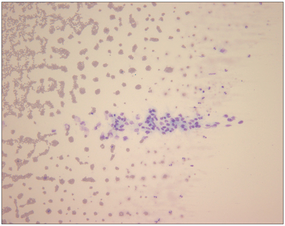
 | Fig. 1Peripheral blood smear finding. Cluster of atypical large cells with convoluted nuclei, coarse chromatin, occasional nucleoli and some cytoplasmic vacuoles in the peripheral blood smear (Wright stain, ×100). |
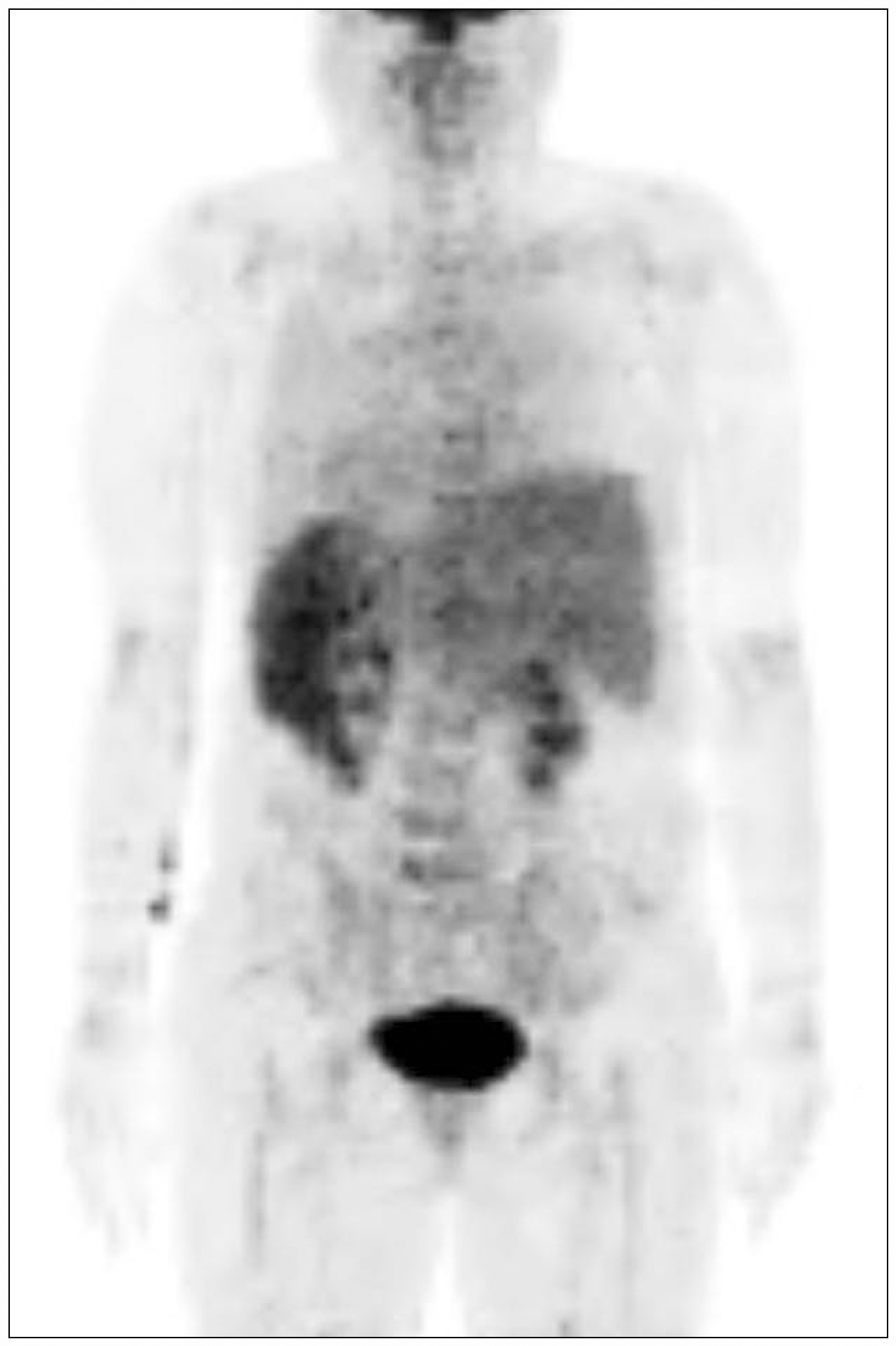
 | Fig. 2PET-CT finding. Diffuse hypermetabolic infiltration in the bone marrow of spine suggesting FDG-avid malignancy. |
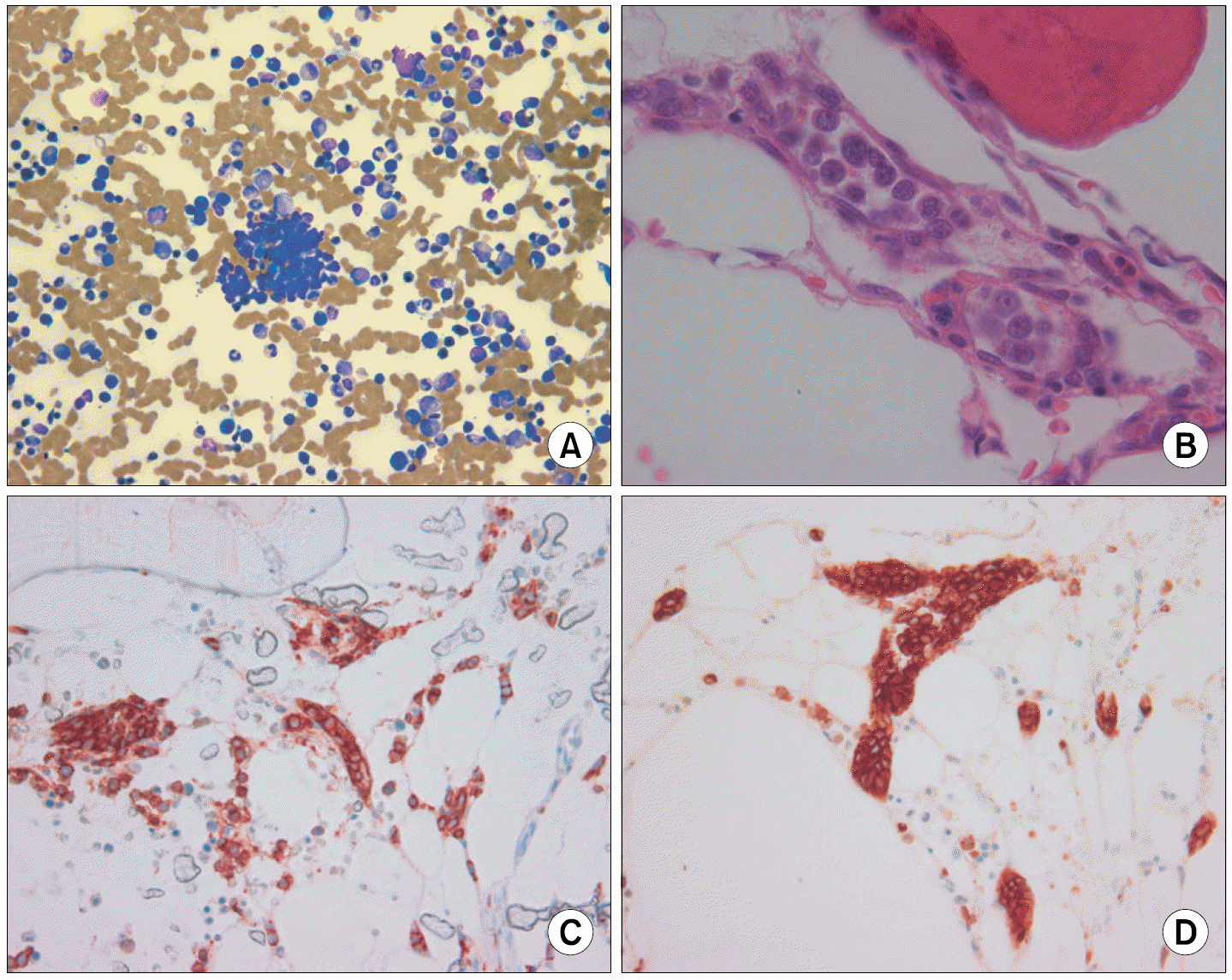
 | Fig. 3Bone marrow biopsy. (A) Aspiration smear: tumor cell cluster in the bone marrow smear (Wright stain, ×200). (B) Biopsy section: blood vessels filled with tumor cells which have irregular large nuclei and prominent nu-cleol in the bone marrow biopsy (H&E stain, ×400). (C) CD45 (LCA) stain: immunohistochemically, tumor cells show strong positivity for the leukocyte common antigen (CD45) (×400). (D) CD20 stain: immunohistochemically, tunor cells show strong positivity for the B-cell marker (CD20) (×400). |


 PDF
PDF ePub
ePub Citation
 Print
Print


 XML Download