Abstract
Although indwelling central venous catheters guarantee a reliable vascular access, and are essential for the management of children undergoing anticancer chemotherapy or stem cell transplantation, these catheters may cause serious mechanical, infectious and thrombotic complications. Central venous catheter-related thrombosis is one of the most important complications that may interfere with the course of treatment. A number of regimens utilizing urokinase have been used but the optimum management of this common problem remains undetermined. We report an 8 year-old boy, who had catheter-related atrial thrombus treated successfully with urokinase. A short course treatment with the use of low-dose urokinase was feasible for the thrombolysis of catheter-related right atrial thrombus in this boy diagnosed with neuroblastoma and undergoing high-dose chemotherapy with autologous peripheral blood stem cell rescue. This treatment was not associated with bleeding.
Go to : 
REFERENCES
1). Fratino G., Molinari AC., Parodi S, et al. Central venous catheter-related complications in children with oncological/hematological diseases: an observational study of 418 devices. Ann Oncol. 2005. 16:648–54.

2). Fratino G., Mazzola C., Buffa P, et al. Mechanical complications related to indwelling central venous catheter in pediatric hematology/oncology patients. Pediatr Hematol Oncol. 2001. 18:317–24.

3). Cesaro S., Paris M., Corro R, et al. Successful treatment of a catheter-related right atrial thrombosis with recombinant tissue plasminogen activator and heparin. Support Care Cancer. 2002. 10:253–5.

5). Revel-Vilk S. Central venous line-related thrombosis in children. Acta Haematol. 2006. 115:201–6.

6). Chan AK., Deveber G., Monagle P., Brooker LA., Massicotte PM. Venous thrombosis in children. J Thromb Heamost. 2003. 1:1443–55.

7). Massicotte MP., Dix D., Monagle P., Adams M., Andrew M. Central venous catheter related thrombosis in children: analysis of the Canadian Registry of Venous Thromboembolic Complications. J Pediatr. 1998. 133:770–6.

8). Nowak-Gottl U., Wermes C., Junker R, et al. Prospective evaluation of the thrombotic risk in children with acute lymphoblastic leukemia carrying the MT-HFR TT 677 genotype, the prothrombin G20210A variant, and further prothrombotic risk factors. Blood. 1999. 93:1595–9.
9). Basford TJ., Poenaru D., Silva M. Comparison of delayed complications of central venous catheters placed surgically or radiologically in pediatric oncology patients. J Pediatr Surg. 2003. 38:788–92.

10). Korones DN., Buzzard CJ., Asselin BL., Harris JP. Right atrial thrombi in children with cancer and indwelling catheters. J Pediatr. 1996. 128:841–6.

11). Ruud E., Holmstrom H., Natvig S., Wesenberg F. Prevalence of thrombophilia and central venous catheter-associated neck vein thrombosis in children with cancer - a prospective study. Med Pediatr Oncol. 2002. 38:405–10.
12). Male C., Chait P., Andrew M, et al. Central venous line-related thrombosis in children: association with central venous line location and insertion technique. Blood. 2003. 101:4273–8.

13). Randolph AG., Cook DJ., Gonzales CA., Andrew M. Benefit of heparin in central venous and pulmonary artery catheters: a meta-analysis of randomized controlled trials. Chest. 1998. 113:165–71.
Go to : 
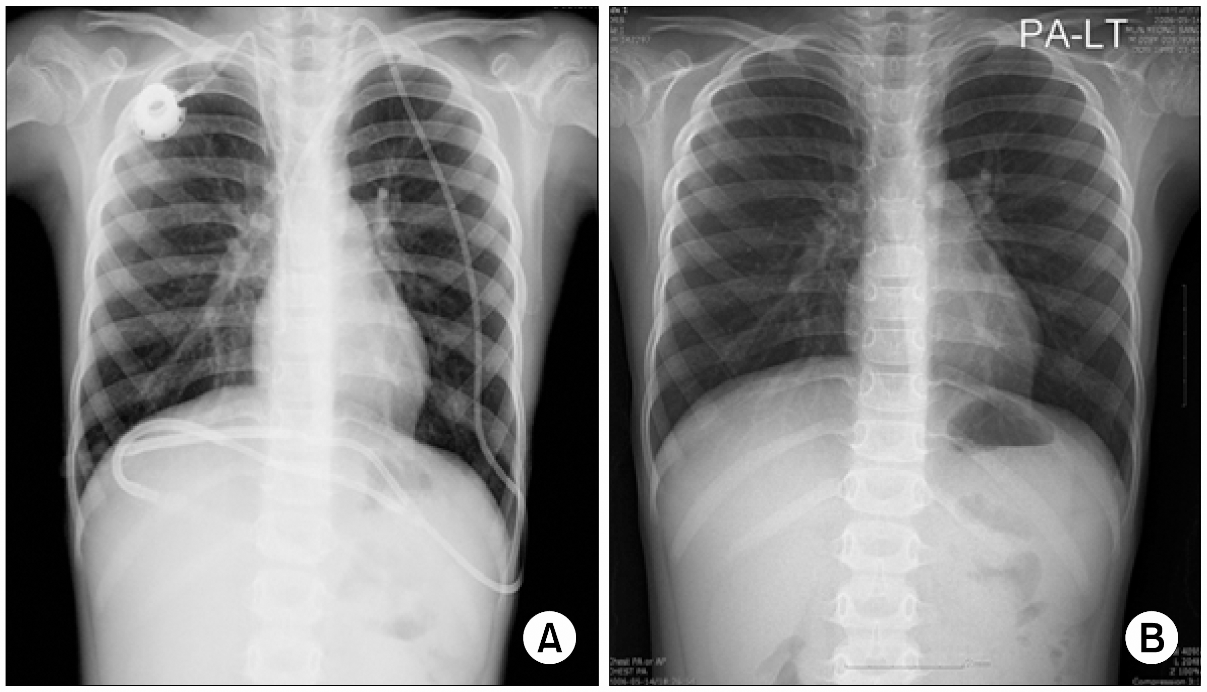
 | Fig. 1Chest PA. (A) Chest PA at the time of diagnosis showed no definite abnormality. Che-moport tip was located in the superior vena cava and Hickman catheter was located in the right atrium. (B) After thrombolytic therapy, che-moport and Hickman catheter were removed state and otherwise was unremarkable. |
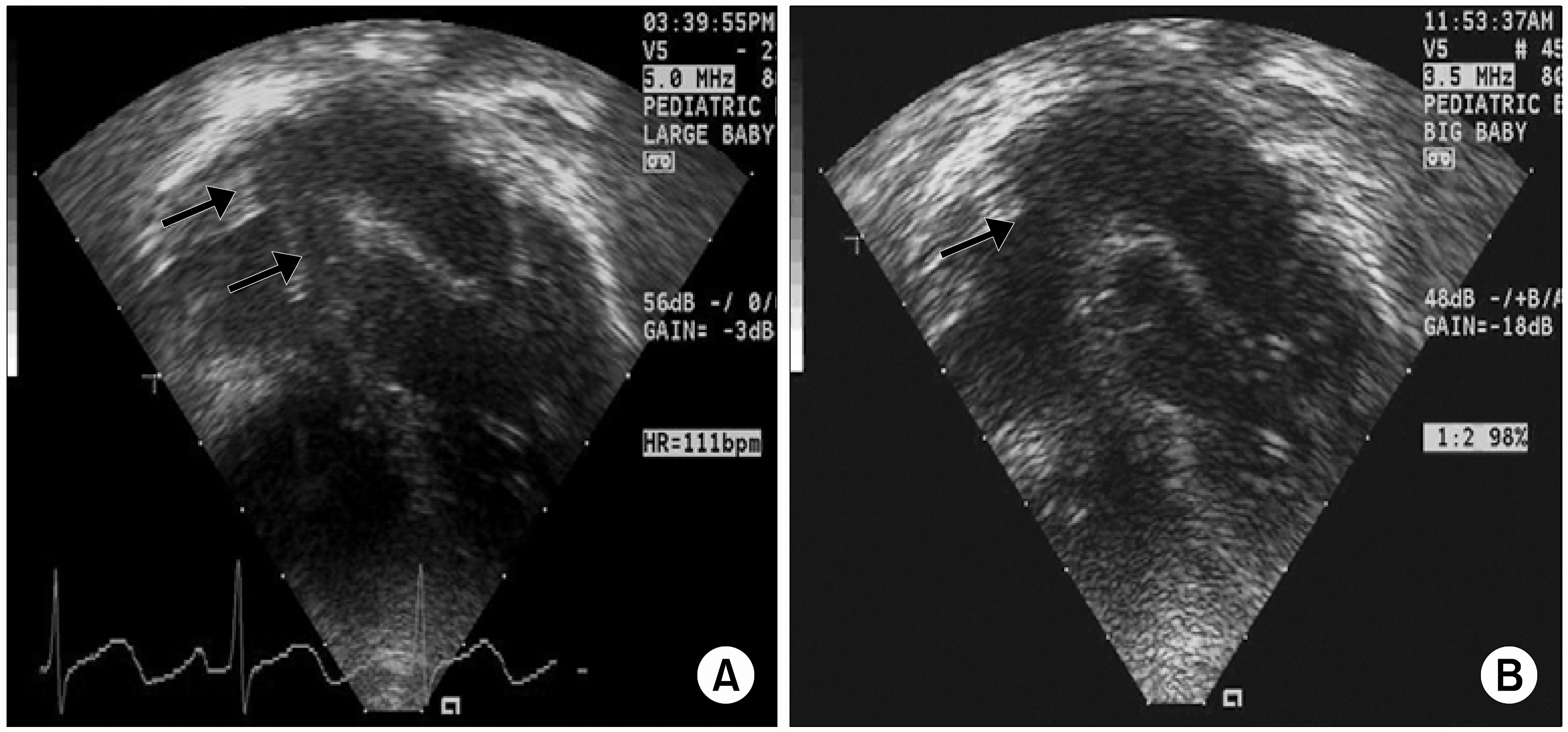
 | Fig. 2Echocardiogram. (A) The echocardiogram at the time of diagnosis showed a normal heart function (EF 71%), but the tip of the Hickman catheter was located deep in the right atrium, close to the tricuspid valve leaflets. Moreover, a large atrial thrombus was observed around the tip of the catheter and adhering to the atrial wall (the upper arrow indicates Hickman catheter while the lower shows the thrombus). (B) After thrombolytic treatment for 3 days with strict monitoring, the thrombus was progressly lyzed and, had shrunk to a small residue adhering to the atrial wall (the arrow indicates Hickman catheter). |


 PDF
PDF ePub
ePub Citation
 Print
Print


 XML Download