

 PDF
PDF ePub
ePub Citation
 Print
Print


Abstract
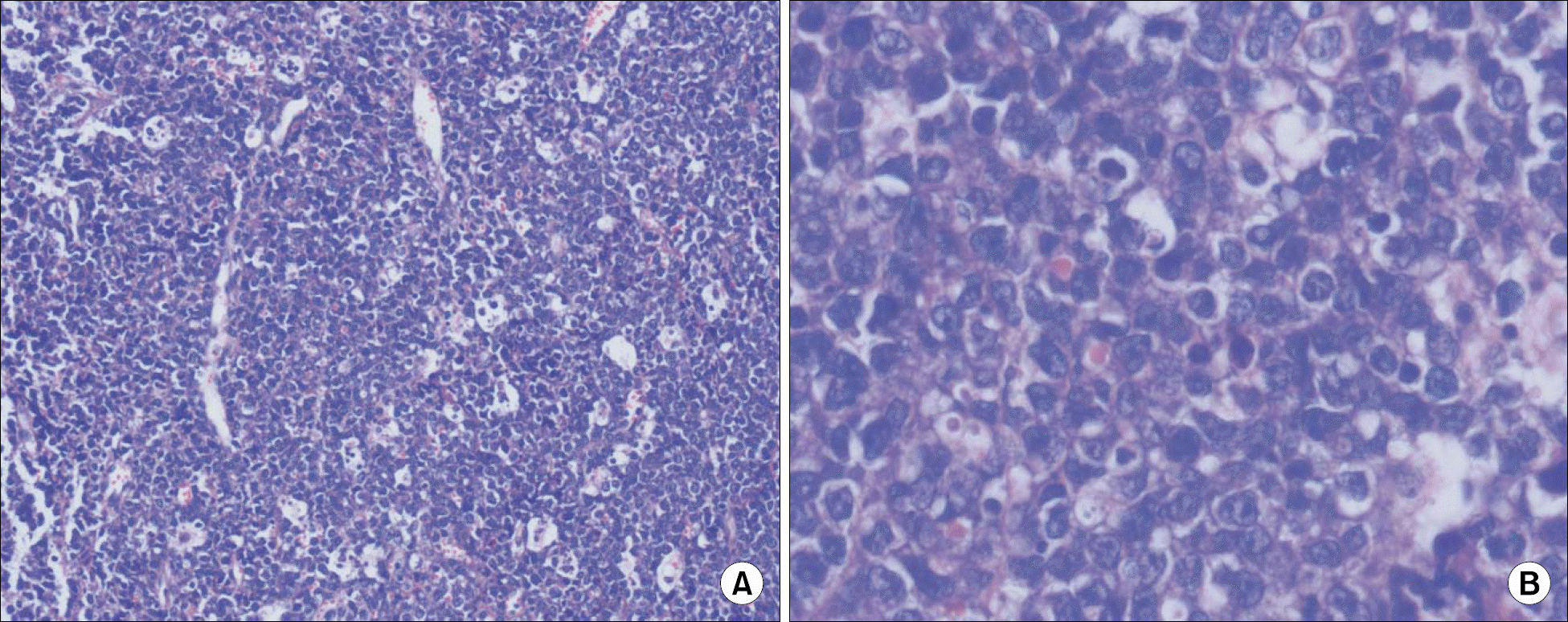
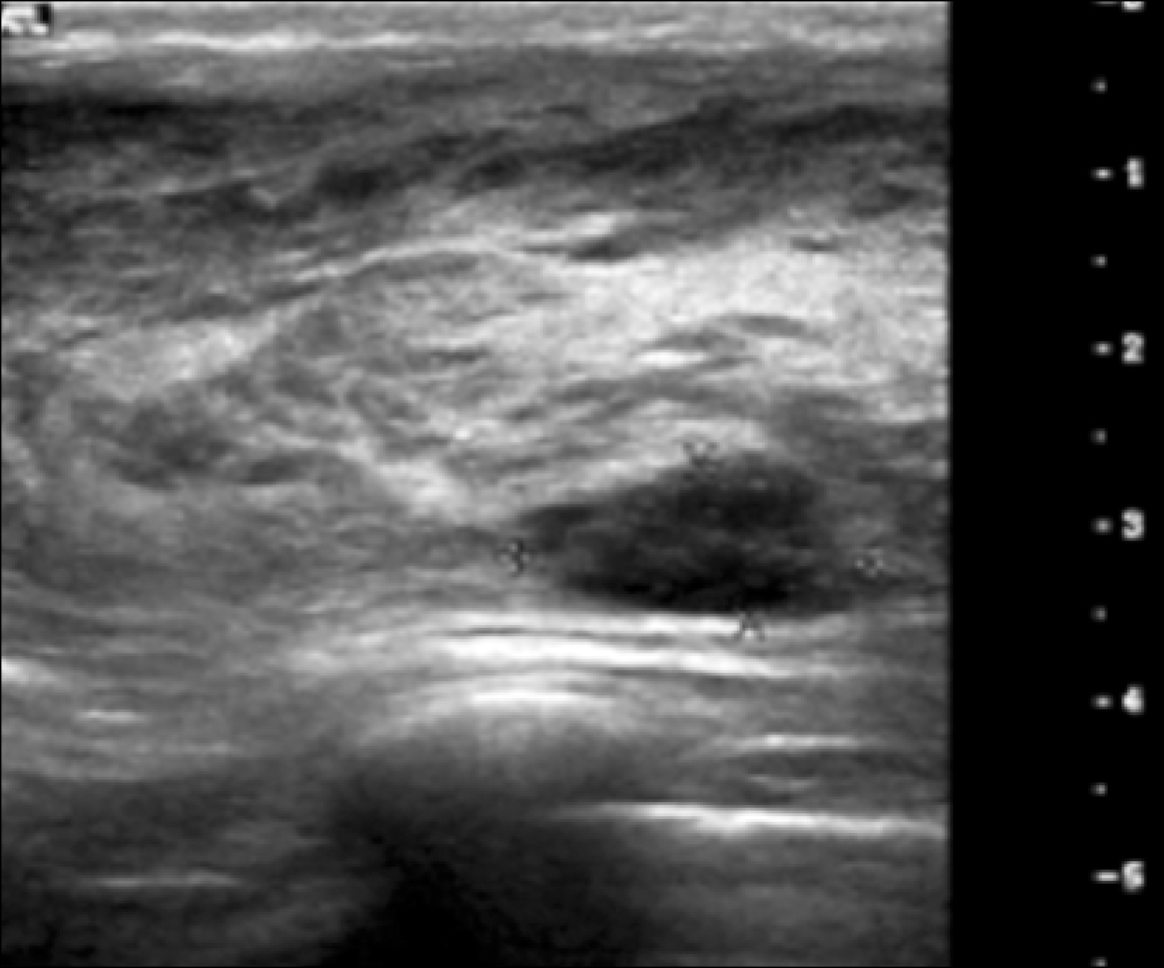
Primary breast lymphoma (PBL) is a rare clinical presentation of localized non-Hodgkin's lymphoma (NHL), and it makes up 0.04~1.1% of all breast tumors and it is 0.38~0.7% of all NHLs. The prognosis and patterns of relapse of PBL are still not clearly defined. The clinical features of PBL are different from those of breast carcinoma and the usual form of lymphoma. These features are a rapidly enlarging breast mass, multiple lesions, the absence of nipple discharge and retraction, and softer axillary lymph nodes as compared to the metastatic lymph nodes from breast carcinoma. B symptoms are unusual in PBL. A 30-year-old pregnant woman was admitted due to dysarthria and right side weakness that she had experienced for 7 days. She had several medical problems: intrauterine pregnancy at 34 weeks, some neurologic deficits and enlargement of both breasts. A biopsy from the breast and a brain magnetic resonance image (MRI) revealed diffuse large B cell lymphoma and multiple brain metastases, respectively. After delivery of a healthy, premature infant by Cesarean section, whole brain radiation therapy and combination chemotherapy (rituximab, cyclophosphamide, adriamycin, vincristine and prednisone) were started. She showed good response to therapy. We report here on this unusual case and we review the related literature.
REFERENCES
1). Jung TH., Chung KS., Kim WM, et al. A case of primary breast lymphoma. Korean J Hematol. 1992. 27:409–13.
2). Gholam D., Bibeau F., El Weshi A., Bosq J., Ribrag V. Primary breast lymphoma. Leuk Lymphoma. 2003. 44:1173–8.

3). Domchek SM., Hecht JL., Fleming MD., Pinkus GS., Canellos GP. Lymphomas of the breast: primary and secondary involvement: primary and secondary involvement. Cancer. 2002. 94:6–13.
4). Ryan GF., Roos DR., Seymour JF. Primary non-Hodg-kin's lymphoma of the breast: retrospective analysis of prognosis and patterns of failure in two Australian centers. Clin Lymphoma Myeloma. 2006. 6:337–41.
5). Yamazaki H., Hanada M., Kitada M, et al. Four cases of central nervous system involvement of breast malignant lymphoma. Jpn J Clin Oncol. 2003. 33:399–403.
6). Wara WM., Bauman GS., Sneed PK., Larson D., Karlsson U. Brain, brain stem, and cerebellum. Perez CA, Brady LW, editors. Principles and practice of radiation oncology. 3rd ed.Philadelphia and New York: Lippincott-Raven;1998. p. 817–20.
7). Grubstein A., Givon-Madhala O., Morgenstern S., Cohen M. Extranodal primary B-cell non-Hodgkin lymphoma of the breast mimicking acute mastitis. J Clin Ultrasound. 2005. 33:140–42.
8). Illes A., Banyai A., Jenei K, et al. Bilateral primary lymphoma of the breasts detected in pregnancy. Orv Hetil. 1996. 137:1315–7.
9). Park YH., Kim SH., Choi SJ., Ryoo BY., Kang YK., Lee SS. Primary malignant lymphoma of the breast: clinicopathological study of nine cases. Leuk Lymphoma. 2004. 45:327–30.
10). Uesato M., Miyazawa Y., Gunji Y., Ochiai T. Primary non-Hodgkin's lymphoma of the breast: report of a case with special reference to 380 cases in the Japanese literature. Breast Cancer. 2005. 12:154–8.
11). Liu MT., Hsieh CY., Wang AY, et al. Primary breast lymphoma: a pooled analysis of prognostic factors and survival in 93 cases. Ann Saudi Med. 2005. 25:288–93.
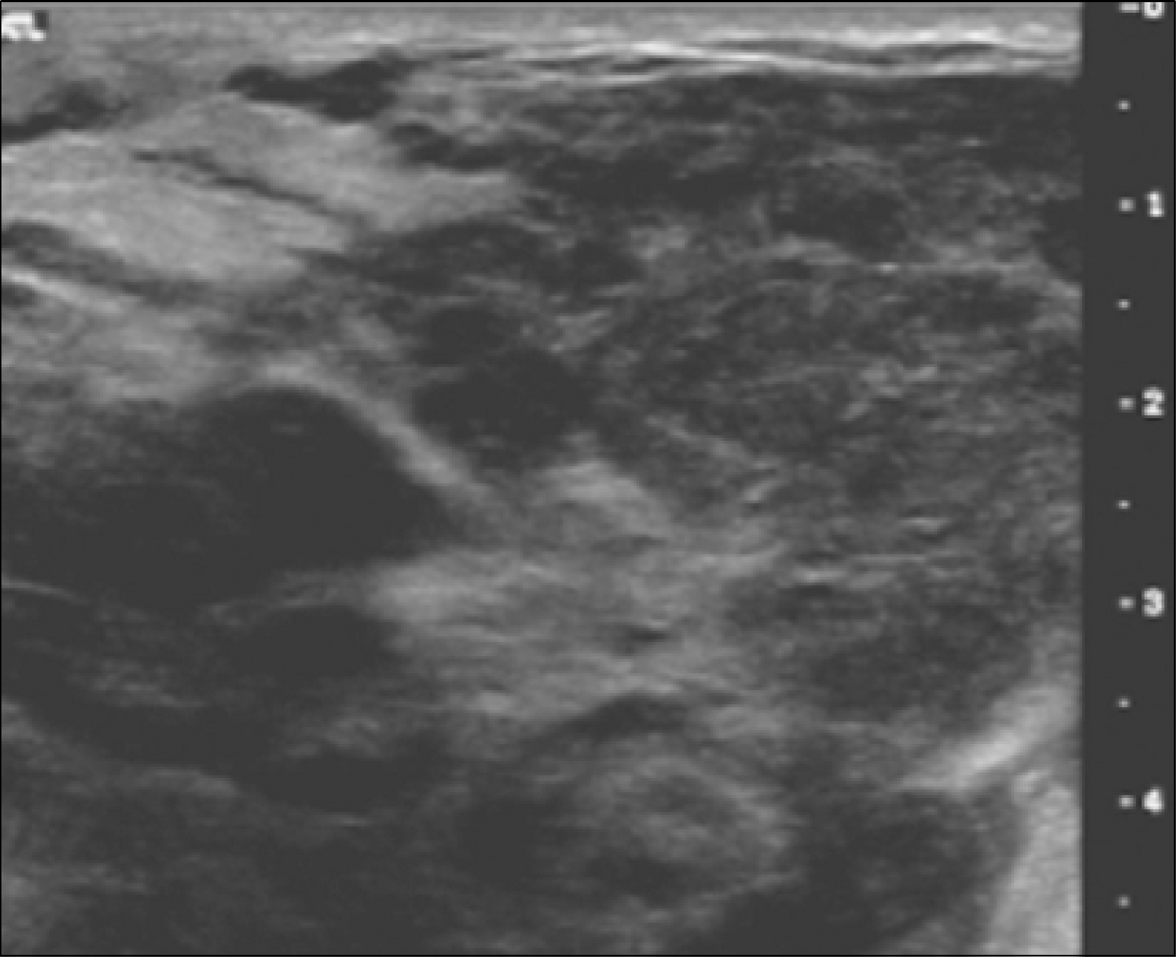
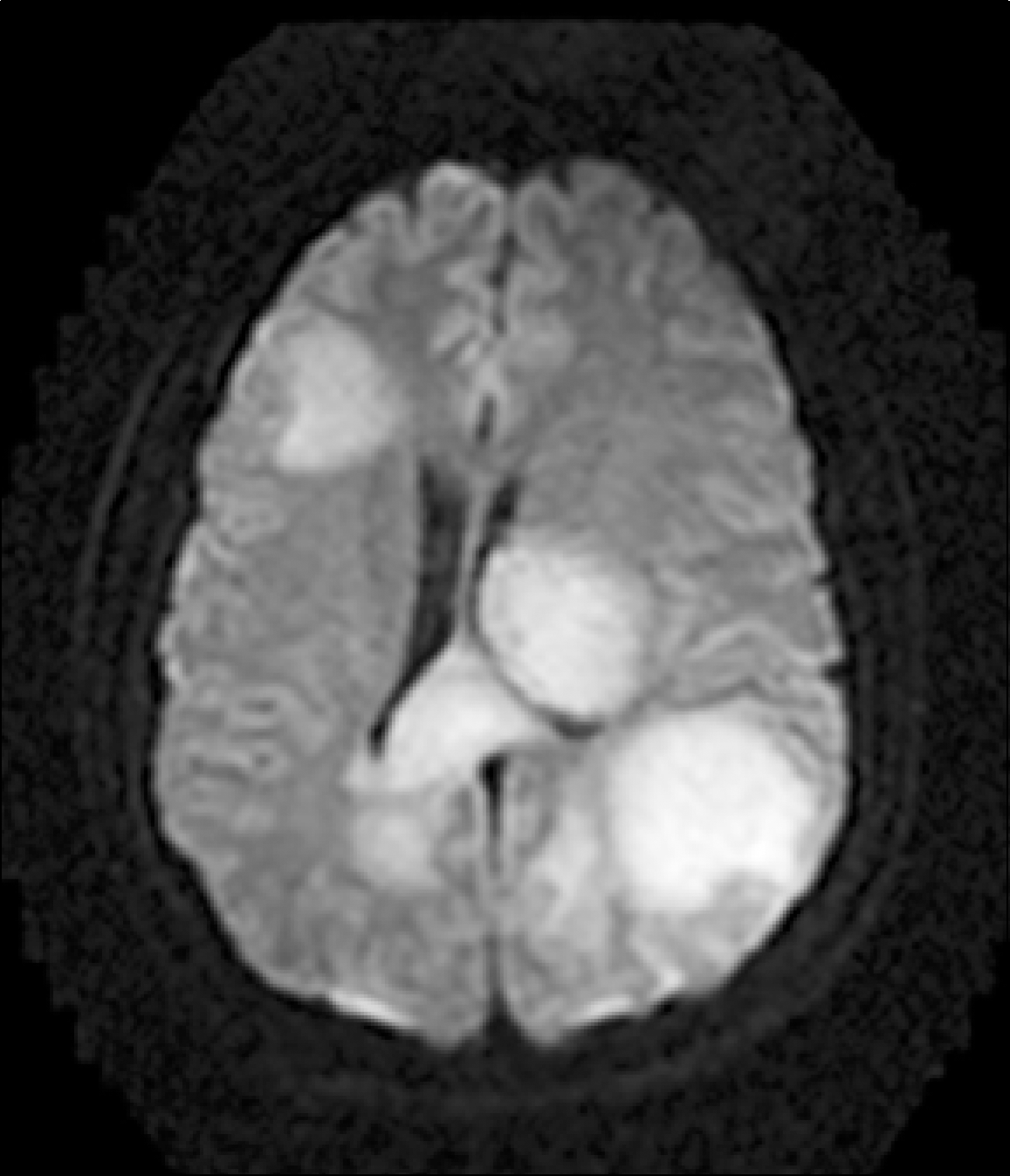
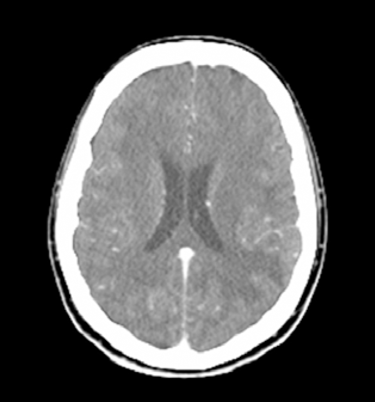
Fig. 2
Brain diffusion MRI shows multiscattered diffuse inflammatory reactive change in near entire brain parenchyme.
 XML Download