Abstract
T-cell prolymphocytic leukemia (T-PLL) is a rare mature post-thymic T-cell malignancy with infiltration to the blood, bone marrow, lymph node, liver, spleen and skin; this disease has a poor prognosis and an aggressive clinical course. We report here on a case of CD56+ T-PLL that was diagnosed by hematological examination, immunophenotyping and molecular studies including determining the TCL1 expression by using reverse-transcriptase polymerase chain reaction (RT-PCR), and direct sequencing of the RT-PCR product.
Go to : 
REFERENCES
1). Jaffe ES., Harris NL., Stein H., Vardiman JW. Pathology and genetic of tumor of hematopoietic and lymphoid tissues. Kleihues P, Sobin L, editors. World Health Organization classification of tumors. vol 3. Lyon: IARC Press;2001.
2). Matutes E., Brito-Babapulle V., Swansbury J, et al. Clinical and laboratory features of 78 cases of T-prolymphocytic leukemia. Blood. 1991. 78:3269–74.

4). Catovsky D., Galetto J., Okos A., Galton DA., Wiltshaw E., Stathopoulos G. Prolymphocytic leukaemia of B and T cell type. Lancet. 1973. 2:232–4.
5). Kojima K., Kobayashi H., Imoto S, et al. 14q11 abnormality and trisomy 8q are not common in Japanese T-cell prolymphocytic leukemia. Int J Hematol. 1998. 68:291–6.
6). Park JE., Kim KM., Kim WY, et al. A Case of Small Cell Variant of T-Cell Prolymphocytic Leukemia. Korean J Hematol. 2005. 40:177–82.

7). Herling M., Khoury JD., Washington LT., Duvic M., Keating MJ., Jones D. A systematic approach to diagnosis of mature T-cell leukemias reveals heterogeneity among WHO categories. Blood. 2004. 104:328–35.

9). Virgilio L., Narducci MG., Isobe M, et al. Identification of the TCL1 gene involved in T-cell malignancies. Proc Natl Acad Sci USA. 1994. 91:12530–4.

Go to : 
 | Fig. 1Morphology of T-PLL cells. Peripheral blood (A) and bone marrow aspirate (B) show many small variants of prolymphocytes (Wright stain, ×1000). Bone marrow section (C) shows infiltration of monotonous leukemic cells and hypercellular marrow for age (50%) (H-E stain, ×100). |
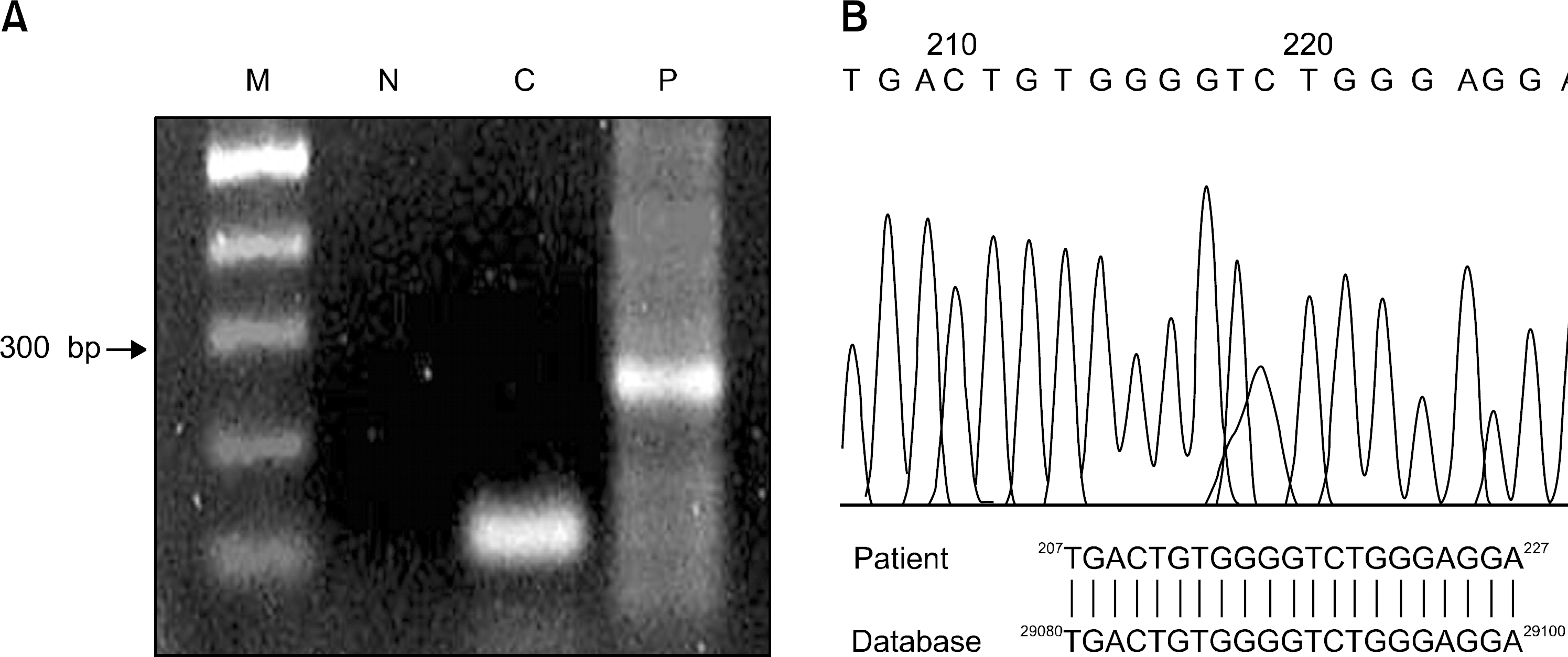
 | Fig. 2Expression of the TCL1 gene. The TCL1 gene was abnormally expressed (A) in patient leukemic cells (P), but not in normal blood cells (N); C, β-actin expression; M, marker (100 bp ladder). Partial sequencing chromatogram (B) of RT-PCR products from the patient and NCBI BLAST search show the identical results for the T-cell receptor alpha delta locus (accession No. 000662.1) that juxtapose TCL1 gene. The TCL1 gene maps at chromosome 14q32.1 and is activated in T cell leukemia and lymphomas by either chromosome translocations or inversions that juxtapose the TCL1 gene to the alpha/delta or the beta locus of the T cell receptor. |


 PDF
PDF ePub
ePub Citation
 Print
Print


 XML Download