Abstract
We experienced a case of pyridoxine refractory hereditary sideroblastic anemia (HSA) in a 4 year-old girl and; therefore, conducted a study of her family. She was admitted to hospital for anemia, which was uncorrected by iron treatment. The peripheral blood smears showed hypochromic microcytic anemia. The results of the biochemical study indicated serum iron of 80μg/dL, TIBC of 275μg/dL and serum ferritin of 67ng/dL. The bone marrow smears showed 80% cellularity, with mild dyserythropoiesis. Many ringed sideroblasts, 45% of normoblasts and an increased amount of hemosiderin particles were observed with iron staining. Despite high-dose pyridoxine therapy, the anemia was not corrected. In the peripheral blood and iron studies conducted on her family members, the mother, maternal aunt and aunt's son showed microcytic hypochromic anemia and normal iron metabolism. Her mother's brother had died of acute myeloid leukemia that had transformed from myelodysplastic syndrome. From a search of the Korean literature, this is the first reported case of HSA with pedigree.
Go to : 
REFERENCES
3). Cooley TB. A severe type of hereditary anemia with elliptocytosis: interesting sequence of splenectomy. Am J Med Sci. 1945; 209:561–8.
4). Amos RJ, Miller AL, Amess JA. Autosomal inheritance of sideroblastic anaemia. Clin Lab Haematol. 1988; 10:347–53.

5). Kasturi J, Basha HM, Smeda SH, Swehli M. Hereditary sideroblastic anaemia in 4 siblings of a Libyan family–autosomal inheritance. Acta Haematol. 1982; 68:321–4.
7). Bottomley SS, May BK, Cox TC, Cotter PD, Bishop DF. Molecular defects of erythroid 5-aminolevulinate synthase in x-linked sideroblastic anemia. J Bioenerg Biomembr. 1995; 27:161–8.

8). Shimada Y, Okuno S, Kawai A, et al. Cloning and chromosomal mapping of a novel abc transporter gene (habc7), a candidate for x-linked sideroblastic anemia with spinocerebellar ataxia. J Hum Genet. 1998; 43:115–22.

9). Takeda Y, Sawada H, Sawai H, et al. Acquired hypochromic and microcytic sideroblastic anaemia responsive to pyridoxine with low value of free erythrocyte protoporphyrin: a possible subgroup of idiopathic acquired sideroblastic anaemia (IASA). Br J Haematol. 1995; 90:207–9.

10). Tuckfield A, Ratnaike S, Hussein S, Metz J. A novel form of hereditary sideroblastic anaemia with macrocytosis. Br J Haematol. 1997; 97:279–85.

11). Jang JH, Kang MC, Sohn CH, Park SJ. A case of hereditary sideroblastic anemia. Korean J hematol. 1984; 19:285–91.

12). Lee GY, Lee YS, Chung IB, et al. The prenatal care and delivery in a pregnant woman complicated by hereditary sideroblastic anemia. Korean J Obstet Gynecol. 2001; 44:1744–60.
13). Roy CN, Andrews NC. Recent advances in disorders of iron metabolism: mutations, mechanisms and modifiers. Hum Mol Genet. 2001; 10:2181–6.

14). Harigae H, Nakajima O, Suwabe N, et al. Aberrant iron accumulation and oxidized status of erythroid-specific delta-aminolevulinate synthase (ALAS2)-deficient definitive erythroblasts. Blood. 2003; 101:1188–93.
16). Cazzola M, May A, Bergamaschi G, Cerani P, Rosti V, Bishop DF. Familial-skewed x-chromosome inactivation as a predisposing factor for late-onset x-linked sideroblastic anemia in carrier females. Blood. 2000; 96:4363–5.

17). Horrigan DL, Harris JW. Pyridoxine-responsive anemia: analysis of 62 cases. Adv Intern Med. 1964; 12:103–74.
18). Hall R, Losowsky MS. The distribution of erythroblast iron in sideroblastic anaemias. Br J Haematol. 1966; 12:334–40.

19). Cotter PD, May A, Li L, et al. Four new mutations in the erythroid-specific 5-aminolevulinate synthase (ALAS2) gene causing x-linked sideroblastic anemia: Increased pyridoxine responsiveness after removal of iron overload by phlebotomy and coinheritance of hereditary hemochromatosis. Blood. 1999; 93:1757–69.

20). Hellstrom-Lindberg E, Ahlgren T, Beguin Y, et al. Treatment of anemia in myelodysplastic syndromes with granulocyte colony-stimulating factor plus erythropoietin: results from a randomized phase ii study and longterm follow-up of 71 patients. Blood. 1998; 92:68–75.
21). Urban C, Binder B, Hauer C, Lanzer G. Congenital sideroblastic anemia successfully treated by allogeneic bone marrow transplantation. Bone Marrow Transplant. 1992; 10:373–5.
Go to : 
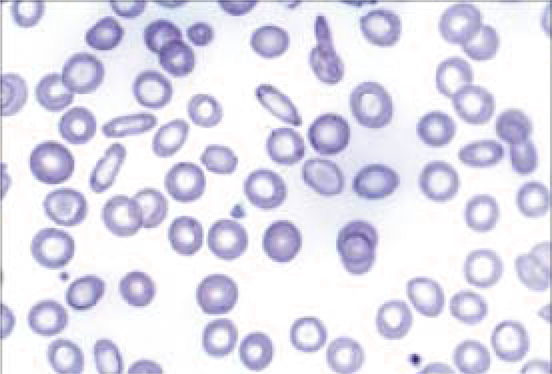
 | Fig. 1.Hypochromic microcytic red blood cells and anisopoikilocytosis (Wright stain in peripheral blood, ×1,000). |
 | Fig. 2.A histiocyte with abundant cytoplasm surrounded by numerous normoblasts (Wright stain in bone marrow aspirate, ×1,000). |
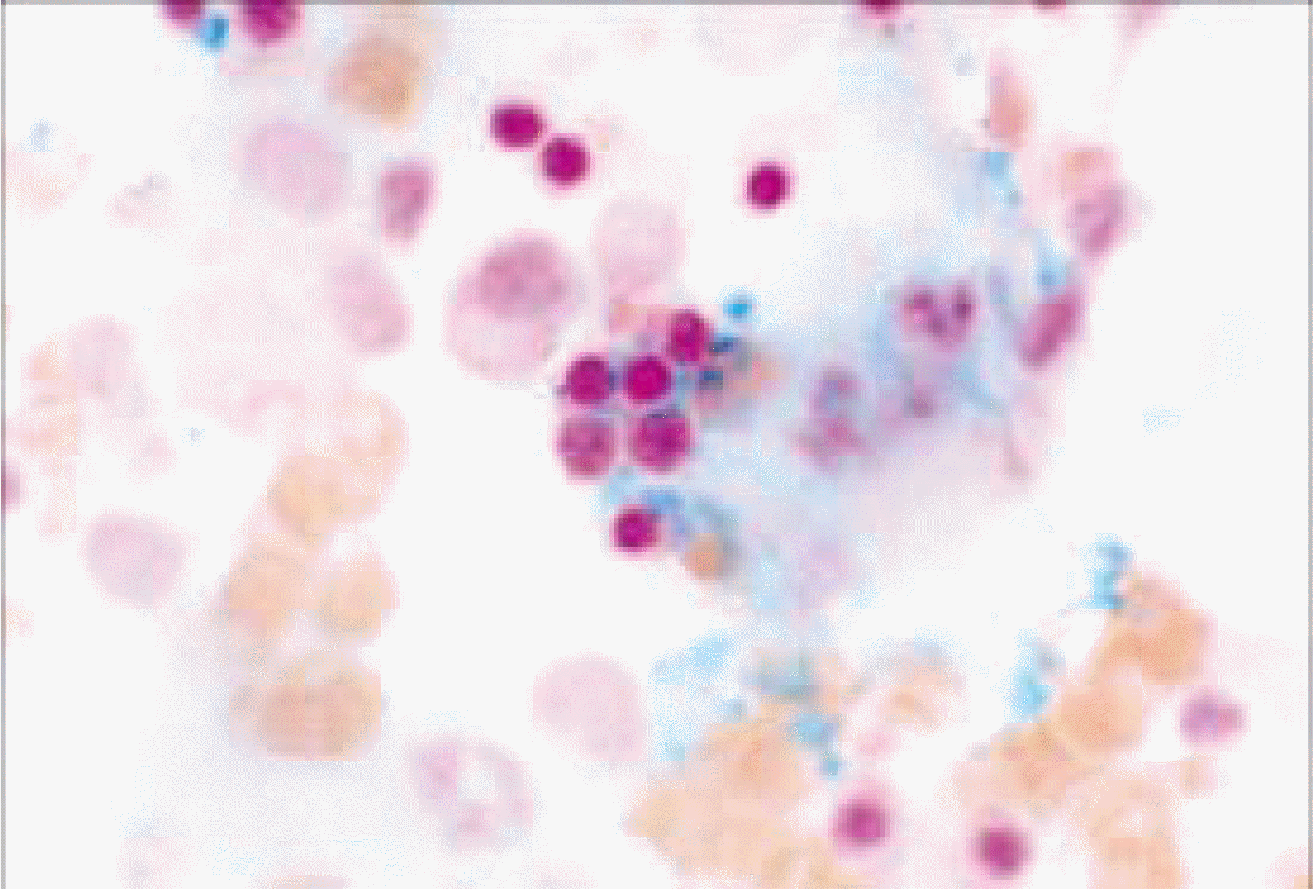
 | Fig. 3.Ringed sideroblasts with minute hemosiderin particles arranged in the perinuclear zone (Iron stain in bone marrow aspirate, ×1,000). |
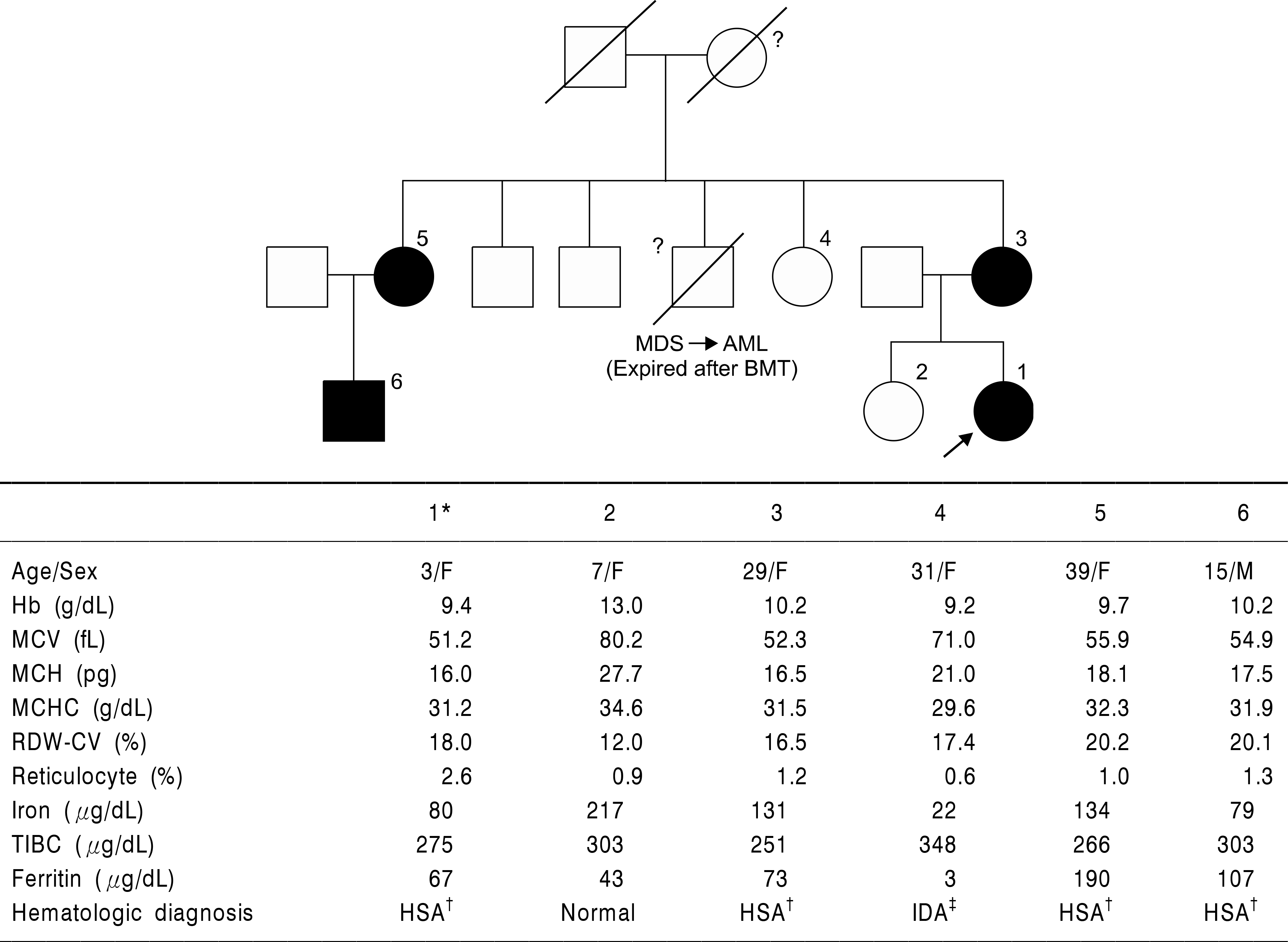
 | Fig. 4.Pedigree of the patient family and hematologic test results of family members. Circles denote female family members; squares, male family members; fulled circle or square, HSA cases; and symbols with diagonal lines, deceased members. The patient of this case is denoted by an arrow. Mother's brother of patient which died due to acute leukemia transformed †Hereditary sideroblastic from myelodysplastic syndrome is denoted by symbol of question. ∗The patient of this case, anemia, ‡Iron deficient anemia. |


 PDF
PDF ePub
ePub Citation
 Print
Print


 XML Download