

 PDF
PDF ePub
ePub Citation
 Print
Print


Abstract
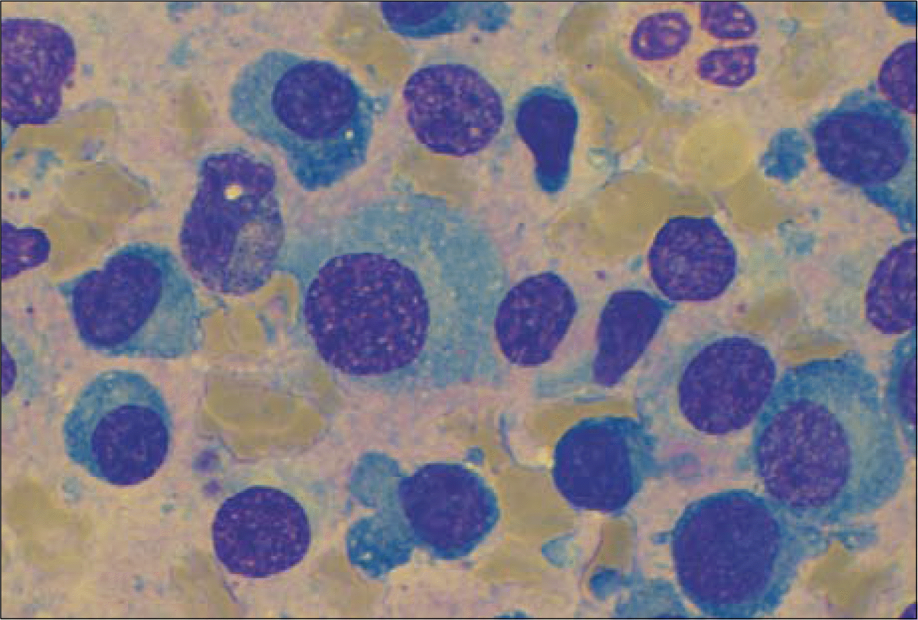
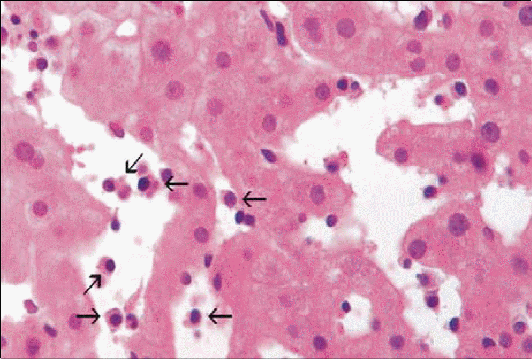
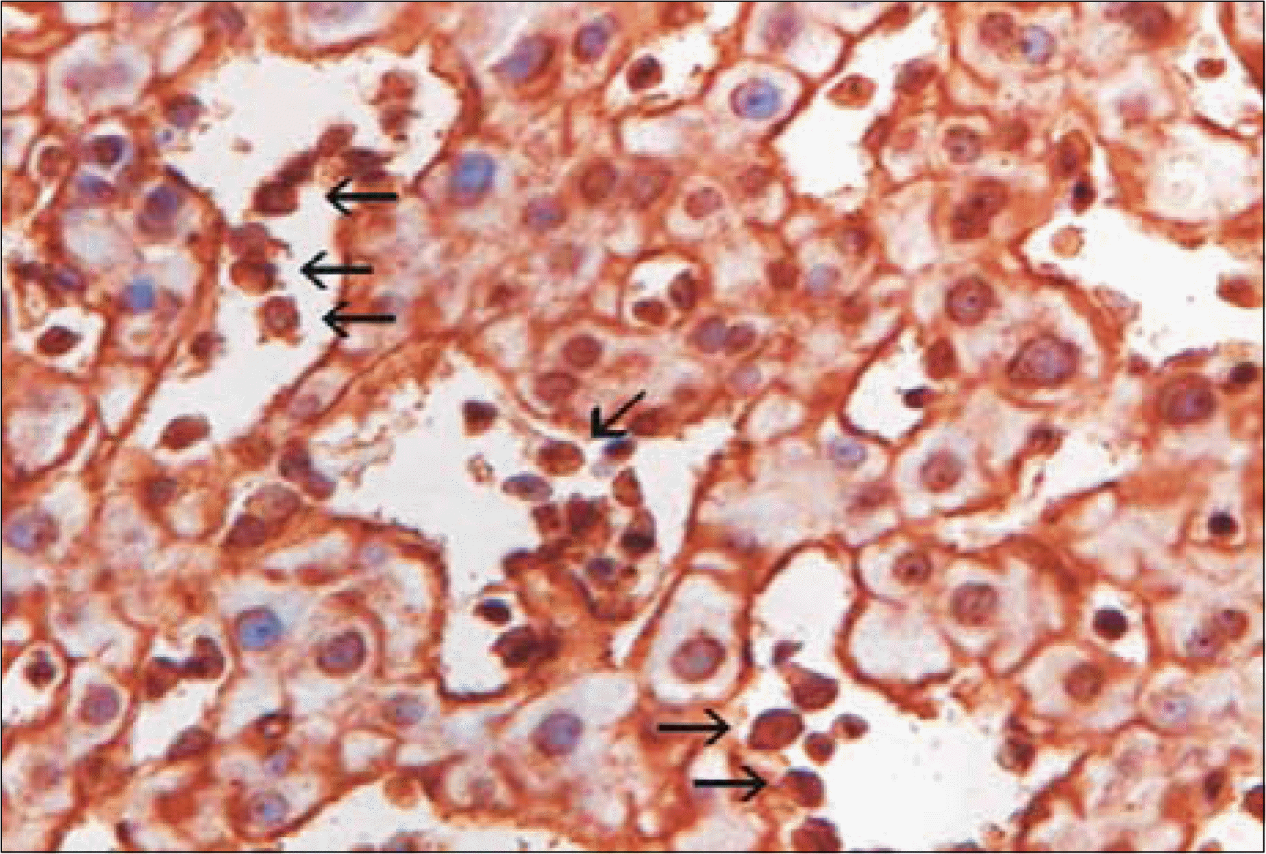
Extraosseous manifestations are found in less than 5% of the patients with multiple myeloma. We reported here on a rare case of multiple myeloma presenting as non-obstructive jaundice due to diffuse plasma cell infiltration of the liver. A 70-year-old man was referred to our hospital because of general weakness, weight loss, jaundice, anemia and proteinuria. The laboratory studies showed: hemoglobin 8.5g/dL, calcium 10.3mg/dL, creatinine 1.3mg/dL, AST 41IU/L, ALT 26IU/L, alkaline phosphatase 304IU/L, total bilirubin 4.0mg/dL, direct bilirubin 2.3mg/dL and 24 hour urinary protein 1,120mg. The serologic tests for hepatitis B and C virus were negative. The abdominal CT scans were normal. The urinary protein studies revealed a M component of the lamda type light chain. The bone marrow biopsy showed atypical plasma cells, and the liver biopsy showed a diffuse sinusoidal infiltration of plasma cells.
Go to : 
REFERENCES
1). Kapadia SB. Multiple myeloma: a clinicopathologic study of 62 consecutively autopsied cases. Medicine (Baltimore). 1980; 59:380–92.
2). Moulopoulos LA, Granfield CA, Dimopoulos MA, Kim EE, Alexanian R, Libshitz HI. Extraosseous multiple myeloma: imaging features. ARJ. 1993; 161:1083–7.

3). Thomas FB, Clausen KP, Greenberger NJ. Liver disease in multiple myeloma. Arch Intern Med. 1973; 132:195–202.
4). Fischer A, Suhrland M, Vogl SE. Myeloma of the head of the pancreas: a case report. Cancer. 1991; 67:681–3.
5). Michopoulos S, Petraki K, Petraki C, Dimopoulos MA. Light chain deposition disease of the liver without renal involvement in a patient with multiple myeloma related to liver failure and rapid fatal outcome. Digestive Disease and Sciences. 2002; 47:730–4.
6). Yoon YS, Yoo HM, Chon CY, et al. Liver involvement in multiple myelola proven by peritoneoscopy: a case report. Yonsei Med J. 1993; 34:90–7.
7). Pastor E, Perella M, Gomez A, Grau E, Perez A, Escandon J. Multiple myeloma of the liver pre-senting as nonobstructive jaundice. Am J Hematol. 1996; 53:205–6.
8). Arebi N, Patel B, Aqel NM, Pitcher MCL. IgA multiple myeloma presenting as non-obstructive jaundice. Postgrad Med J. 2004; 80:489–90.
9). Stegman R, Alexanian R. Solid tumors in multiple myeloma. Ann Intern Med. 1979; 90:780–2.
Go to :
 XML Download